

Parkinson's disease: leucine-rich repeat kinase 2 and autophagy, intimate enemies
- PMID: 22970411
- PMCID: PMC3437299
- DOI: 10.1155/2012/151039
Parkinson's disease: leucine-rich repeat kinase 2 and autophagy, intimate enemies
Abstract
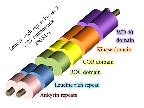
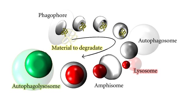
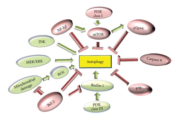
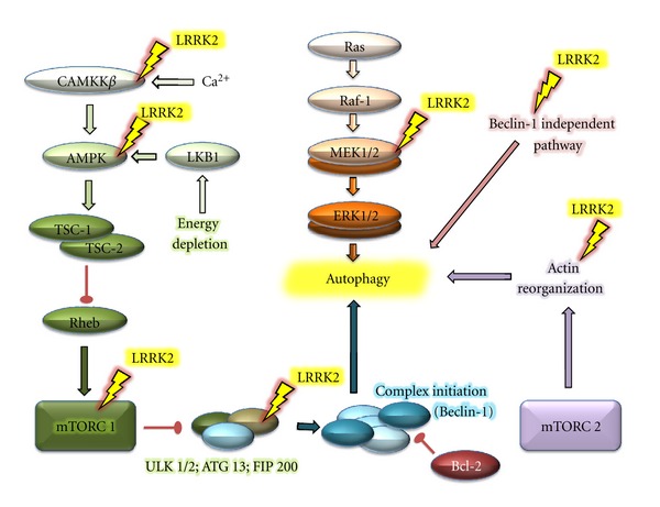
Parkinson's disease is the second common neurodegenerative disorder, after Alzheimer's disease. It is a clinical syndrome characterized by loss of dopamine-generating cells in the substancia nigra, a region of the midbrain. The etiology of Parkinson's disease has long been through to involve both genetic and environmental factors. Mutations in the leucine-rich repeat kinase 2 gene cause late-onset Parkinson's disease with a clinical appearance indistinguishable from Parkinson's disease idiopathic. Autophagy is an intracellular catabolic mechanism whereby a cell recycles or degrades damage proteins and cytoplasmic organelles. This degradative process has been associated with cellular dysfunction in neurodegenerative processes including Parkinson's disease. We discuss the role of leucine-rich repeat kinase 2 in autophagy, and how the deregulations of this degradative mechanism in cells can be implicated in the Parkinson's disease etiology.
Figures
Similar articles
-
Possible involvement of the relationship of LRRK2 and autophagy in Parkinson's disease.Biochem Soc Trans. 2012 Oct;40(5):1129-33. doi: 10.1042/BST20120095. Biochem Soc Trans. 2012. PMID: 22988877 Review.
-
DNL151, DNL201, and BIIB094: experimental agents for the treatment of Parkinson's disease.Expert Opin Investig Drugs. 2023 Jul-Dec;32(9):787-792. doi: 10.1080/13543784.2023.2263357. Epub 2023 Oct 13. Expert Opin Investig Drugs. 2023. PMID: 37755071 Review.
-
LRRK2 at the Crossroad of Aging and Parkinson's Disease.Genes (Basel). 2021 Mar 29;12(4):505. doi: 10.3390/genes12040505. Genes (Basel). 2021. PMID: 33805527 Free PMC article. Review.
-
LRRK2-mediated Rab10 phosphorylation in immune cells from Parkinson's disease patients.Mov Disord. 2019 Mar;34(3):406-415. doi: 10.1002/mds.27601. Epub 2018 Dec 30. Mov Disord. 2019. PMID: 30597610
-
G2019S LRRK2 mutant fibroblasts from Parkinson's disease patients show increased sensitivity to neurotoxin 1-methyl-4-phenylpyridinium dependent of autophagy.Toxicology. 2014 Oct 3;324:1-9. doi: 10.1016/j.tox.2014.07.001. Epub 2014 Jul 10. Toxicology. 2014. PMID: 25017139
Cited by
-
Protein Translation in the Pathogenesis of Parkinson's Disease.Int J Mol Sci. 2024 Feb 18;25(4):2393. doi: 10.3390/ijms25042393. Int J Mol Sci. 2024. PMID: 38397070 Free PMC article. Review.
-
The autophagy-lysosome pathway: a potential target in the chemical and gene therapeutic strategies for Parkinson's disease.Neural Regen Res. 2025 Jan 1;20(1):139-158. doi: 10.4103/NRR.NRR-D-23-01195. Epub 2024 Jan 31. Neural Regen Res. 2025. PMID: 38767483 Free PMC article.
-
Molecular targets for modulating the protein translation vital to proteostasis and neuron degeneration in Parkinson's disease.Transl Neurodegener. 2019 Feb 4;8:6. doi: 10.1186/s40035-019-0145-0. eCollection 2019. Transl Neurodegener. 2019. PMID: 30740222 Free PMC article. Review.
References
-
- Rajput AH. Frequency and cause of Parkinson’s disease. Canadian Journal of Neurological Sciences. 1992;19(1, supplement):103–107. - PubMed
-
- Hughes AJ, Daniel SE, Blankson S, Lees AJ. A clinicopathologic study of 100 cases of Parkinson’s disease. Archives of Neurology. 1993;50(2):140–148. - PubMed
-
- Ehringer H, Hornykiewicz O. Distribution of noradrenaline and dopamine (3-hydroxytyramine) in the human brain and their behavior in diseases of the extrapyramidal system. Parkinsonism & Related Disorders. 1998;4(2):53–57. - PubMed
-
- Lee CS, Schulzer M, Mak EK, et al. Clinical observations on the rate of progression of idiopathic Parkinsonism. Brain. 1994;117, part 3:501–507. - PubMed
-
- Benmoyal-Segal L, Soreq H. Gene-environment interactions in sporadic Parkinson’s disease. Journal of Neurochemistry. 2006;97(6):1740–1755. - PubMed
LinkOut - more resources
Full Text Sources
