Review
. 2012 Nov;117(4):355-69.
doi: 10.3109/03009734.2012.724732. Epub 2012 Sep 13.

Cyclic AMP dynamics in the pancreatic β-cell

Affiliations
Review

Cyclic AMP dynamics in the pancreatic β-cell

Anders Tengholm. Ups J Med Sci. 2012 Nov.

Abstract

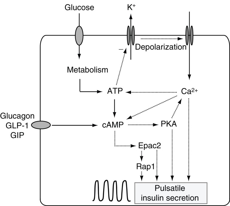
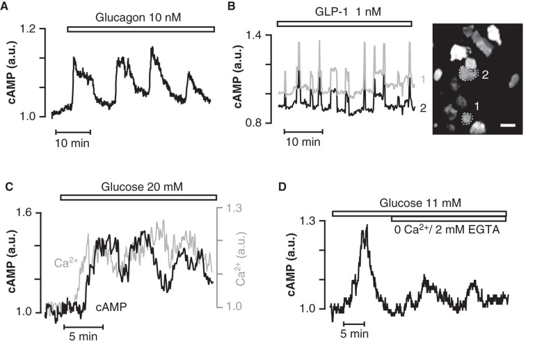
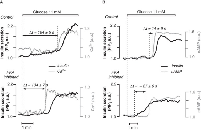
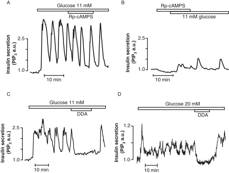
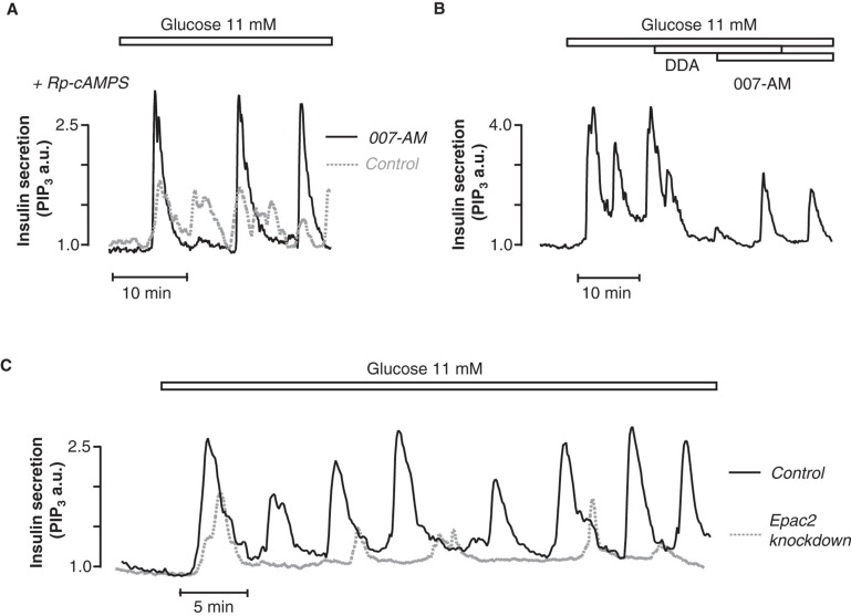
Insulin secretion from pancreatic β-cells is tightly regulated by glucose and other nutrients, hormones, and neural factors. The exocytosis of insulin granules is triggered by an elevation of the cytoplasmic Ca(2+) concentration ([Ca(2+)](i)) and is further amplified by cyclic AMP (cAMP). Cyclic AMP is formed primarily in response to glucoincretin hormones and other G(s)-coupled receptor agonists, but generation of the nucleotide is critical also for an optimal insulin secretory response to glucose. Nutrient and receptor stimuli trigger oscillations of the cAMP concentration in β-cells. The oscillations arise from variations in adenylyl cyclase-mediated cAMP production and phosphodiesterase-mediated degradation, processes controlled by factors like cell metabolism and [Ca(2+)](i). Protein kinase A and the guanine nucleotide exchange factor Epac2 mediate the actions of cAMP in β-cells and operate at multiple levels to promote exocytosis and pulsatile insulin secretion. The cAMP signaling system contains important targets for pharmacological improvement of insulin secretion in type 2 diabetes.


Figures

Figure 1.
Figure 1.
Cyclic AMP signaling in insulin secretion. Schematic drawing of a β-cell and the involvement of cAMP in insulin secretion stimulated by glucose and amplified by hormones. Glucose metabolism generates ATP, which inhibits ATP-sensitive K+ channels and causes voltage-dependent Ca2+ influx. Elevation of [Ca2+]i triggers exocytotic release of insulin granules. ATP also promotes formation of cAMP, which amplifies secretion via Epac2 and protein kinase A (PKA). Activation of Gs-coupled receptors by e.g. glucagon, GLP-1, or GIP leads to cAMP formation and enhancement of insulin release. Cyclic variations in metabolism, [Ca2+]i and cAMP concentration caused by incompletely understood feedback circuits result in pulsatile insulin secretion.
Figure 2.
Figure 2.
Cyclic AMP oscillations in hormone- and glucose-stimulated β-cells. Total internal reflection fluorescence (TIRF) microscopy recordings of the sub-membrane cAMP concentration in mouse β-cells within intact pancreatic islets. A, B: Cyclic AMP oscillations evoked by 10 nM glucagon and 1 nM GLP-1 in β-cells exposed to 3 mM glucose. Oscillations are synchronized among different β-cells within the islet as illustrated by graphs from the numbered cells in the TIRF image (B). C, D: Elevation of the glucose concentration from 3 to 11 or 20 mM evokes co-ordinated oscillations of cAMP and Ca2+ beneath the plasma membrane. The cAMP oscillations are amplified by Ca2+ but are maintained also when Ca2+ entry is prevented (D).
Figure 3.
Figure 3.
Temporal relationship between glucose-induced Ca2+ and cAMP signals and insulin secretion. A: TIRF microscopy recordings of sub-membrane Ca2+ concentration (dotted curves) and the insulin secretory response (solid curves) in MIN6 β-cells show that inhibition of PKA markedly shortens the delay between glucose stimulation and the initial Ca2+ elevation triggering secretion. B: Simultaneous recordings of cAMP (dotted curve) and insulin secretion (solid curve) showing that PKA inhibition shifts the timing such that secretion, which normally follows the amplifying cAMP signal, instead precedes the cAMP elevation.
Figure 4.
Figure 4.
Cyclic AMP dependence of glucose-induced pulsatile insulin secretion. TIRF microscopy recordings of the insulin secretory response from individual MIN6 β-cells. A: The PKA inhibitor Rp-8-CPT-cAMPS (100 µM) barely affects pulsatile insulin secretion triggered by glucose. B: In contrast, if added prior to glucose stimulation, the inhibitor markedly suppresses the subsequent secretory response. C, D: Glucose-induced pulsatile insulin secretion critically depends on cAMP generation as 50 µM of the AC inhibitor dideoxyadenosine (DDA) inhibits secretion in both MIN6 (C), and primary mouse pancreatic β-cells (D).
Figure 5.
Figure 5.
Involvement of Epac in glucose-induced pulsatile insulin secretion. TIRF microscopy recordings of the insulin secretory response from individual MIN6 β-cells. A, B: The Epac-selective cAMP analogue 8-pCPT-2’-O-Me-cAMP-AM (007-AM, 1 µM) restores the magnitude of insulin secretion initiated by glucose in the presence of the PKA inhibitor Rp-8-CPT-cAMPS (A), as well as that of established glucose-induced insulin pulses suppressed by AC inhibition with dideoxyadenosine (DDA) (B). C: Knock-down of Epac2 with siRNA reduces the magnitude of both initial and subsequent pulsatile insulin secretion in response to glucose.

References

    1. Pørksen N. The in vivo regulation of pulsatile insulin secretion. Diabetologia. 2002;45:3–20. - PubMed
    1. Hellman B. Pulsatility of insulin release—a clinically important phenomenon. Ups J Med Sci. 2009;114:193–205. - PMC - PubMed
    1. Schmitz O, Rungby J, Edge L, Juhl CB. On high-frequency insulin oscillations. Ageing Res Rev. 2008;7:301–5. - PubMed
    1. Tengholm A, Gylfe E. Oscillatory control of insulin secretion. Mol Cell Endocrinol. 2009;297:58–72. - PubMed
    1. Gilon P, Ravier MA, Jonas JC, Henquin JC. Control mechanisms of the oscillations of insulin secretion in vitro and in vivo. Diabetes. 2002;51:S144–51. - PubMed

Publication types

LinkOut - more resources