. 2013 May;137(5):668-84.
doi: 10.5858/arpa.2012-0263-RA. Epub 2012 Sep 12.

Diagnosis of lung cancer in small biopsies and cytology: implications of the 2011 International Association for the Study of Lung Cancer/American Thoracic Society/European Respiratory Society classification

Affiliations

Diagnosis of lung cancer in small biopsies and cytology: implications of the 2011 International Association for the Study of Lung Cancer/American Thoracic Society/European Respiratory Society classification

William D Travis et al. Arch Pathol Lab Med. 2013 May.

Abstract

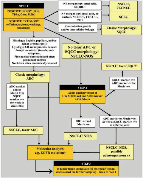
The new International Association for the Study of Lung Cancer/American Thoracic Society/European Respiratory Society lung adenocarcinoma classification provides, for the first time, standardized terminology for lung cancer diagnosis in small biopsies and cytology; this was not primarily addressed by previous World Health Organization classifications. Until recently there have been no therapeutic implications to further classification of NSCLC, so little attention has been given to the distinction of adenocarcinoma and squamous cell carcinoma in small tissue samples. This situation has changed dramatically in recent years with the discovery of several therapeutic options that are available only to patients with adenocarcinoma or NSCLC, not otherwise specified, rather than squamous cell carcinoma. This includes recommendation for use of special stains as an aid to diagnosis, particularly in the setting of poorly differentiated tumors that do not show clear differentiation by routine light microscopy. A limited diagnostic workup is recommended to preserve as much tissue for molecular testing as possible. Most tumors can be classified using a single adenocarcinoma marker (eg, thyroid transcription factor 1 or mucin) and a single squamous marker (eg, p40 or p63). Carcinomas lacking clear differentiation by morphology and special stains are classified as NSCLC, not otherwise specified. Not otherwise specified carcinomas that stain with adenocarcinoma markers are classified as NSCLC, favor adenocarcinoma, and tumors that stain only with squamous markers are classified as NSCLC, favor squamous cell carcinoma. The need for every institution to develop a multidisciplinary tissue management strategy to obtain these small specimens and process them, not only for diagnosis but also for molecular testing and evaluation of markers of resistance to therapy, is emphasized.


Figures

Figure 1
Figure 1
Step 1: When positive biopsies (fiberoptic bronchoscopy [FOB] or transbronchial [TBBx], core, or surgical lung biopsy [SLBx]) or cytology (effusion, aspirate, washings, brushings) show clear adenocarcinoma (ADC) or squamous cell carcinoma (SQCC) morphology, the diagnosis can be firmly established. If there is neuroendocrine morphology, the tumor may be classified as small cell carcinoma (SCLC) or non–small cell lung carcinoma (NSCLC), probably large cell neuroendocrine carcinoma (LCNEC) according to standard criteria. If there is no clear ADC or SQCC morphology, the tumor is regarded as NSCLC, not otherwise specified (NOS). Step 2: NSCLC NOS can be further classified based on (1) immunohistochemical stains, (2) mucin (diastase–periodic acid-Schiff or mucicarmine) stains, or (3) molecular data. If the stains all favor ADC, with positive ADC marker(s) (ie, thyroid transcription factor 1 [TTF-1] and/or mucin positive) and negative SQCC markers, then the tumor is classified as NSCLC, favor ADC. If SQCC markers (ie, p63 and/or cytokeratin [CK] 5/6) are positive with negative ADC markers, the tumor is classified as NSCLC, favor SQCC. If the ADC and SQCC markers are both strongly positive in different populations of tumor cells, the tumor is classified as NSCLC-NOS, with a comment it may represent adenosquamous carcinoma. If all markers are negative, the tumor is classified as NSCLC-NOS. See text for recommendations on NSCLCs with marked pleomorphic and overlapping ADC/SQCC morphology. † Epidermal growth factor receptor (EGFR) mutation testing should be performed in (1) classic ADC; (2) NSCLC, favor ADC; (3) NSCLC-NOS; and (4) NSCLC-NOS, possible adenosquamous carcinoma. In these cases, if EGFR mutation testing is negative, testing for EML4-anaplastic lymphoma kinase (ALK) should be performed. In NSCLC-NOS, if either EGFR mutation or ALK rearrangements are positive, the tumor is more likely to be ADC than SQCC. Step 3: If clinical management requires a more specific diagnosis than NSCLC-NOS, additional biopsies may be indicated. Abbreviations: ca, carcinoma; IHC, immunohistochemistry; NE, neuroendocrine; +, positive; −, negative; +/−, positive or negative; −ve, negative; +ve, positive.
Figure 2
Figure 2
Adenocarcinoma. This small biopsy shows fragments of adenocarcinoma with a papillary configuration (hematoxylin-eosin, original magnification ×40).
Figure 3
Figure 3
Squamous cell carcinoma. This small biopsy shows squamous cell carcinoma with nests of tumor cells that have keratinization and pearls (hematoxylin-eosin, original magnification X20).
Figure 4
Figure 4
Adenocarcinoma, cytology. A, A flat, cohesive sheet of rather uniform-appearing glandular cells is characterized by mild variability in nuclear sizes, inconspicuous nucleoli, very delicate cytoplasm, and a low level of disruption of polarity (nuclear crowding). B, This flat, cohesive sheet of uniform-appearing glandular cells has abundant clear cytoplasm filled with mucin and irregularly arranged nuclei in the “drunken honeycombing” pattern characteristic of invasive mucinous adenocarcinoma. C, A luminal space is surrounded by glandular cells with delicate cytoplasm and clearly malignant and often eccentrically located nuclei, each with a well-developed nucleolus. Note the mitotic figure (Papanicolaou, original magnification X40 [A]; Diff-Quik, original magnification X40 [B and C]).
Figure 5
Figure 5
Squamous cell carcinoma, cytology. A, Many of the tumor cells manifest cytoplasmic keratin as a dense, almost glassy red to orange coloration. Each cell houses a hyperchromatic nucleus, many of which possess jagged outlines. Nonkeratinized neoplastic cells with cyanophilic cytoplasm are also present. B, A flat mosaic sheet of malignant epithelial cells that are characterized by dense (or opaque) cyanophilic cytoplasm. Their nuclei are obviously hyperchromatic with small chromocenters and/or nucleoli. A mitotic figure is present (Papanicolaou, original magnification X40 [A]; Diff-Quik, original magnification X40 [B]).
Figure 6
Figure 6
Non–small cell lung carcinoma, favor adenocarcinoma. A, This tumor shows a solid pattern of growth with no clear squamous acinar, papillary, or lepidic growth and no intracytoplasmic mucin. The tumor was thought to have a pseudosquamous morphology and was initially diagnosed as a squamous cell carcinoma. B, A thyroid transcription factor 1 (TTF-1) stain is positive, favoring an adenocarcinoma. This tumor had an epidermal growth factor receptor exon 21 L858R mutation (hematoxylin-eosin, original magnification X20 [A]; immunohistochemistry for TTF-1, original magnification X40 [B]).
Figure 7
Figure 7
Non–small cell lung carcinoma, favor squamous cell carcinoma. A, This biopsy shows a solid nest of tumor cells with no clear glandular or squamous differentiation. B, p40 shows strong nuclear staining (hematoxylin-eosin, original magnification ×20 [A]; immunohistochemistry for p40, original magnification ×40 [B]).
Figure 8
Figure 8
Non–small cell carcinoma, not otherwise specified. This poorly differentiated carcinoma does not show any morphologic features of glandular or squamous differentiation, and both TTF-1 and p40 stains were negative (hematoxylin-eosin, original magnification ×20).
Figure 9
Figure 9
Adenocarcinoma with lepidic pattern. A, This core biopsy shows an adenocarcinoma with a pure lepidic pattern. No clear invasive areas are identified. B, Atypical pneumocytes line the alveolar walls in a crowded manner consistent with a lepidic pattern of adenocarcinoma. The few structures that have a somewhat papillary or acinar appearance are most likely tangential cuts of alveolar walls rather than definite invasion. The differential diagnosis includes adenocarcinoma in situ, minimally invasive adenocarcinoma, and invasive adenocarcinoma with a lepidic component (hematoxylin-eosin, original magnifications ×4 [A] and ×40 [B]).
Figure 10
Figure 10
Invasive mucinous adenocarcinoma. A, This adenocarcinoma is composed of columnar tumor cells with abundant apical mucin and small, basally oriented nuclei. Tumor cells line alveolar walls and are so crowded they form small papillary protrusions into some air spaces. B, The computed tomography scan from this patient shows bilateral nodules of consolidation with some air bronchograms, indicating this is not mucinous adenocarcinoma in situ or minimally invasive adenocarcinoma, but invasive mucinous adenocarcinoma (hematoxylin-eosin, original magnification ×20).
Figure 11
Figure 11
Non–small cell carcinoma, favor sarcomatoid carcinoma. This poorly differentiated tumor consists of spindle-shaped cells in the pattern of a spindle cell carcinoma. The tumor stained positively for AE1/AE3 pancytokeratin and showed focal weak staining for thyroid transcription factor 1 (hematoxylin-eosin, original magnification ×20).
Figure 12
Figure 12
Non–small cell carcinoma, favor large cell neuroendocrine carcinoma. A, This core biopsy shows a poorly differentiated carcinoma with neuroendocrine morphology consisting of organoid nesting arrangements of the tumor cells with some rosettelike structures. The tumor cells have relatively abundant cytoplasm and some nucleoli, suggesting a non–small cell carcinoma. B, The tumor cells stain strongly with the neuroendocrine marker CD56 showing a membranous pattern of staining (hematoxylin-eosin, original magnification ×20 [A]; CD56 immunostain, original magnification ×20 [B]).
Figure 13
Figure 13
Adenocarcinoma with colloid pattern. A, Initial core biopsy shows fibrous tissue and focal pools of mucin in air spaces (arrows), but no clear adenocarcinoma. B, Higher magnification shows pools of alveolar mucin, but no tumor cells can be seen. C, Deeper sections of same core show larger pools of mucin in air spaces (thin arrows), but in addition foci of adenocarcinoma are revealed (thick arrows). D, Along fibrotic connective tissue are glandular tumor cells with abundant apical mucin and small, basally oriented nuclei, diagnostic of adenocarcinoma. The overall pattern is suggestive of a colloid adenocarcinoma pattern (hematoxylin-eosin, original magnifications ×4 [A and C], ×10 [B], and ×20 [D]).

Comment in

References

    1. Travis WD, Brambilla E, Noguchi M, et al. The New IASLC/ATS/ERS international multidisciplinary lung adenocarcinoma classification. J Thorac Oncol. 2011;6(2):244–285. - PMC - PubMed
    1. Travis WD, Brambilla E, Noguchi M, et al. Diagnosis of lung adenocarcinoma in resected specimens: implications of the 2011 International Association for the Study of Lung Cancer/American Thoracic Society/European Respiratory Society Classification. Arch Pathol Lab Med. 2012;137(5):685–705. - PubMed
    1. Travis WD, Brambilla E, Müller-Hermelink HK, Harris CC. Pathology and Genetics: Tumours of the Lung, Pleura, Thymus and Heart. IARC; Lyon, France: 2004.
    1. Travis WD, Colby TV, Corrin B, et al. Histological Typing of Lung and Pleural Tumors. 3rd ed Springer; Berlin, Germany: 1999.
    1. Schunemann HJ, Oxman AD, Brozek J, et al. Grading quality of evidence and strength of recommendations for diagnostic tests and strategies. BMJ. 2008;336(7653):1106–1110. - PMC - PubMed