. 2012 Nov 9;423(5):677-86.
doi: 10.1016/j.jmb.2012.09.001. Epub 2012 Sep 10.

Phosphatidylethanolamine and cardiolipin differentially affect the stability of mitochondrial respiratory chain supercomplexes

Affiliations

Phosphatidylethanolamine and cardiolipin differentially affect the stability of mitochondrial respiratory chain supercomplexes

Lena Böttinger et al. J Mol Biol. .

Abstract

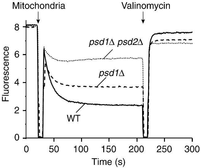
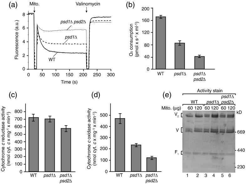
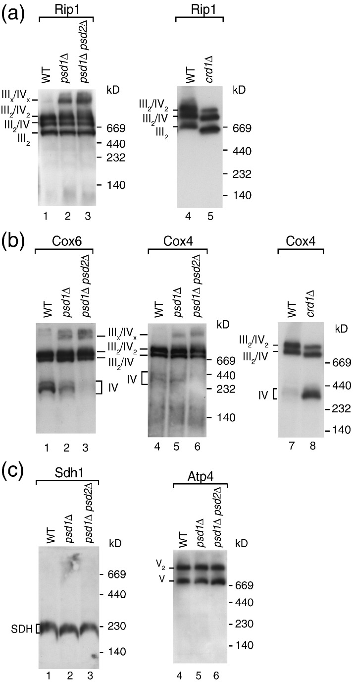
The mitochondrial inner membrane contains two non-bilayer-forming phospholipids, phosphatidylethanolamine (PE) and cardiolipin (CL). Lack of CL leads to destabilization of respiratory chain supercomplexes, a reduced activity of cytochrome c oxidase, and a reduced inner membrane potential Δψ. Although PE is more abundant than CL in the mitochondrial inner membrane, its role in biogenesis and assembly of inner membrane complexes is unknown. We report that similar to the lack of CL, PE depletion resulted in a decrease of Δψ and thus in an impaired import of preproteins into and across the inner membrane. The respiratory capacity and in particular the activity of cytochrome c oxidase were impaired in PE-depleted mitochondria, leading to the decrease of Δψ. In contrast to depletion of CL, depletion of PE did not destabilize respiratory chain supercomplexes but favored the formation of larger supercomplexes (megacomplexes) between the cytochrome bc(1) complex and the cytochrome c oxidase. We conclude that both PE and CL are required for a full activity of the mitochondrial respiratory chain and the efficient generation of the inner membrane potential. The mechanisms, however, are different since these non-bilayer-forming phospholipids exert opposite effects on the stability of respiratory chain supercomplexes.


Figures

None
Graphical abstract
Fig. 1
Fig. 1
PE is selectively depleted in inner membrane vesicles from psd1Δ and psd1Δ psd2Δ mitochondria. The S. cerevisiae strains crd1Δ, psd1Δ, and psd1Δ psd2Δ in the BY4741 background were grown in YPLac medium at 30 °C to early logarithmic growth phase. Mitochondria were isolated by differential centrifugation, the protein concentrations were adjusted, and inner membrane vesicles were isolated by sucrose gradient centrifugation as previously described. Phospholipids were extracted, separated by thin-layer chromatography, and analyzed as reported previously. The amounts of individual phospholipid classes were determined using a phosphate solution with 1 mg/ml phosphor as standard. Shown are the mean values of two determinations with range. DMPE, dimethylphosphatidylethanolamine; LP, lysophospholipids; PA, phosphatidic acid; PC, phosphatidylcholine; PG, phosphatidylglycerol; PI, phosphatidylinositol; PS, phosphatidylserine.
Fig. 2
Fig. 2
PE-depleted mitochondria are impaired in import of preproteins into and across the inner membrane. (a) Isolated mitochondria from wild-type, psd1Δ, and psd1Δ psd2Δ yeast strains were incubated with the 35S-labeled precursors of cytochrome c1 (Cyt1), F1β, and Su9-DHFR in import buffer [3% (w/v) bovine serum albumin, 250 mM sucrose, 80 mM KCl, 5 mM MgCl2, 5 mM methionine, 2 mM KH2PO4, 10 mM Mops/KOH, pH 7.2, 2 mM NADH, 5 mM creatine phosphate, 0.1 mg/ml creatine kinase, and 2 mM ATP] at 25 °C for the indicated periods. In control reactions, the membrane potential (Δψ) was dissipated prior to import by addition of 8 μM antimycin A, 1 μM valinomycin, and 20 μM oligomycin. The import reactions were stopped by adding 8 μM antimycin A, 1 μM valinomycin, and 20 μM oligomycin. After washing with SEM buffer (250 mM sucrose, 1 mM ethylenediaminetetraacetic acid, and 10 mM Mops/KOH, pH 7.2), the mitochondria were lysed under denaturing conditions and subjected to SDS-PAGE followed by digital autoradiography. p, precursor; i, intermediate; m, mature. (b) Isolated mitochondria from wild-type, psd1Δ, and psd1Δ psd2Δ yeast strains were incubated with 35S-labeled AAC at 25 °C as indicated in the presence or absence of Δψ. The mitochondria were lysed with 1% (w/v) digitonin in digitonin buffer [20 mM Tris/HCl, pH 7.4, 50 mM NaCl, 0.1 mM ethylenediaminetetraacetic acid, and 10% (v/v) glycerol] and protein complexes were separated by blue native electrophoresis.35S-labeled proteins were detected by digital autoradiography. (c) Wild-type, psd1Δ, and psd1Δ psd2Δ mitochondria were lysed with 1% (w/v) digitonin in digitonin buffer and subjected to blue native electrophoresis. Protein complexes were detected by Western blotting using the indicated antisera.
Fig. 3
Fig. 3
PE is required for the activity of the respiratory chain. (a) The membrane potential (Δψ) of wild-type, psd1Δ, and psd1Δ psd2Δ yeast mitochondria was assessed at 25 °C by fluorescence quenching using the Δψ-sensitive dye DiSC3(5) (3,3′-dipropylthiadicarbocyanine iodide) in membrane potential buffer [0.6 M sorbitol, 0.1% (w/v) bovine serum albumin, 10 mM MgCl2, 0.5 mM ethylenediaminetetraacetic acid, and 20 mM KPi, pH 7.2] as described previously. (b) The oxygen consumption of isolated wild-type, psd1Δ, and psd1Δ psd2Δ mitochondria was analyzed by oxygraph measurements at 25 °C. Isolated yeast mitochondria (100 μg protein) were added to 2 ml of buffer (10 mM Mops/KOH, pH 7.2, 250 mM sucrose, 5 mM MgCl2, 80 mM KCl, 5 mM KPi, 1 mM ADP, and 1 mM NADH) and oxygen consumption was measured. The oxygen flux (negative time derivative of oxygen concentration) corrected for instrumental background flux was expressed in picomoles per second per milliliter. Shown are the mean values with standard error of the mean (n = 3). (c and d) The activity of the cytochrome bc1 complex (c) and the cytochrome c oxidase (d) was determined in submitochondrial particles prepared from wild-type, psd1Δ, and psd1Δ psd2Δ yeast cells as described earlier. Ubiquinol-dependent cytochrome c reduction was measured as described by Palsdottir and Hunte using 3 μg protein (submitochondrial particles), 50 μM horse heart cytochrome c, and 80 μM decylubiquinol for 1 ml assay volume (40 mM potassium phosphate buffer, pH 7.4, 1 mM NaN3, and 0.05 % β-d-undecylmaltoside). Reduction of cytochrome c was monitored at 550 nm and the activity was calculated with an extinction coefficient of 19.4 mM− 1 cm− 1. The activity was fully sensitive to the specific inhibitor stigmatellin (1 μM). Cytochrome c oxidase activity was measured as described by Horvath et al. with 50 μM reduced horse heart cytochrome c and 3–50 μg of protein (submitochondrial particles) in 1 ml assay volume (75 mM potassium phosphate buffer, pH 7.4, 1 mM antimycin A, and 0.05% β-d-dodecylmaltoside). Oxidation of cytochrome c was monitored and quantified as for the cytochrome bc1 complex. The activity was fully sensitive to the specific inhibitor sodium azide (1 μM). Specific enzyme activities are based on total protein determined by bicinchoninic acid assay (Pierce). Three preparations per strain were used and the activity measurements were repeated five times for each sample. Mean values with standard error of the mean are shown. (e) The activity of the mitochondrial ATPase was assessed by in‐gel calcium phosphate precipitation upon ATP hydrolysis. Mitochondria isolated from wild-type, psd1Δ, or psd1Δ psd2Δ strains were lysed with 1% (w/v) digitonin in digitonin buffer and protein complexes were separated by blue native electrophoresis. Subsequently, the gel was washed with water and incubated with ATP-containing buffer (50 mM glycine, pH 8.4, 5 mM MgCl2, and 20 mM ATP) for 20 min and transferred into 10% (w/v) CaCl2 solution. Incubation was performed until calcium phosphate precipitation became visible and the reaction was stopped by transfer into water. V2, ATP synthase dimer; V, ATP synthase monomer; F1, F1 part of the ATP synthase.
Fig. 4
Fig. 4
Depletion of PE stabilizes respiratory chain supercomplexes. (a–c) Wild-type, psd1Δ, psd1Δ psd2Δ, and crd1Δ mitochondria were lysed with 1% (w/v) digitonin in digitonin buffer and subjected to blue native electrophoresis, followed by Western blotting using the indicated antisera. III, cytochrome bc1 complex; IV, cytochrome c oxidase; V2, ATP synthase dimer; V, ATP synthase monomer; Atp4, ATP synthase subunit 4 (subunit b); Cox4, cytochrome c oxidase subunit 4; Cox6, cytochrome c oxidase subunit 6; Rip1, Rieske iron–sulfur protein; SDH, succinate dehydrogenase (complex II).

Similar articles

Cited by

References

    1. Schuiki I., Daum G. Phosphatidylserine decarboxylase, key enzymes of lipid metabolism. IUBMB Life. 2009;61:151–162. - PubMed
    1. Osman C., Voelker D., Langer T. Making heads or tails of phospholipids in mitochondria. J. Cell Biol. 2011;192:7–16. - PMC - PubMed
    1. Claypool S.M., Koehler C.M. The complexity of cardiolipin in health and disease. Trends Biochem. Sci. 2012;37:32–41. - PMC - PubMed
    1. Zinser E., Sperka-Gottlieb C.D.M., Fasch E.V., Kohlwein S.D., Paltauf F., Daum G. Phospholipid synthesis and lipid composition of subcellular membranes in the unicellular eukaryote Saccharomyces cerevisiae. J. Bacteriol. 1991;173:2026–2034. - PMC - PubMed
    1. Storey M.K., Clay K.L., Kutateladze T., Murphy R.C., Overduin M., Voelker D.R. Phosphatidylethanolamine has an essential role in Saccharomyces cerevisiae that is independent of its ability to form hexagonal phase structures. J. Biol. Chem. 2001;276:48539–48548. - PubMed

Publication types

MeSH terms

LinkOut - more resources