. 2012 Nov 1;72(21):5516-28.
doi: 10.1158/0008-5472.CAN-12-0775. Epub 2012 Sep 12.

RhoJ regulates melanoma chemoresistance by suppressing pathways that sense DNA damage

Affiliations

RhoJ regulates melanoma chemoresistance by suppressing pathways that sense DNA damage

Hsiang Ho et al. Cancer Res. .

Abstract

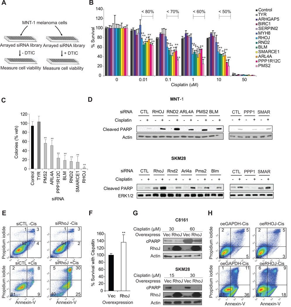
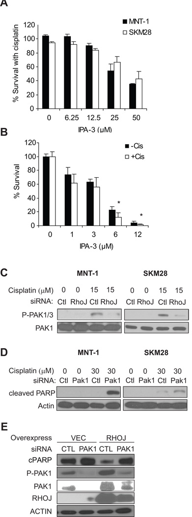
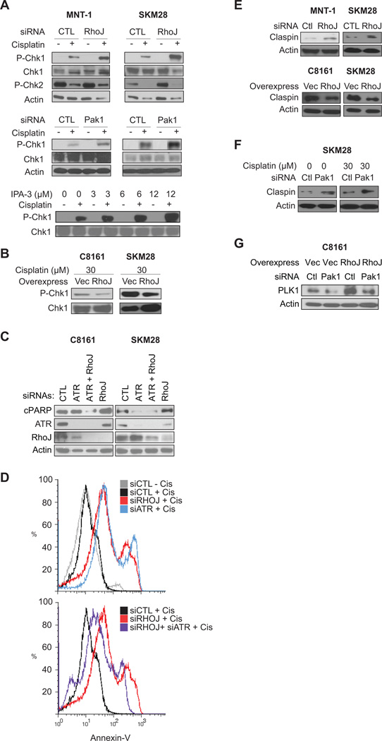
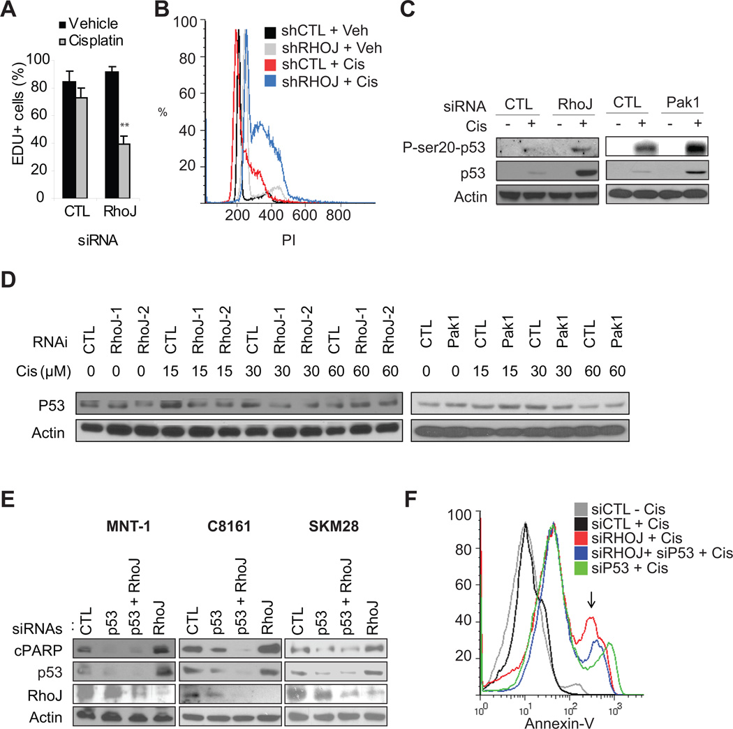
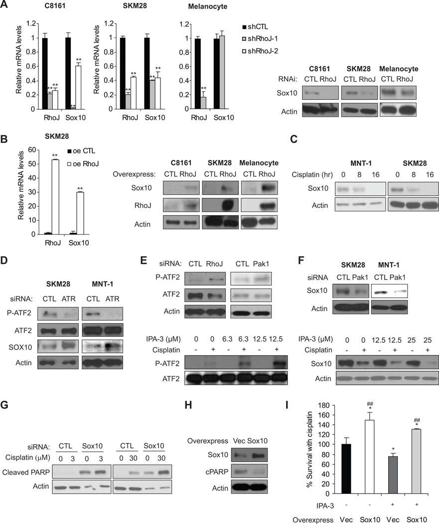
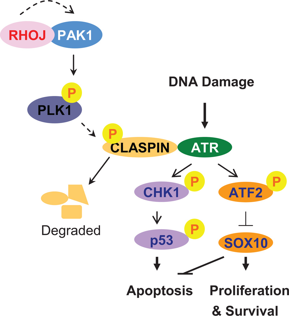
Melanomas resist conventional chemotherapeutics, in part, through intrinsic disrespect of apoptotic checkpoint activation. In this study, using an unbiased genome-wide RNA interference screen, we identified RhoJ and its effector PAK1, as key modulators of melanoma cell sensitivity to DNA damage. We find that RhoJ activates PAK1 in response to drug-induced DNA damage, which then uncouples ATR from its downstream effectors, ultimately resulting in a blunted DNA damage response (DDR). In addition, ATR suppression leads to the decreased phosphorylation of ATF2 and consequent increased expression of the melanocyte survival gene Sox10 resulting in a higher DDR threshold required to engage melanoma cell death. In the setting of normal melanocyte behavior, this regulatory relationship may facilitate appropriate epidermal melanization in response to UV-induced DNA damage. However, pathologic pathway activation during oncogenic transformation produces a tumor that is intrinsically resistant to chemotherapy and has the propensity to accumulate additional mutations. These findings identify DNA damage agents and pharmacologic inhibitors of RhoJ/PAK1 as novel synergistic agents that can be used to treat melanomas that are resistant to conventional chemotherapies.


Conflict of interest statement

The authors report no conflicts of interest

Figures

Figure 1
Figure 1. Genome-wide siRNA screen identifies core regulators of melanoma chemoresistance
A. Identification of regulators of dacarbazine resistance in MNT-1 cells MNT-1 melanoma cells were transfected with a genome-wide siRNA library. 72 hours post transfection, duplicate plates were incubated in the presence and absence of a sublethal dose of dacarbazine for an additional 48 hours. Cell titer glo values were determined for each well and normalized to internal reference samples on each plate, followed by normalization to the experimental mean for each well calculated from the full data set. Similarly adjusted luminescence values from drug treated samples were generated and used to calculate a normalized ratio (drug treated/untreated). B. Identification of Gene Targets that Potently Sensitize Melanoma Cells to Cisplatin. SKM28 melanoma cells were transfected with the indicated siRNA and incubated with cisplatin as indicated. Relative cell number was determined using a Cell TiterGlo assay. The fraction of surviving cells is indicated to highlight siRNAs that significantly sensitize cells to cisplatin. **, p< 0.01 comparing to siControl, determined by Student’s t-t-test. C. Rho GTPases sensitize Melanoma Cells to Chemotherapy Induced Apoptosis. SKM28 and MNT-1 melanoma cells were transfected with 50 nM target siRNAs and incubated in the presence and absence of the 30µM cisplatin. 24 hours after drug treatment, lysates were prepared and subjected to immunoblotting with cleaved PARP and actin loading control antibodies. Representative blot is shown. D. Clonogenicity assays identify genes that regulate cell survival in the presence of cisplatin. SKM28 melanoma cells were transfected with the indicated siRNAs and incubated with 10 µM cisplatin for 72 hours. Wells were then washed and media was repleted. Relative cell number in cisplatin treated and untreated samples were quantified using a sulforhodamine B assay and used to calculate the ratio of surviving cells. **, p< 0.01 comparing to siControl, determined by Student’s t-t-test. E. RhoJ Depletion Sensitizes Melanoma Cells To Cisplatin-induced Apoptosis. MNT-1 cells were transfected with the indicated siRNAs for 48 hours and incubated in the presence and absence of 30 µM cisplatin for 48 hours. Apoptotic cells were defined as Annexin V-positive, while necrotic cells were defined as PI-positive Annexin V-negative population by flow cytometry. F. Overexpression of RhoJ promotes melanoma chemoresistance. The relative sensitivity of vector infected or RhoJ overexpressing C8161 cells to 30 µM cisplatin for 48hrs was measured using a Cell-titer-glo ATP accumulation assay. **, t-test p-value<0.01 versus vector. Bar represents mean ± STD (left panel). G. RhoJ Overexpression Inhibits Cisplatin-induced PARP cleavage. C8161 and SK-MEL-28 RhoJ overexpressing or control vector infected cells were incubated in the presence of the indicated doses of cisplatin for 24hrs. The relative accumulation of cleaved PARP was measured by immunoblotting. H. RhoJ overexpression inhibits Cisplatin induced Apoptosis. C8161 RhoJ overexpressing or control vector infected cells were incubated in the presence of 60 µM cisplatin for 24 hours. Apoptotic cells were defined as Annexin V-positive, while necrotic cells were defined as PI-positive Annexin V-negative by flow cytometry.
Figure 2
Figure 2. RhoJ Regulates the Activation of Group I Pak kinases
A. Group I Pak inhibitors sensitize melanoma cells to cisplatin. SK-MEL-28 cells were treated with 30 µM cisplatin and the indicated dose of IPA-3 for 48 hours. The ratio of surviving cells in cisplatin+IPA-3 treated wells was normalized to wells treated with cisplatin alone. B. Group I Pak inhibitors slightly sensitizes melanocytes to cisplatin. Deeply pigmented human epidermal melanocytes are treated with indicated doses of the Pak1 inhibitor IPA-3 in the presence or absence of 20µM cisplatin for 48hrs. The ratios of Cell-Titer-Glo values were normalized to the value in vehicle-treated cells. *, p-value from Student’s t-test < 0.05 compared to IPA-3 alone in the same dose. C. RhoJ Activates Pak1/Pak3 in Response to DNA Damage. SK-MEL-28 and MNT-1 melanoma cells were transfected with the indicated siRNAs and incubated with or without cisplatin for 24hrs. The accumulation of phosphorylated Pak1, Pak2, or Pak3 was determined. The accumulation of phosphorylated Pak2, which migrates at a lower molecular weight than Pak1/Pak3, was not observed. D. Pak1 depletion sensitizes melanoma cells to cisplatin induced PARP cleavage. MNT-1 or SK-MEL-28 cells were transfected with Pak1 siRNAs. 48 hours after transfection cells were incubated with the indicated doses of cisplatin for 20 hours. Cell lysates were prepared and subjected to immunoblotting with the indicated antibodies. E. Pak1 depletion sensitizes RhoJ overexpressing cells to cisplatin-induced PARP cleavage. Vector or RhoJ-overexpressing C8161 melanoma cells are transfected with control or Pak1 siRNAs for 48hrs, and were incubated with 30 µM cisplatin for 24hrs. The relative levels of cleaved PARP, P-Ser199-Pak1, Pak1, RhoJ and Actin were measured.
Figure 3
Figure 3. RhoJ and Pak kinases Suppress ATR Activation
A. Depletion of RhoJ/Pak1 or Group I Pak inhibition Enhances Chk1 Activation. MNT-1 or SK-MEL-28 melanoma cells were transfected with the indicated siRNAs for 48hrs and incubated in the presence of 30 µM cisplatin (RhoJ depleted cells) or 15 µM cisplatin (Pak1 depleted cells) for 24hrs. Phospho-ser 345 Chk1 (ATR dependent site), Chk1, Phospho-Thr 68 Chk2 (ATM dependent site) and actin accumulation was measured by immunoblotting. SK-MEL-28 cells were also treated with the indicated dose of the group I Pak inhibitor IPA-3 and cisplatin and phospho-ser 345 Chk1 accumulation was measured. B. RhoJ Suppresses Chk-1 activation upon cisplatin treatment. C8161 and SK-MEL-28 cells overexpressing RhoJ or infected with the control vector were incubated in the presence of the indicated doses of cisplatin for 24hrs. The relative accumulation of pChk1 was measured by immunoblotting. C. ATR Depletion Mitigates the Effects of RhoJ Depletion on Cisplatin induced PARP cleavage. C8161 and SK-MEL-28 melanoma cells were treated with indicated siRNA for 48hrs and then incubated with 30µM cisplatin for 24hrs. Relative accumulation of cleaved PARP was measured. D. ATR Depletion Mitigates the Effects of RhoJ Depletion on Cisplatin induced Apoptosis. MNT-1 melanoma cells were transfected with the indicated siRNAs for 48 hours. Subsequently cells were incubated with 30 µM cisplatin for 48 hours. Apoptosis was quantified by measuring Annexin V staining. E. RhoJ Modulates Claspin accumulation. MNT-1/ SK-MEL-28 melanoma cells were transfected with the indicated siRNAs for 72hrs and the relative accumulation of Claspin was measured by immunoblotting. C8161 and SK-MEL-28 melanoma cells overexpressing RhoJ expressed lower levels of Claspin when compared to cells that express a control vector (right panel). F. RhoJ Modulates Claspin Accumulation via a mechanism involving Pak1. SK-MEL-28 cells transfected with control or Pak1 siRNAs for 48hrs, then treated with or without 30 µM cisplatin for 24hrs. The relative accumulation of claspin was measured by by immunoblotting. G. Pak1 depletion restores RhoJ overexpressing-induced Plk1 accumulation. Vector or RhoJ overexpressing C8161 cells treated with control or Pak1 siRNA for 72hrs and analyzed by immunoblotting.
Figure 4
Figure 4. RhoJ Inhibits Drug-Induced Cell Cycle Arrest and Apoptosis
A. RhoJ Depletion Synergizes with Chemotherapy to Inhibit Proliferation. SK-MEL-28 cells were transfected with 50nM siRNAs and incubated with 1µM cisplatin or vehicle for 48 hours. EdU incoporation was quantified. Representative images are contained in Figure S4D. B. RhoJ Depletion Synergizes with Cisplatin to Induce S phase arrest. C8161 melanoma cells expressing control or RhoJ shRNAs were incubated in the presence and absence of 15µM cisplatin for 24hrs and subjected to FACS analysis as described. Note the accumulation of cells in the peak between G1 (200) and G2 (400). C. RhoJ/Pak1 Modulates Cisplatin-induced p53 accumulation in p53 wild type cells. MNT-1 cells were transfected with the indicated siRNAs and incubated with 30µM cisplatin for 24hr. The accumulation of pSer20-p53 (Chk1 dependent phosphorylation site) and p53 was measured. D. RhoJ/Pak1 does not modulate Cisplatin-induced p53 accumulation in p53 mutant cells. SK-MEL-28 cells expressing the indicated shRNA (left panel) or transfected with the indicated siRNAs (right panel) were incubated with the indicated doses of cisplatin for 24 hours. As pSer20-p53 could not be detected in these cells (data not shown), the accumulation of p53 was measured. E. p53 Depletion Mitigates the Effects of RhoJ Depletion on Cisplatin induced PARP cleavage in p53 wild type cells. P53 wild type (C8161, MNT-1) and p53 mutant (SK-MEL-28) melanoma cells were incubated with the indicated siRNAs for 48 hours followed by 30 µM cisplatin for 24 hours. Relative accumulation of cleaved PARP was measured. F. p53 Depletion partially mitigates the effects of RhoJ Depletion on Cisplatin induced Apoptosis. MNT-1 melanoma cells were transfected with the indicated siRNAs for 48 hours. Subsequently cells were incubated with 30 mM cisplatin for 48 hours. Apoptosis was quantified by measuring the intensity of Annexin V staining.
Figure 5
Figure 5. RhoJ Inhibits Sox10 Expression in Melanoma Cells
A. RhoJ is Required for Sox10 Expression in Melanoma Cells but not Melanocytes. The relative mRNA expression of RhoJ and SOX10 was measured by RT-qPCR in C8161, SK-MEL-28 melanoma cells and melanocytes expressing control or two different RhoJ shRNAs (left panel). **, p< 0.01 comparing to shCTL, determined by Student’s t-t-test.Sox10 protein accumulation in RhoJ deficient melanocytes and melanoma cells was measured via immunoblotting (right panel). B. RhoJ Modulates the Expression of Sox10 in Melanoma cells and Melanocytes. Relative mRNA expression of RhoJ and Sox10 in RhoJ overexpressing SK-MEL-28 melanoma cells was quantified using RT-qPCR (left panel). **, p< 0.01 comparing to shCTL, determined by Student’s t-t-test. Lysates from C8161, SK-MEL-28 melanoma cells and melanocytes overexpresing RhoJ were subjected to western blotting with Sox10 and actin antibodies to measure the relative accumulation of RhoJ (right panel). C. Sox10 expression is modulated by the DNA damage response. MNT-1 and SK-MEL-28 cells were treated with 30µM cisplatin at the indicated time points. The relative amount of Sox10 protein was measured via immunoblotting. D. ATR modulates ATF2 phosphorylation. SK-MEL-28 and MNT-1 cells were transfected with control or ATR siRNAs followed by incubation with 30 µM cisplatin for 24hrs. The accumulation of phospho-Thr 490/498 ATF2, a downstream target of ATR, and Sox10 was measured by immunoblotting. E. RhoJ/Pak1 Depletion or Group I Pak inhibition Modulates ATF2 phosphorylation. SK-MEL-28 cells were either transfected with the indicated siRNAs followed by incubation with 30 µM cisplatin for 24hrs or were incubated in the presence or absence 30 µM cisplatin combined with indicated doses of IPA-3 for 24hrs. The accumulation of phospho-Thr-490/498 ATF2 and ATF2 was measured via immunoblotting. F. Pak1 depletion or Inhibition Modulates Sox10 accumulation. SK-MEL-28 cells were transfected with the indicated siRNAs followed by incubation with 30 µM cisplatin for 24hrs or were incubated in the presence or absence of 30 µM cisplatin and the indicated dose of IPA-3 for 24 hours. Relative accumulation of Sox10 and Actin was measured by western blotting. G. Sox10 Depletion Sensitizes Melanoma Cells to Chemotherapy Induced Apoptosis. MNT-1 cells were treated with control or Sox10 siRNA and the indicated dose of cisplatin for 24hrs. Relative accumulation of cleaved PARP and actin was measured via immunoblotting. H. Sox10-overexpression melanoma cells are more resistant to cisplatin-induced cell death. SK-MEL-28 cells overexpressing Sox10 or vector were treated with 30 µM cisplatin for 24hrs before immunolblotting analysis. I. Sox10 Overexpression Partially Mitigates the Effect of Pak Inhibitors on Cisplatin-induced Apoptosis. SK-MEL-28 cells overexpressing Sox10 or infected with virus encoding the empty vector were treated with or without 3 µM IPA-3 in the presence of 30 µM cisplatin for 72 hrs. Cell survival was quantified using a Cell-titer-Glo assay. *, p≤0.05 by Student’s t-test; ##, p≤0.01 using a Student’s t-test.
Figure 6
Figure 6. RhoJ/Pak1 Regulate Melanoma Chemoresistance by Suppressing Cellular Mechanisms that Sense DNA Damage
Our studies revealed that RhoJ/ Pak kinases activate Plk1, resulting in the phosphorylation and degradation of Claspin. In the absence of Claspin, ATR is not able to activate its downstream effectors Chk1 and ATF2. In p53 wild type cells, Chk1 is no longer able to phosphorylate p53 and induce apoptosis while ATF2 is no longer able to regulate the expression of Sox10, which also suppresses apoptosis and stimulates cell proliferation.

Similar articles

Cited by

References

    1. Tsao H, Atkins MB, Sober AJ. Management of cutaneous melanoma. N Engl J Med. 2004;351:998–1012. - PubMed
    1. Bradbury PA, Middleton MR. DNA repair pathways in drug resistance in melanoma. Anticancer Drugs. 2004;15:421–426. - PubMed
    1. Solit DB, Rosen N. Resistance to BRAF inhibition in melanomas. N Engl J Med. 2011;364:772–774. - PubMed
    1. Soengas MS, Lowe SW. Apoptosis and melanoma chemoresistance. Oncogene. 2003;22:3138–3151. - PubMed
    1. Lee JT, Herlyn M. MEK'ing the most of p53 reactivation therapy in melanoma. J Invest Dermatol. 2012;132:263–265. - PubMed

Publication types