

Review
doi: 10.1007/s00018-012-1151-x.
Epub 2012 Sep 13.
Genetic visualization of notch signaling in mammalian neurogenesis
Affiliations
- PMID: 22971775
- PMCID: PMC3663255
- DOI: 10.1007/s00018-012-1151-x
Item in Clipboard
Review
Genetic visualization of notch signaling in mammalian neurogenesis
Cell Mol Life Sci.
2013 Jun.
Abstract
Notch signaling plays crucial roles in fate determination and the differentiation of neural stem cells in embryonic and adult brains. It is now clear that the notch pathway is under more complex and dynamic regulation than previously thought. To understand the functional details of notch signaling more precisely, it is important to reveal when, where, and how notch signaling is dynamically communicated between cells, for which the visualization of notch signaling is essential. In this review, we introduce recent technical advances in the visualization of notch signaling during neural development and in the adult brain, and we discuss the physiological significance of dynamic regulation of notch signaling.
Figures

Notch signaling pathway. The proneural genes Mash1 and Ngn2 induce expression of notch ligands such as Dll1, which activate notch signaling in neighboring cells. Upon activation, the notch intracellular domain (NICD) is released from the transmembrane region and transferred into the nucleus, where NICD forms a complex with RBPj and induces Hes1 and Hes5 expression. Hes1 and Hes5 repress proneural gene expression. During maturation and trafficking to the cell surface, notch receptors undergo furin processing and glycosylation, which can impact their responsiveness to their ligands. The ligand-induced activation of notch signaling is dynamically regulated by endocytic trafficking, which can be modulated by the different ubiquitin ligases, such as Mind bom and Neuralized

Neurogenesis in the developing and adult forebrain. a Differentiation of NSCs in the embryonic dorsal telencephalon. NSCs initially undergo symmetric cell division and proliferate extensively. Then, these cells give rise to neurons or intermediate neural progenitors (INPs) by asymmetric cell division. Neurons and INPs migrate into the cortical plate (CP) and the subventricular zone (SVZ), respectively. INPs further divide in the SVZ and produce more neurons. Some NSCs become outer SVZ (OSVZ) or outer VZ (OVZ) progenitors, which have radial fibers that extend to the pial surface but lack apical processes. After production of neurons, NSCs finally differentiate into glial cells. b Neurogenic niche of the subventricular zone (SVZ) of the lateral ventricle (LV) of the adult brain. In this region, NSCs (type B cells) exist and new neurons are continuously generated. Type B cells are GFAP-positive cells with the structural and molecular characteristics of astrocytes. Type B cells divide to generate transit-amplifying cells (type C cells), which in turn differentiate into neuroblasts (type A cells) that migrate into the olfactory bulb. c Adult neurogenesis in the dentate gyrus (DG) of the hippocampus. Type 1 and type 2a progenitor cells in the subgranular zone (SGZ) are shown. These progenitor cells give rise to transit-amplifying cells (type 2b/type 3 cells). Then, transit-amplifying cells differentiate into dentate granule cells through several maturation steps

Transgenic constructs for monitoring the notch signaling activity. a TNR mouse. b Hes5-nlsLacZ knock-in mouse. c Hes1 BAC-EGFP mouse. d pHes1-d2EGFP mouse. e pHes5-EGFP mouse. f pHes5-d2EGFP mouse. g The LCI reporter for monitoring notch activation. h pHes1-Ub-Luc mouse. More detailed information is described in the text
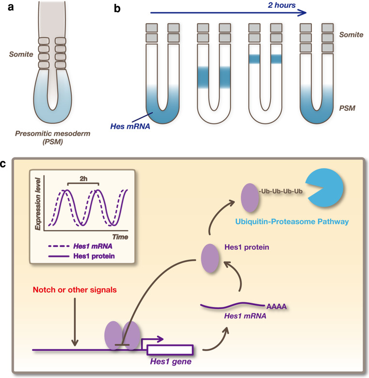
Oscillatory expression of Hes genes. a Somites form periodically by segmentation of the anterior region of the presomitic mesoderm (PSM). b
Hes expression is periodically propagated, like a wave, from the posterior end to the anterior region of the PSM, and each wave leads to the generation of a pair of somites. c Oscillatory expression of Hes1 is regulated by negative feedback. Promoter activation induces the production of Hes1 protein, which represses expression of its own gene. Then, both Hes1 mRNA and Hes1 protein disappear rapidly because they have very short half-lives, allowing the next round of expression. In this way, Hes1 expression autonomously oscillates
References
Publication types
MeSH terms
Substances
LinkOut - more resources
Full Text Sources
Other Literature Sources
