. 2012 Nov;18(11):1682-7.
doi: 10.1038/nm.2964.

SAMHD1 restricts HIV-1 infection in resting CD4(+) T cells

Affiliations

SAMHD1 restricts HIV-1 infection in resting CD4(+) T cells

Hanna-Mari Baldauf et al. Nat Med. 2012 Nov.

Abstract

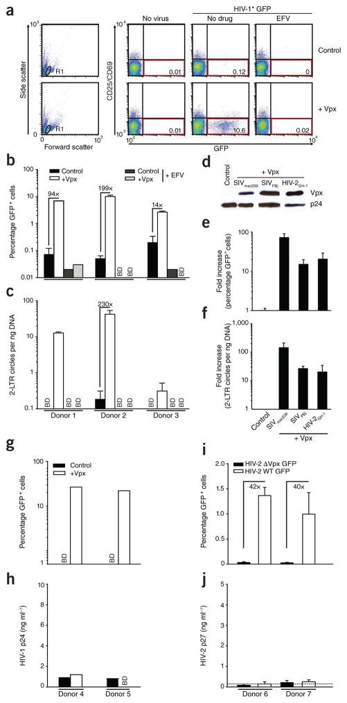
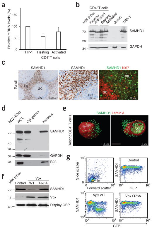
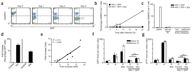
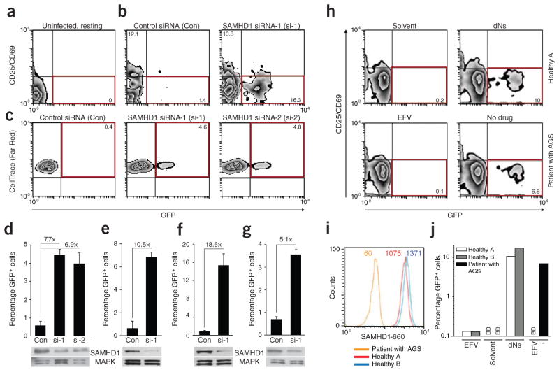
Unlike activated CD4(+) T cells, resting CD4(+) T cells are highly resistant to productive HIV-1 infection. Early after HIV-1 entry, a major block limits reverse transcription of incoming viral genomes. Here we show that the deoxynucleoside triphosphate triphosphohydrolase SAMHD1 prevents reverse transcription of HIV-1 RNA in resting CD4(+) T cells. SAMHD1 is abundantly expressed in resting CD4(+) T cells circulating in peripheral blood and residing in lymphoid organs. The early restriction to infection in unstimulated CD4(+) T cells is overcome by HIV-1 or HIV-2 virions into which viral Vpx is artificially or naturally packaged, respectively, or by addition of exogenous deoxynucleosides. Vpx-mediated proteasomal degradation of SAMHD1 and elevation of intracellular deoxynucleotide pools precede successful infection by Vpx-carrying HIV. Resting CD4(+) T cells from healthy donors following SAMHD1 silencing or from a patient with Aicardi-Goutières syndrome homozygous for a nonsense mutation in SAMHD1 were permissive for HIV-1 infection. Thus, SAMHD1 imposes an effective restriction to HIV-1 infection in the large pool of noncycling CD4(+) T cells in vivo. Bypassing SAMHD1 was insufficient for the release of viral progeny, implicating other barriers at later stages of HIV replication. Together, these findings may unveil new ways to interfere with the immune evasion and T cell immunopathology of pandemic HIV-1.


Conflict of interest statement

COMPETING FINANCIAL INTERESTS

The authors declare no competing financial interests.

Figures

Figure 1
Figure 1
Vpx overcomes a restriction to HIV-1 and HIV-2 infection in resting human CD4+ T cells. (ac) Resting CD4+ T cells were challenged with equivalent infectious units of HIV-1* GFP virions with (+Vpx) or without (Control) incorporated Vpx from SIVmac239 and analyzed on day 3 after infection. The reverse transcriptase inhibitor efavirenz (EFV) served as specificity control. (a) Dot plots of flow cytometric analysis for 1 out of 23 donors. The percentages of viable (gate R1, left graphs) resting (CD25CD69) and infected (GFP+) CD4+ T cells are shown in the bottom right quadrants. (b,c) Percentage of GFP+ cells (b) and relative copy numbers of HIV-1 2-LTR circles (c). Bars represent means of triplicates + s.d., with the factors of increase by Vpx indicated. (d) Immunoblotting of HIV-1 virions for incorporated epitope-tagged Vpx proteins from SIVmac239, SIVPBj or HIV-2GH-1. HIV-1 p24 served as a loading control. (e,f) Percentage of GFP+ cells (e) and relative 2-LTR circle copy numbers (f) of resting CD4+ T cells challenged with the Vpx-carrying viruses relative to the Vpx-negative control virus. Bars represent means + s.e.m. of the factors of increase (n = 4). Percentages of GFP+ cells ranged from 0.5% to 3.1% for Vpx of SIVmac239, from 0.2% to 0.7% for Vpx of SIVPBj and from 0.1% to 0.9% for Vpx of HIV-2GH-1. (g–j) Resting CD4+ T cells from the same donors were challenged with equivalent infectious units of HIV-1* GFP virions with (+Vpx) or without (Control) incorporated Vpx (g,h) or HIV-2ROD9 GFP or HIV-2ROD9 ΔVpx GFP (i,j) and analyzed for the percentage of GFP+ cells (g,i) and the amount of HIV-1 p24 or HIV-2 p27, respectively, released into the cell culture supernatant on day 3 after infection (h,j). Bars in i,j represent means of triplicates + s.d. The dotted line in j indicates the cut-off of the p27 ELISA. BD, below detection.
Figure 2
Figure 2
SAMHD1 is expressed in resting CD4+ T cells and depleted by Vpx. (a) RT-PCR–based quantification of SAMHD1 mRNA levels in CD4+ T cells. RNaseP-normalized mRNA levels in resting and PHA- and IL-2-activated CD4+ T cells are presented relative to those from THP-1 cells that were arbitrarily set to 100%. Means + s.e.m. of four donors. (b) Immunoblotting for endogenous expression of SAMHD1 in CD4+ T cells (two donors). GAPDH: loading control. (c) In situ expression of SAMHD1 in explants of human tonsil. Left and center, immunohistochemical detection of SAMHD1. Nuclei were counterstained with hematoxylin. GC, germinal center; LW, lymphocyte wall; PR, perifollicular region. Right, immunofluorescence analysis of SAMHD1 expression (green) relative to the proliferation marker Ki67 (red). (d) SAMHD1 immunoblot analysis of a nuclear-cytoplasmic fractionation of resting CD4+ T cells. WCL, whole cell lysate. GAPDH and B23 served as cytoplasmic and nuclear markers, respectively. (e) Three-dimensional reconstructions of deconvolution confocal microscopy images of a resting CD4+ T cell co-stained for SAMHD1 (green) and the nuclear envelope protein lamin A (red). At right is an optical section across the cell to show nuclear and cytoplasmic SAMHD1. (f) Immunoblots of endogenous SAMHD1 in nucleofected resting CD4+ T cells expressing WT Vpx, Vpx Q76A or an empty vector control. Co-expression of surface-exposed Display-GFP was used to purify a homogenous population of nucleofected cells. Similar results were obtained for two other donors. (g) Quantification of intracellular SAMHD1 amounts in nucleofected resting CD4+ T cells. Cells were nucleofected to co-express GFP together with WT Vpx, Vpx Q76A or an empty control and 16 h later assessed for SAMHD1 expression in relation to GFP by flow cytometry.
Figure 3
Figure 3
Susceptibility of resting CD4+ T cells to Vpx-carrying HIV-1 is paralleled by proteasomal degradation of SAMHD1 and increased dNTP levels. (a) Time course of SAMHD1 and GFP expression in resting CD4+ T cells after challenge with HIV-1* GFP + Vpx (SIVmac239). The percentages of cells in the respective quadrants are shown. (b) Quantification of SAMHD1 expression in resting CD4+ T cells within the first 22 h after exposure to HIV-1* GFP with (+ Vpx) or without (Control) incorporated Vpx. Data points mark the percentages for cells with low SAMHD1 levels (corresponding to the bottom left quadrants in FACS panels shown in a). (c) Effect of proteasome or HIV-1 entry inhibitors on SAMHD1 levels in resting CD4+ T cells after exposure to HIV-1* GFP ± Vpx. Resting CD4+ T cells were pretreated for 1 h with either the proteasome inhibitor (PI) MG132 (10 μM), the fusion inhibitor T20 (50 μM) or the CXCR4 antagonist AMD3100 (5 μM) before infection with HIV-1* GFP with or without virion-packaged Vpx. Drugs were removed 20 h later. Shown are the percentages of resting CD4+ T cells with low levels of SAMHD1 24 h after infection for one of three donors. (d) Relative factor of increase of infection of resting CD4+ T cells with HIV-1* GFP by virion-packaged Vpx (n = 15) or by incubation with deoxynucleosides (dNs) (n = 8). Means + s.e.m. (e) Correlation between the enhancement of infection of resting CD4+ T cells with HIV-1* GFP either by virion-packaged Vpx or by dN treatment. Depicted is the relative factor of increase in infection for both parameters obtained in parallel experiments for cells from eight different donors. (f,g) Quantification of dATP (f) and dTTP (g) levels in primary CD4+ T cells under different experimental conditions determined by single-nucleotide incorporation assay. R, resting CD4+ T cells; A, activated CD4+ T cells. dNs, cultivation in the presence of dNs (2 mM) for 3 h. Challenge with HIV-1* GFP + Vpx had resulted in depletion of SAMHD1 in 17% (donor 12) or 31% (donor 13) of resting CD4+ T cells 24 h after challenge, respectively, at which time cells were harvested for subsequent dNTP analyses. See also Supplementary Figure 10 for primary data of the single-nucleotide incorporation assay. *P < 0.05; ***P < 0.005.
Figure 4
Figure 4
Silencing of SAMHD1 by RNA interference or a homozygous nonsense mutation renders resting CD4+ T cells permissive for HIV-1 infection. (ac) Flow cytometric analysis of freshly isolated, uninfected resting CD4+ T cells (staining reference) (a) or of post-activated resting CD4+ T cells infected with HIV-1 GFP after treatment with the indicated siRNAs (b,c). Red boxes indicate populations of infected cells with relative percentages indicated. Cells’ activation state was assessed by surface expression of CD25 and CD69 (a,b) or CellTrace (Far Red) dilution (c). (dg) Quantification of the percentage of GFP+ cells and the relative increase of HIV-1 GFP infection in resting CD4+ T cells from four donors (each corresponding to a panel) following silencing with SAMHD1-specific siRNAs (si-1 or si-2) relative to a nontargeting control siRNA (Con). Shown are means + s.d. of triplicate infections. SAMHD1 levels in corresponding cell lysates were determined by immunoblotting. MAPK, loading control. See also Supplementary Figure 11b–e for results with cells from two additional donors, including the use of SAMHD1 si-3. (h) Flow cytometric analyses of PBMCs from healthy donor A (solvent- or dN-treated, top graphs) or PBMCs from a patient with AGS with a homozygous nonsense mutation in the SAMHD1 gene (EFV-treated or untreated, bottom graphs) 3 d after challenge with HIV-1 GFP (multiplicity of infection = 1). The percentages of resting (CD25CD69), infected (GFP+) CD3+CD4+ PBMCs are shown in the bottom right quadrants boxed in red, with the percentage of infected cells indicated. (i) Intracellular SAMHD1 expression in resting CD4+ T cells of PBMCs from the patient with AGS and from healthy donors A and B, as determined by flow cytometry. The mean fluorescence intensity of SAMHD1 levels is indicated. (j) Quantification of the percentage of infected (GFP+) resting CD4+ T cells from the patient with AGS and from healthy donors (see panel h for primary data). Shown are arithmetic means of duplicates. BD, below detection.

Comment in

References

    1. Doitsh G, et al. Abortive HIV infection mediates CD4 T cell depletion and inflammation in human lymphoid tissue. Cell. 2010;143:789–801. - PMC - PubMed
    1. Ganesh L, et al. The gene product Murr1 restricts HIV-1 replication in resting CD4+ lymphocytes. Nature. 2003;426:853–857. - PubMed
    1. Korin YD, Zack JA. Progression to the G1b phase of the cell cycle is required for completion of human immunodeficiency virus type 1 reverse transcription in T cells. J Virol. 1998;72:3161–3168. - PMC - PubMed
    1. Pierson TC, et al. Molecular characterization of preintegration latency in human immunodeficiency virus type 1 infection. J Virol. 2002;76:8518–8531. - PMC - PubMed
    1. Stevenson M, Stanwick TL, Dempsey MP, Lamonica CA. HIV-1 replication is controlled at the level of T cell activation and proviral integration. EMBO J. 1990;9:1551–1560. - PMC - PubMed

Publication types

MeSH terms

Substances

Supplementary concepts