. 2013 May;20(5):575-80.
doi: 10.1038/gt.2012.75. Epub 2012 Sep 13.

Inhibition of TGF-β signaling in genetically engineered tumor antigen-reactive T cells significantly enhances tumor treatment efficacy

Affiliations

Inhibition of TGF-β signaling in genetically engineered tumor antigen-reactive T cells significantly enhances tumor treatment efficacy

L Zhang et al. Gene Ther. 2013 May.

Abstract

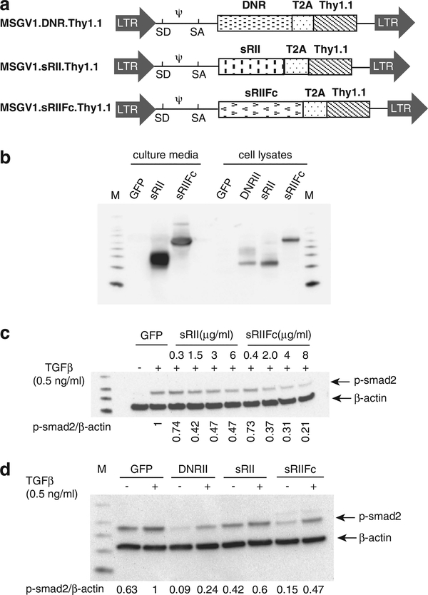
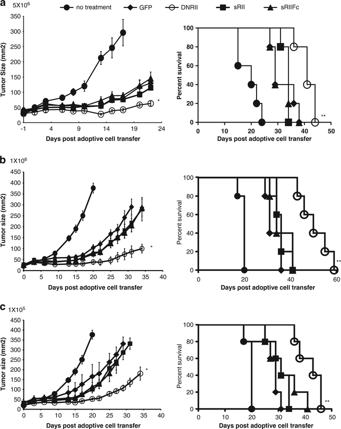
Transforming growth factor β (TGF-β) is a cytokine with complex biological functions that may involve tumor promotion or tumor suppression. It has been reported that multiple types of tumors secrete TGF-β, which can inhibit tumor-specific cellular immunity and may represent a major obstacle to the success of tumor immunotherapy. In this study, we sought to enhance tumor immunotherapy using genetically modified antigen-specific T cells by interfering with TGF-β signaling. We constructed three γ-retroviral vectors, one that expressed TGF-β-dominant-negative receptor II (DNRII) or two that secreted soluble TGF-β receptors: soluble TGF-β receptor II (sRII) and the sRII fused with mouse IgG Fc domain (sRIIFc). We demonstrated that T cells genetically modified with these viral vectors were resistant to exogenous TGF-β-induced smad-2 phosphorylation in vitro. The functionality of antigen-specific T cells engineered to resist TGF-β signaling was further evaluated in vivo using the B16 melanoma tumor model. Antigen-specific CD8+ T cells (pmel-1) or CD4+ T cells (tyrosinase-related protein-1) expressing DNRII dramatically improved tumor treatment efficacy. There was no enhancement in the B16 tumor treatment using cells secreting soluble receptors. Our data support the potential application of the blockade of TGF-β signaling in tumor-specific T cells for cancer immunotherapy.


Conflict of interest statement

CONFLICT OF INTEREST

The authors declare no conflict of interest.

Figures

Figure 1.
Figure 1.
DNRII-, sRII-, sRIIFc-transduced T cells were resistant to TGF-β-mediated smad2 phosphorylation. (a) Schematic representation of retroviral vectors: MSGV1.DNRII, MSGV1.sRII and MSGV1.sRIIFc. LTR, long terminal repeat; SD, splice donor; SA, splice acceptor; T2A, ribosomal skip peptide. (b) Mouse splenocytes were transduced with the MSGV1.GFP, MSGV1.DNRII, MSGV1.sRII and MSGV1.sRIIFc. The cells and culture supernatant were harvested 48 h later. The DNRII, sRII and sRIIFc expression were measured by immunoblotting with anti-TGF-β-RII antibody. (c) Different amount of partially concentrated conditioned media was added to T cells treated with exogenous TGF-β1 (0.5 ng ml−1) for 1 h. Phosphorylation smad2 (p-smad2) was measured by western blot. The relative level of p-smad2 was normalized by β-actin. The p-smad2 level in the cells treated with TGF-β1 and the supernatant from GFP-transduced cells was set as 1. (d) The T cells were transduced with GFP, DNRII, sRII or sRIIFc individually and treated without or with exogenous TGF-β1 (0.5 ng ml−1, 1 h). The smad2 phosphorylation was measured by western blot. The relative level of p-smad2 was normalized by β-actin. The relative p-smad2 level in the GFP-transduced cells treated with TGF-β1 and was set as 1.
Figure 2.
Figure 2.
Pmel-1 T cells expressing DNRII, sRII or sRIIFc did not affect cell proliferation or antigen recognition. (a) The pmel-1 cells were transduced with GFP, DNRII, sRII or sRIIFc, and analyzed by fluorescence-activated cell sorting using Thy1.1-FITC and CD8-PE antibody. (b). The transduced cells were enumerated every 2 days by trypan blue exclusion. (c) The cells transduced with DNRII, sRII or sRIIFc vector were co-cultured with various concentration (from 10−6 M to 10−12 M) of hgp10025–33 peptide-pulsed cells for 16 h. The interferon-γ level in the culture was measured by enzyme-linked immunosorbent assay (shown are the mean values of duplicate determinations). NP, negative control peptide.
Figure 3.
Figure 3.
DNRII expressing pmel-1 cells had enhanced antitumor activity against B16 melanoma tumor. Pmel-1 cells were transduced with vector-expressing GFP, DNRII, sRII or sRIIFc. B16 tumor-bearing mice (n = 5) were adoptively transferred with 5 × 10⁶ (a), 1 × 10⁶ (b) or 1 × 10⁵ (c) cells genetically modified by pmel-1 cells as described in Materials and methods. Tumor sizes were assessed with serial measurements. Error bars represent s.e.m. (*P = 0.009, DNRII compared with GFP). The survival of tumor-bearing mice that received 5 × 10⁶ (a), 1 × 10⁶ (b) or 1 × 10⁵ (c) of genetic-modified cell transfer were determined as shown (**P<0.05, DNRII compared with GFP).
Figure 4.
Figure 4.
TRP1 CD4 cells co-expressing DNRII dramatically augmented the tumor treatment in B16 melanoma tumor model. CD4 T cells were isolated from normal mouse splenocytes and stimulated with anti-CD3 and anti-CD28 in vitro. The cells were than cotransduced with TRP1-TCR and GFP, DNRII, sRII or sRIIFc. B16 tumor-bearing mice (n = 5) were adoptively transferred with 1 × 10⁵ (a) or 1 × 10⁶ (b) double-engineered CD4 T cells. Tumor sizes were assessed with serial measurements. Error bars represent s.e.m. (*P<0.05, TRP1 + DNRII compared with TRP1 + GFP). The survival of tumor-bearing mice that received 1 × 10⁵ (a) or 1 × 10⁶ (b) of genetically modified cell transfer were determined as shown (**P<0.05, TRP1 + DNRII compared with TRP1 + GFP).

References

    1. Dudley ME, Wunderlich JR, Robbins PF, Yang JC, Hwu P, Schwartzentruber DJ et al. Cancer regression and autoimmunity in patients after clonal repopulation with antitumor lymphocytes. Science 2002; 298: 850–854. - PMC - PubMed
    1. Rosenberg SA, Yang JC, Sherry RM, Kammula US, Hughes MS, Phan GQ et al. Durable complete responses in heavily pretreated patients with metastatic melanoma using T-cell transfer immunotherapy. Clin Cancer Res 2011; 17: 4550–4557. - PMC - PubMed
    1. Yingling JM, Blanchard KL, Sawyer JS. Development of TGF-beta signalling inhibitors for cancer therapy. Nat Rev Drug Discov 2004; 3: 1011–1022. - PubMed
    1. Penafuerte C, Galipeau J. TGF beta secreted by B16 melanoma antagonizes cancer gene immunotherapy bystander effect. Cancer Immunol Immunother 2008; 57: 1197–1206. - PMC - PubMed
    1. Gorelik L, Flavell RA. Transforming growth factor-beta in T-cell biology. Nat Rev Immunol 2002; 2: 46–53. - PubMed

Publication types

MeSH terms

Substances