. 2012 Nov 15;303(10):C1034-44.
doi: 10.1152/ajpcell.00175.2012. Epub 2012 Sep 12.

Pannexin1 contributes to pathophysiological ATP release in lipoapoptosis induced by saturated free fatty acids in liver cells

Affiliations

Pannexin1 contributes to pathophysiological ATP release in lipoapoptosis induced by saturated free fatty acids in liver cells

Feng Xiao et al. Am J Physiol Cell Physiol. .

Abstract

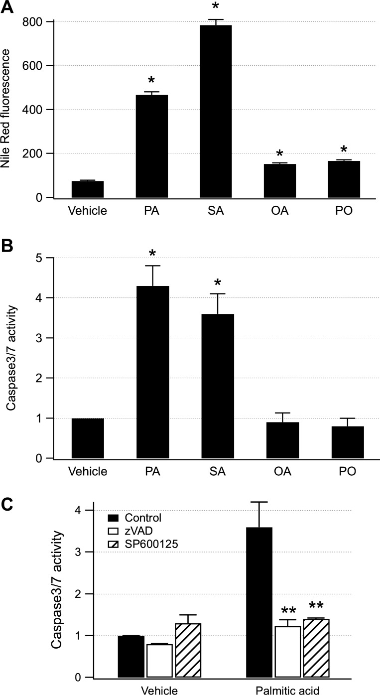
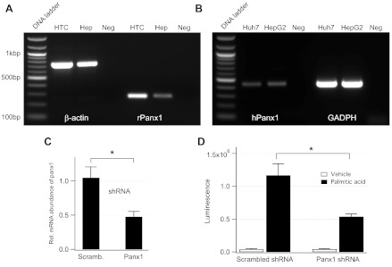
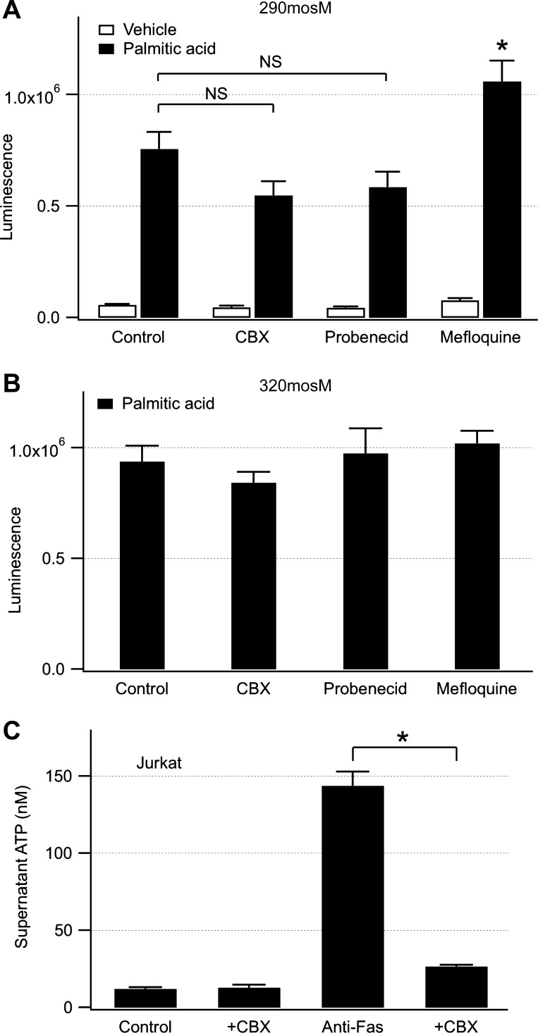
Hepatocyte lipoapoptosis induced by saturated free fatty acids (FFA) contributes to hepatic inflammation in lipotoxic liver injury, and the cellular mechanisms involved have not been defined. Recent studies have shown that apoptosis in nonhepatic cells stimulates ATP release via activation of pannexin1 (panx1), and extracellular ATP functions as a proinflammatory signal for recruitment and activation of the inflammatory cells. However, it is not known whether lipoapoptosis stimulates ATP release in liver cells. We found that lipoapoptosis induced by saturated FFA stimulated ATP release in liver cells that increased extracellular ATP concentration by more than fivefold above the values observed in healthy cells. This sustained pathophysiological ATP release was not dependent on caspase-3/7 activation. Inhibition of c-Jun NH(2)-terminal kinase (JNK), a key mediator of lipoapoptosis, with SP600125 blocked pathophysiological ATP release in a dose-dependent manner. RT-PCR analysis indicated that panx1 is expressed in hepatocytes and multiple liver cell lines. Notably, inhibition of panx1 expression with short hairpin (sh)RNA inhibited in part pathophysiological ATP release. Moreover, lipoapoptosis stimulated uptake of a membrane impermeable dye YoPro-1 (indicative of panx1 activation), which was inhibited by panx1 shRNA, probenecid, and mefloquine. These results suggest that panx1 contributes to pathophysiological ATP release in lipoapoptosis induced by saturated FFA. Thus panx1 may play an important role in hepatic inflammation by mediating an increase in extracellular ATP concentration in lipotoxic liver injury.


Figures

Fig. 1.
Fig. 1.
Saturated free fatty acids (FFA) stimulate apoptosis and inhibit cell proliferation. HTC cells were treated overnight with 0.5% isopropanol (vehicle) or FFA (500 μM). A: cells were fixed and processed by a Click-iT transferase-mediated dUTP nick end-label (TUNEL) assay. Graph shows relative TUNEL fluorescence after treatment with palmitic acid (PA), stearic acid (SA), oleic acid (OA), and palmitoleic acid (PO). Saturated but not unsaturated FFA increased TUNEL fluorescence (*P < 0.001; NS, not significant from vehicle). Number of cells analyzed was from 60 to 131. B: number of cells in a dish was measured after overnight treatment with different FFA. Saturated FFA inhibited and unsaturated FFA stimulated cell proliferation by ∼50% (*P < 0.01 for all FFA). Number of dishes analyzed was from 7 to 13. C: cell diameter was measured after overnight treatment with different FFA (500 μM) or 24-h treatment with Fas ligand (FasL; 100 ng/ml). Number of cells used to generate histograms was from ∼20,000 to 40,000. Percentage of shrunken cells from 3 to 9 μm in diameter is indicated for each condition. Note that saturated FFA markedly increased the population of shrunken cells.
Fig. 2.
Fig. 2.
Saturated FFA stimulate intracellular lipid accumulation and JNK-dependent caspase-3/7 activity. A: Nile red fluorescence was measured after overnight treatment of HTC cells with different FFA (500 μM). All FFA significantly increased Nile red fluorescence (*P < 0.001). Number of cells analyzed was form 66 to 86. B: caspase-3/7 activity was measured after overnight treatment with FFA (500 μM). Palmitic (n = 15 dishes) and stearic acid (n = 3 dishes) potently increased caspase-3/7 activity (*P < 0.001). Oleic and palmitoleic acid did not significantly increase caspase-3/7 activity (n = 7 dishes for both). C: caspase-3/7 activity was measured after overnight treatment with palmitic acid (500 μM), and addition of a pancaspase inhibitor zVAD, or a JNK inhibitor SP600125 (both at 100 μM). zVAD was added 1 h before addition of palmitic acid, and SP600125 was added at the same time with palmitic acid. Note that both zVAD and SP600125 inhibited caspase-3/7 activation in HTC cells (**P < 0.03). Experiments were done in triplicate.
Fig. 3.
Fig. 3.
Saturated FFA stimulate pathophysiological ATP release in liver cells. A: HTC cells were treated overnight with palmitic acid (500 μM). Luminescence was measured from dishes treated with 0.5% isopropanol (vehicle, n = 8 dishes) or palmitic acid (2× wash, n = 7 dishes) after washing extracellular media with OptiMEM 2 times to remove palmitic acid and ATP released from the dead cells. Palmitic acid stimulated ATP release, and additional 2 times washing did not change ATP release (palmitic acid, 4× wash, n = 6 dishes). B: fluorescence was measured in HTC cells loaded with calcein-AM. Note that the majority of cells treated with palmitic acid retained calcein. Calcein leaked out in ∼10% of cells as indicated by triangles. C: luminescence was measured from dishes treated overnight with 500 μM FFA. Palmitic acid (n = 40 dishes) and stearic acid (n = 6 dishes) stimulated pathophysiological ATP release (*P < 0.001 for both) compared with vehicle (n = 56 dishes). Oleic and palmitoleic acid inhibited pathophysiological ATP release by ∼40% (n = 6 dishes, **P < 0.05 for both). D: luminescence was measured from dishes treated overnight with palmitic acid (500 μM) and different concentrations of a JNK inhibitor SP600125 as indicated. Note that SP600125 inhibited pathophysiological ATP release in a dose-dependent manner (n = 4 dishes for all conditions, *P < 0.05).
Fig. 4.
Fig. 4.
Apoptosis induced by FasL stimulates pathophysiological ATP in liver cells. Luminescence was measured from dishes with HTC cells under control conditions (n = 3 dishes), and after 24-h treatment with FasL (50 or 100 ng/ml, n = 3 dishes). FasL is a canonical apoptotic stimulus. Note that FasL significantly increased luminescence in a dose-dependent manner (*P < 0.05).
Fig. 5.
Fig. 5.
Pathophysiological ATP release in lipoapoptosis is not dependent on caspase-3/7 activation. A and B: caspase-3/7 activity and luminescence were measured from dishes with HTC cells after varying the exposure to palmitic acid (500 μM) from 0 to 12 h. Exposures to palmitic acid for up to 8 h did not activate caspase-3/7 (NS, not significant). During this time, pathophysiological ATP release increased by ∼3-fold (*P < 0.02). Number of dishes was 3 for each condition (**P < 0.001). C: luminescence was measured from dishes with HTC cells incubated overnight with 0.5% isopropanol (vehicle) or palmitic acid (500 μM), and in the absence or presence of caspase-3/7 inhibitor zVAD (100 μM). zVAD was added 1 h before addition of palmitic acid. Note that zVAD did not inhibit pathophysiological ATP release in HTC cells. Number of dishes was from 6 to 8.
Fig. 6.
Fig. 6.
Brefeldin A does not inhibit pathophysiological ATP in liver cells. HTC cells were treated overnight with palmitic acid (500 μM). Luminescence was measured under control conditions and after a 1-h incubation with 10 μM brefeldin A (n = 3 for all conditions). Note that brefeldin A did not inhibit pathophysiological ATP release induced by palmitic acid (NS, not significant).
Fig. 7.
Fig. 7.
Pannexin1 (panx1) contributes to pathophysiological ATP release in lipoapoptosis. A: RT-PCR bands for rat panx1 (rPanx1) were obtained from HTC cells and rat hepatocytes (Hep). β-actin was used as an endogenous control and water as a negative control (Neg). B: human panx1 (hPanx1) bands were obtained from Huh7 and HepG2 cells. GADPH was used as an endogenous control. Note that panx1 is expressed in all cells studied. C: HTC cells were transfected with scrambled and panx1 shRNA, and panx1 expression was measured using real-time PCR. Panx1 short hairpin (sh)RNA significantly decreased panx1 mRNA (*P < 0.002, n = 4). D: luminescence was measured from dishes with the HTC cell clones obtained with scrambled and panx1 shRNA, and after overnight treatment with 0.5% isopropanol (vehicle, n = 8 dishes) or palmitic acid (500 μM, n = 14 dishes). Note that panx1 shRNA decreased pathophysiological ATP release by ∼50% (*P < 0.001). Dishes were obtained from 4 independent experiments.
Fig. 8.
Fig. 8.
Effects of panx1 inhibitors on pathophysiological ATP release. A: luminescence was measured from dishes with HTC cells treated overnight with palmitic acid (500 μM), and after 15-min incubation with panx1 inhibitors CBX (100 μM), probenecid (2 mM), or mefloquine (10 μM). Panx1 inhibitors were also present during measurement of luminescence in OptiMEM (290 mosM). CBX and probenecid did not significantly inhibit pathophysiological ATP release (NS, not significant), and mefloquine increased pathophysiological ATP release by ∼30% (*P < 0.05). Number of dishes was from 4 to 11. B: pathophysiological ATP release evoked by palmitic acid was measured in hyperosmolar solution. Osmolarity of OptiMEM was increased to 320 mosM by addition of 30 mM d-sucrose. Panx1 inhibitors failed to inhibit pathophysiological ATP release under these conditions (n = 5 dishes for all inhibitors). C: to assess whether CBX is an inhibitor of canonical panx1-dependent ATP release, supernatant ATP concentration was measured from Jurkat T cells treated for 4 h with anti-Fas (250 ng/ml) in the absence or presence of 500 μM CBX. Note that anti-Fas stimulated ATP release in Jurkat cells, and CBX significantly inhibited ATP release by (*P < 0.001). Experiments were done in triplicate.
Fig. 9.
Fig. 9.
Lipoapoptosis stimulates YoPro-1 uptake in liver cells. A: fluorescence was measured from HTC cells that were treated overnight with palmitic acid (500 μM), and exposed to 1 μM YoPro-1 at arrow. YoPro-1 fluorescence rapidly increased within ∼3 min in cells treated with palmitic acid (n = 51 cells) but not in vehicle (n = 24 cells). B: YoPro-1 fluorescence was measured after overnight treatment with different FFA (500 μM). Palmitic and stearic acid but not oleic and palmitoleic acid increased YoPro-1 fluorescence (*P < 0.001). Number of cells analyzed was from 94 to 190. C: YoPro-1 fluorescence was measured in cells transfected with scrambled (n = 67 cells) or panx1 shRNA (n = 71 cells), and after overnight treatment with palmitic acid (500 μM). Panx1 shRNA inhibited YoPro-1 uptake by ∼50% (*P < 0.01). D: YoPro-1 fluorescence was measured in cells treated overnight with palmitic acid (500 μM), and after a 15-min incubation with panx1 inhibitors probenecid (PB, 2 mM), mefloquine (MFQ, 10 μM), or CBX (100 μM). Panx1 inhibitors were present during the fluorescence measurement. Probenecid and mefloquine inhibited YoPro-1 uptake (*P < 0.001, **P < 0.05), and CBX had no effect on YoPro-1 uptake. Number of cells was from 67 to 97.
Fig. 10.
Fig. 10.
Panx1 mRNA is increased in lipoapoptosis. A: panx1 mRNA was measured after treatment of HTC cells with different doses of palmitic acid (0–500 μM) using real-time PCR. Palmitic acid significantly increased panx1 mRNA only at 500 μM (*P < 0.001). B: luminescence was measured from dishes with HTC cells treated overnight with different doses of palmitic acid. Significant pathophysiological ATP release was observed at the doses <500 μM (*P < 0.001). Number of dishes was from 4 to 64.

Similar articles

Cited by

References

    1. Akazawa Y, Cazanave S, Mott JL, Elmi N, Bronk SF, Kohno S, Charlton MR, Gores GJ. Palmitoleate attenuates palmitate-induced Bim and PUMA up-regulation and hepatocyte lipoapoptosis. J Hepatol 52: 586– 593, 2010 - PMC - PubMed
    1. Belfort R, Harrison SA, Brown K, Darland C, Finch J, Hardies J, Balas B, Gastaldelli A, Tio F, Pulcini J, Berria R, Ma JZ, Dwivedi S, Havranek R, Fincke C, DeFronzo R, Bannayan GA, Schenker S, Cusi K. A placebo-controlled trial of pioglitazone in subjects with nonalcoholic steatohepatitis. N Engl J Med 355: 2297– 2307, 2006 - PubMed
    1. Blum AE, Joseph SM, Przybylski RJ, Dubyak GR. Rho-family GTPases modulate Ca2+-dependent ATP release from astrocytes. Am J Physiol Cell Physiol 295: C231– C241, 2008 - PMC - PubMed
    1. Blum AE, Walsh BC, Dubyak GR. Extracellular osmolarity modulates G protein-coupled receptor-dependent ATP release from 1321N1 astrocytoma cells. Am J Physiol Cell Physiol 298: C386– C396, 2010 - PMC - PubMed
    1. Bunse S, Locovei S, Schmidt M, Qiu F, Zoidl G, Dahl G, Dermietzel R. The potassium channel subunit Kvbeta3 interacts with pannexin 1 and attenuates its sensitivity to changes in redox potentials. FEBS J 276: 6258– 6270, 2009 - PubMed

MeSH terms