. 2012 Oct 9;126(15):1828-37.
doi: 10.1161/CIRCULATIONAHA.112.096388. Epub 2012 Sep 12.

Multiple reaction monitoring to identify site-specific troponin I phosphorylated residues in the failing human heart

Affiliations

Multiple reaction monitoring to identify site-specific troponin I phosphorylated residues in the failing human heart

Pingbo Zhang et al. Circulation. .

Abstract

Background: Human cardiac troponin I is known to be phosphorylated at multiple amino acid residues by several kinases. Advances in mass spectrometry allow sensitive detection of known and novel phosphorylation sites and measurement of the level of phosphorylation simultaneously at each site in myocardial samples.

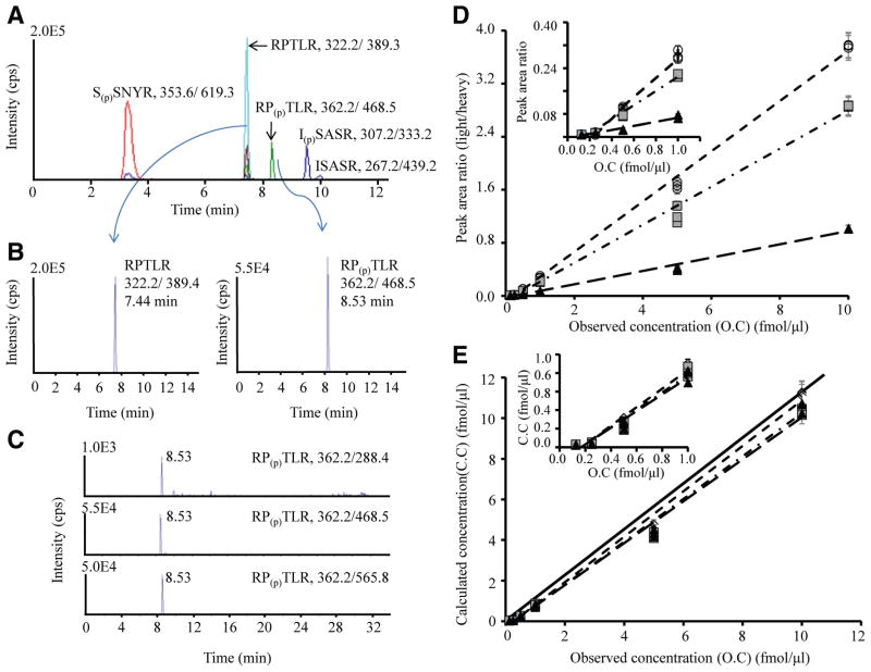
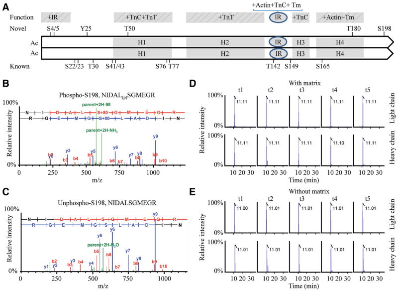
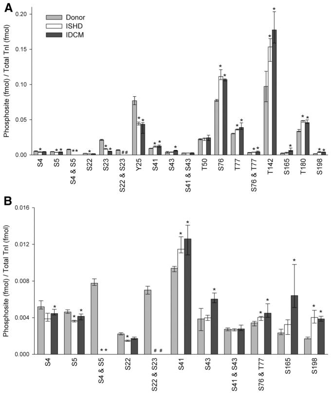
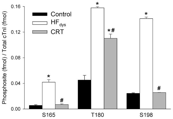
Methods and results: On the basis of in silico prediction and liquid chromatography/mass spectrometry data, 14 phosphorylation sites on cardiac troponin I, including 6 novel residues (S4, S5, Y25, T50, T180, S198), were assessed in explanted hearts from end-stage heart failure transplantation patients with ischemic heart disease or idiopathic dilated cardiomyopathy and compared with samples obtained from nonfailing donor hearts (n=10 per group). Thirty mass spectrometry-based multiple reaction monitoring quantitative tryptic peptide assays were developed for each phosphorylatable and corresponding nonphosphorylated site. The results show that in heart failure there is a decrease in the extent of phosphorylation of the known protein kinase A sites (S22, S23) and other newly discovered phosphorylation sites located in the N-terminal extension of cardiac troponin I (S4, S5, Y25), an increase in phosphorylation of the protein kinase C sites (S41, S43, T142), and an increase in phosphorylation of the IT-arm domain residues (S76, T77) and C-terminal domain novel phosphorylation sites of cardiac troponin I (S165, T180, S198). In a canine dyssynchronous heart failure model, enhanced phosphorylation at 3 novel sites was found to decline toward control after resynchronization therapy.

Conclusions: Selective, functionally significant phosphorylation alterations occurred on individual residues of cardiac troponin I in heart failure, likely reflecting an imbalance in kinase/phosphatase activity. Such changes can be reversed by cardiac resynchronization.


Figures

Figure 1
Figure 1
Development of multiple reaction monitoring assays for the representative cardiac troponin I (cTnI) peptide and its corre-sponding phosphorylated peptide. A typical total ion chromatogram is shown for the multiplex consisting of the peptides S(p)SNYR, RPTLR, RP(p)TLR, ISASR, and I(p)SASR (A). The extracted ion chromatogram for the unphosphorylated peptide (RPTLR with m/z 322.2/389.4) and its corresponding phosphorylated peptide (RP(p)TLR with m/z 362.2/468.5) eluting at 7.44 and 8.53 minutes (B). Three transitions for the unphosphorylated and phosphorylated peptide corresponding to RPTLR at m/z 362.2/288.4, 362.2/468.5, and 362.2/565.8 (C); Table II in the online-only Data Supplement lists the other transitions used. The plot of peak area ratio vs observed concentration (D) or calculated versus observed concentration (E) with linear calibration curve slopes for the 3 transitions of the phosphorylated peptide RP(p)TLR. The ratio is the peak area of phosphorylated peptide RP(p)TLR (unlabeled peptide) divided by the corresponding unphosphorylated (heavy labeled peptide) at 6-point different dilution ratio of concentrations 0.125, 0.25, 0.5, 1, 5, and 10 fmol/μL. The concentration of unlabeled phosphorylation site T142 peptide ranges from subfemtomole to tens of femtomole levels, whereas the concentration of heavy labeled internal standard is kept constant at 1 fmol/μL. Three replicate measurements are represented at each concentration point. Circle indicates transition 1 (m/z 362.2/288.4, y2=0.3789x−0.0931, R2=0.9975); square, transition 2 (m/z 362.2/468.5, y3=0.2868x−0.0745, R2=0.9940); and triangle, transition 3 (m/z 362.2/565.8, y4=0.1005x−0.0267, R2=0.9915). The plots demonstrates good linearity, with slopes falling close to the diagonal black line (theoretical slope=1) and good agreement between the 3 transitions at each concentration point. Inset plots show the lower end of the concentration range. The lower limits of detection and quantification calibration curve generated for this peptide in buffer were 0.1 and 0.3 fmol/μL, respectively.
Figure 2
Figure 2
Alignment of cardiac troponin I (cTnI) sequence and the representative peptide containing a novel phosphorylation site S198 in peptide NIDAL(p)SGMEGR. A schematic of the human cTnI sequence (1–209 amino acid) illustrating function domains and linear positions of the known and novel phosphorylation sites; (A). Residues 1 through 15 interact with the inhibitory region (IR). The domain H1–H4 stands for 4 helixes of cTnI protein. The H1 binds the C terminal of cardiac troponin C (cTnC) and cardiac troponin T (cTnT); H2 binds TnT; H3 binds the N domain of TnC in response to Ca2+ and is referred to as the switch region; and H4 binds actin-tropomyosin. The IR binds both TnC and actin-TM tropomyosin in a Ca2+-dependent manner. The representative mass spectrometry spectrum of the phosphorylated peptide NIDAL(p)SGMEGR (B) containing the site S198 and the corresponding unphosphorylated peptide NIDALSGMEGR (C) of cTnI in left ventricular tissues. Representative multiple reaction monitoring (MRM) spectrum of the phosphorylated peptide NIDAL(p)SGMEGR in matrix (D) or without matrix (E). A 6-point dilution ratio of light/heavy peptide was run for each MRM assay, illustrating the coelution time and relative intensity for the native light peptide phosphorylated S198 peptides, respectively. MRM transitions of t1 to t5 were arranged for the native peptide as in an order (Q1>Q2): t1, 622.8+2>549.7+1 (y5); t2, 622.8+2>573.7+2 (y11–98); t3, 622.8+2>716.8+1 (y6); t4, 622.8+2>918.8+1 (y9–98); and t5, 622.8+2>1016.9+1 (y9); and for the heavy labeled peptide as in an order: t1, 627.8+2>242.4+1(y2); t2, 627.8+2>559.6+1 (y5); t3, 627.8+2>726.8+1 (y6); t4, 627.8+2>839.7+2 (y7); and t5, 627.8+2>928.8+1 (y8), respectively.
Figure 3
Figure 3
Quantification of phosphorylation sites in myocardium obtained from donor and failing hearts. The stoichiometric quantity (fmol phosphorylation/fmol protein) phosphorylation of cardiac troponin I (cTnI) by multiple reaction monitoring assay for donor (light gray), ischemic heart failure (ISHD; white), and dilated cardiomyopathy (IDCM; dark gray; n=10 per group) for all of the sites (A) and the 11 least abundant sites (B). All raw data were calibrated by the synthesized internal standard peptides, converted for femtomole using standard curves, and normalized to the total quantify of TnI in the sample (fmol). Values are median, first quartile, and third quartile. *P<0.05 for ISHD or IDCM vs donor. #Values were below the lower limit of quantification. Phosphorylated T30 and S149 were not detected in any of the 3 groups and were omitted. See Table V and Figure XVIII in the online-only Data Supplement for complementary data.
Figure 4
Figure 4
Quantification of phosphorylated sites in a canine model of heart failure (HF). The stoichiometric quantity (fmol phosphorylation/fmol protein) of phosphorylation sites on cardiac troponin I (cTnI) by multiple reaction monitoring assay in control (black), dyssynchronous HF (HFdys; white), and cardiac resynchronization therapy (CRT; gray; n=4 per group). All raw data were calibrated by the synthesized internal standard peptides and then normalized to total TnI. Error bars indicate the SEM. *P<0.001, HFdys or CRT vs control. #P<0.001, CRT vs HFdys by 1-way ANOVA, followed by Bonferroni multiple-comparisons post hoc test.

Comment in

References

    1. Murphy AM. Heart failure, myocardial stunning, and troponin: a key regulator of the cardiac myofilament. Congest Heart Fail. 2006;12:32–38. - PubMed
    1. Solaro RJ, van der Velden J. Why does troponin I have so many phosphorylation sites? Fact and fancy. J Mol Cell Cardiol. 2010;48:810–816. - PMC - PubMed
    1. Zakhary DR, Moravec CS, Stewart RW, Bond M. Protein kinase A (PKA)-dependent troponin-I phosphorylation and PKA regulatory subunits are decreased in human dilated cardiomyopathy. Circulation. 1999;99:505–510. - PubMed
    1. Kobayashi T, Yang X, Walker LA, Van Breemen RB, Solaro RJ. A non-equilibrium isoelectric focusing method to determine states of phosphorylation of cardiac troponin I: identification of Ser-23 and Ser-24 as significant sites of phosphorylation by protein kinase C. J Mol Cell Cardiol. 2005;38:213–218. - PubMed
    1. Haworth RS, Cuello F, Herron TJ, Franzen G, Kentish JC, Gautel M, Avkiran M. Protein kinase D is a novel mediator of cardiac troponin I phosphorylation and regulates myofilament function. Circ Res. 2004;95:1091–1099. - PubMed

Publication types