. 2012 Dec;108(11):3087-95.
doi: 10.1152/jn.01171.2011. Epub 2012 Sep 12.

Intra- and interhemispheric connectivity between face-selective regions in the human brain

Affiliations

Intra- and interhemispheric connectivity between face-selective regions in the human brain

Jodie Davies-Thompson et al. J Neurophysiol. 2012 Dec.

Abstract

Neuroimaging studies have revealed a number of regions in the human brain that respond to faces. However, the way these regions interact is a matter of current debate. The aim of this study was to use functional MRI to define face-selective regions in the human brain and then determine how these regions interact in a large population of subjects (n = 72). We found consistent face selectivity in the core face regions of the occipital and temporal lobes: the fusiform face area (FFA), occipital face area (OFA), and superior temporal sulcus (STS). Face selectivity extended into the intraparietal sulcus (IPS), precuneus (PCu), superior colliculus (SC), amygdala (AMG), and inferior frontal gyrus (IFG). We found evidence for significant functional connectivity between the core face-selective regions, particularly between the OFA and FFA. However, we found that the covariation in activity between corresponding face regions in different hemispheres (e.g., right and left FFA) was higher than between different face regions in the same hemisphere (e.g., right OFA and right FFA). Although functional connectivity was evident between regions in the core and extended network, there were significant differences in the magnitude of the connectivity between regions. Activity in the OFA and FFA were most correlated with the IPS, PCu, and SC. In contrast, activity in the STS was most correlated with the AMG and IFG. Correlations between the extended regions suggest strong functional connectivity between the IPS, PCu, and SC. In contrast, the IFG was only correlated with the AMG. This study reveals that interhemispheric as well as intrahemispheric connections play an important role in face perception.


Figures
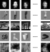
Fig. 1.
Fig. 1.
Examples of stimuli from face and non-face stimulus conditions. From top to bottom: faces, bodies, places, objects, and scrambled images. Face images were taken from the Psychological Image Collection at Stirling (http://www.pics.psych.stir.ac.uk).
Fig. 2.
Fig. 2.
Correlating residual activity between face-selective regions. A: the general linear model (GLM) removes the stimulus-driven activity from the time course. The residual activity was then averaged across participants to ensure there was no remaining stimulus-driven activity. MR, magnetic resonance. B: the residuals were then entered into a correlation analysis to determine functional connectivity between pairs of face-selective regions.
Fig. 3.
Fig. 3.
Average face-selective statistical map thresholded at P < 0.05 (corrected for multiple comparisons). A: location of core face-selective regions (FFA, fusiform face area; OFA, occipital face area; STS, superior temporal sulcus) across subjects in a whole brain analysis. B: location of other regions showing face selectivity (IFG, inferior frontal gyrus; IPS, intraparietal sulcus; PCu, precuneus; SC, superior colliculus; AMG, amygdala).
Fig. 4.
Fig. 4.
A: average correlations (Pearson's r transformed into Fisher's z) in the residual time courses between core face-selective regions within participants. This shows significant interhemispheric correlations between corresponding face regions (lFFA-rFFA, lOFA-rOFA, lSTS-rSTS, where “l” is left and “r” is right) and strong intrahemispheric correlations between the OFA and FFA. B: average correlations in the residual time courses between core face-selective regions across participants. The absence of significant correlations between regions in this analysis shows that the correlations in A are specific to each individual and do not reflect any global trend.
Fig. 5.
Fig. 5.
Effect of stimulus condition on the average correlation between the residual time courses of different core face-selective regions. Correlations between the OFA-FFA were significantly increased when faces were presented.
Fig. 6.
Fig. 6.
Average correlations (Pearson's r transformed into Fisher's z) in the residual time courses between the core regions and extended regions. This shows strong correlations between the OFA or FFA and the IPS, PCu, and SC. In contrast, the STS was more strongly correlated with the AMG and IFG.
Fig. 7.
Fig. 7.
Average correlations (Pearson's r transformed into Fisher's z) in the residual time courses between the extended regions across all subjects. This shows significant correlations between the IPS, PCu, and SC.

Similar articles

Cited by

References

    1. Andrews TJ, Clarke A, Pell P, Hartley T. Selectivity for low-level features of objects in the human ventral stream. Neuroimage 49: 703–711, 2010a - PubMed
    1. Andrews TJ, Davies-Thompson J, Kingstone A, Young AW. Internal and external features of the face are represented holistically in face-selective regions of visual cortex. J Neurosci 30: 3544–3552, 2010b - PMC - PubMed
    1. Andrews TJ, Ewbank MP. Distinct representations for facial identity and changeable aspects of faces in the human temporal lobe. Neuroimage 23: 905–913, 2004 - PubMed
    1. Berman MG, Park J, Gonzalez R, Polk TA, Gehrke A, Knaffla S, Jonides J. Evaluating functional localizers: the case of the FFA. Neuroimage 50: 56–71, 2010 - PMC - PubMed
    1. Biswal B, Yetkin FZ, Haughton VM, Hyde JS. Functional connectivity in the motor cortex of resting human brain using echo-planar MRI. Magn Reson Med 34: 537–541, 1995 - PubMed

Publication types