. 2012 Dec;108(12):3196-205.
doi: 10.1152/jn.00514.2012. Epub 2012 Sep 12.

Morphology, PKCδ expression, and synaptic responsiveness of different types of rat central lateral amygdala neurons

Affiliations

Morphology, PKCδ expression, and synaptic responsiveness of different types of rat central lateral amygdala neurons

Taiju Amano et al. J Neurophysiol. 2012 Dec.

Abstract

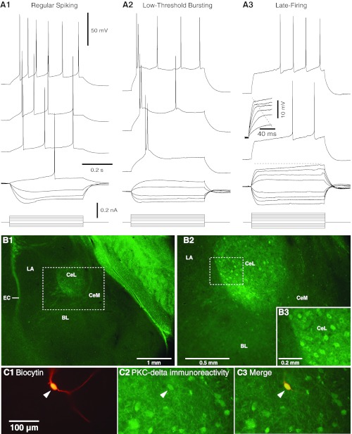
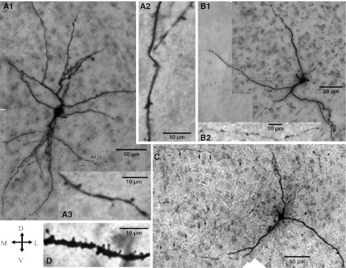
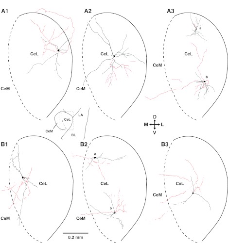
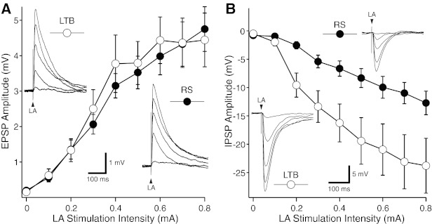
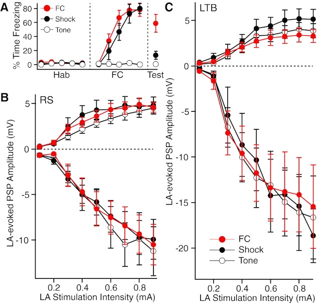
Recent findings implicate the central lateral amygdala (CeL) in conditioned fear. Indeed, CeL contains neurons exhibiting positive (CeL-On) or negative (CeL-Off) responses to fear-inducing conditioned stimuli (CSs). In mice, these cells differ in their expression of protein kinase Cδ (PKCδ) and physiological properties. CeL-Off cells are PKCδ(+) and late firing (LF), whereas CeL-On cells are PKCδ(-) and express a regular-spiking (RS) or low-threshold bursting (LTB) phenotype. However, the scarcity of LF cells in rats raises questions about the correspondence between the organization of CeL in mice and rats. Therefore, we studied the PKCδ expression, morphological properties, synaptic responsiveness, and fear conditioning-induced plasticity of rat CeL neurons. No PKCδ(+) LF cells were encountered, but ≈20-25% of RS and LTB neurons were PKCδ(+). Compared with RS neurons, a higher proportion of LTB cells projected to central medial amygdala (CeM) and they had fewer primary dendritic branches, yet the amplitude of excitatory postsynaptic potentials (EPSPs) evoked by lateral amygdala (LA) stimulation was similar in RS and LTB cells. In contrast, LA-evoked inhibitory postsynaptic potentials (IPSPs) had a higher amplitude in LTB than RS neurons. Finally, fear conditioning did not induce plasticity at LA inputs to RS or LTB neurons. These findings point to major species differences in the organization of CeL. Since rat LTB cells are subjected to stronger feedforward inhibition, they are more likely to exhibit inhibitory CS responses than RS cells. This is expected to cause a disinhibition of CeM fear output neurons and therefore an increase in fear expression.


Figures

Fig. 1.
Fig. 1.
Electroresponsive properties of central lateral (CeL) amygdala neurons. A: voltage responses to depolarizing and hyperpolarizing current pulses in 3 types of CeL cells: regular spiking (RS, A1), low-threshold bursting (LTB, A2), and late firing (LF, A3). When sufficiently depolarized, RS cells generate spike trains that display marked spike frequency accommodation. Although LTB cells also generate accommodating spike trains when depolarized from membrane potentials (Vm) positive to −70 mV, they generate high-frequency (≥100 Hz) spike bursts or doublets followed by single spikes from more negative Vm. In response to suprathreshold depolarizations, LF neurons display a conspicuous delay to firing that is especially pronounced from Vm negative to −75 mV. Also characteristic of LF cells is a marked change in the rising phase of voltage responses to depolarizing current pulses as the stimulus intensity is increased (inset in A3). Prepulse potentials in A1–A3 were −68, −75, and −80 mV, respectively. B: PKCδ immunoreactivity on coronal sections at low (B1) and high (B2) power. Dashed rectangle in B1 is expanded in B2. Dashed rectangle in B2 is expanded in B3. CeM, medial sector of central amygdala; LA, lateral nucleus of amygdala; EC, external capsule; BL, basolateral nucleus of amygdala. C1: CeL neuron labeled with biocytin. C2: post hoc immunohistochemistry revealed that this cell expressed PKCδ (arrowhead). C3: overlay of C1 and C2.
Fig. 2.
Fig. 2.
Morphological properties of LTB and RS neurons recorded in CeL. Cross (bottom left) indicates orientation of the micrographs (D, dorsal; V, ventral; M, medial; L, lateral). A1: RS neuron. The dendritic segments enclosed in the dashed rectangles are shown at a higher magnification in A2 and A3. Although this cell had a low spine density, other RS cells displayed a very high spine density, similar to the LTB cell displayed in D. B and C: LTB neurons. The rectangles in B1 enclose axonal (top) and dendritic (bottom) segments shown at a higher magnification in B2 and D, respectively. Arrows in C point to axonal varicosities.
Fig. 3.
Fig. 3.
Dendritic (black) and axonal (red) patterns of CeL neurons. A: RS cells. B: LTB cells. Scheme between A1 and A2 (bottom) depicts a low-power view of the amygdala; rectangle indicates region depicted in A1–A3 and B1–B3. Cross in center indicates orientation of the drawings.
Fig. 4.
Fig. 4.
LA-evoked responses in LTB and RS CeL neurons. A: average ± SE excitatory postsynaptic potential (EPSP) amplitude (y-axis) as a function of LA stimulation intensity (x-axis) in samples of 27 RS and 16 LTB neurons. In all cases, prestimulus Vm was set to −80 mV. Insets: representative examples of responses seen in LTB (top left) and RS (bottom right) neurons with LA stimulus intensities ranging between 0.2 and 0.8 mA (each response is the average of ≥3 trials). B: average ± SE inhibitory postsynaptic potential (IPSP) amplitude (y-axis) as a function of LA stimulation intensity (x-axis) in samples of 12 RS and 10 LTB neurons. In all cases, prestimulus Vm was set to 0 mV. Insets: representative examples of responses seen in LTB (bottom left) and RS (top right) neurons with LA stimulus intensities ranging between 0.2 and 0.8 mA (each response is the average of ≥3 trials). In all tested cells, LA shocks were 0.1 ms in duration.
Fig. 5.
Fig. 5.
Impact of fear conditioning on the responses of CeL neurons to electrical stimuli delivered in LA. A: after habituation (Hab) to the tone conditioned stimulus (CS), 3 groups of rats were either subjected to fear conditioning (FC) or presented with the same number of shocks (Shock) or tones (Tone). Graph plots % time freezing in the 3 groups. A recall test (Test, right) was performed the next day. These animals were not used for the physiological experiments but were treated exactly as the experimental subjects (see materials and methods). B and C: amplitude (y-axis) of LA-evoked EPSPs (positive values) or IPSPs (negative values) as a function of stimulation intensity (x-axis) in RS (B) or LTB (C) neurons.
Fig. 6.
Fig. 6.
Impact of fear conditioning on the responses of CeL neurons to 2 LA stimuli delivered in rapid succession. Rats were subjected to fear conditioning or control procedures (tone only or shock only). The next day, they were deeply anesthetized, their brain was extracted, and coronal sections of the amygdala were prepared as in the previous experiments. In each tested cell, 2 LA stimuli were applied in rapid succession (50-ms intershock interval) and responses were recorded in voltage-clamp mode from a holding potential of −80 mV. A: responses to paired LA stimuli in representative RS (A1) and LTB (A2) neurons. In both cell types, the second LA stimulus generally elicited EPSCs of greater amplitudes than the first. For all cells, we computed a paired-pulse ratio (EPSC2/EPSC1). B: paired-pulse ratio (y-axis) for RS (left) and LTB (right) cells of the 3 behavioral groups (tone only, open bars; FC, red bars; shock only, black bars).

References

    1. Amano T, Unal CT, Pare D. Synaptic correlates of fear extinction in the amygdala. Nat Neurosci 13: 489–494, 2010 - PMC - PubMed
    1. Amir A, Amano T, Pare D. Physiological identification and infralimbic responsiveness of rat intercalated amygdala neurons. J Neurophysiol 105: 3054–3066, 2011 - PMC - PubMed
    1. Bernard JF, Alden M, Besson JM. The organization of the efferent projections from the pontine parabrachial area to the amygdaloid complex: a Phaseolus vulgaris leucoagglutinin (PHA-L) study in the rat. J Comp Neurol 329: 201–229, 1993 - PubMed
    1. Ciocchi S, Herry C, Grenier F, Wolff SB, Letzkus JJ, Vlachos I, Ehrlich I, Sprengel R, Deisseroth K, Stadler MB, Müller C, Lüthi A. Encoding of conditioned fear in central amygdala circuits. Nature 468: 277–282, 2010 - PubMed
    1. Davis M. Pharmacological analysis of fear-potentiated startle. Braz J Med Biol Res 26: 235–260, 1993 - PubMed

Publication types

Substances