. 2012 Dec 6;120(24):4859-68.
doi: 10.1182/blood-2012-01-401893. Epub 2012 Sep 12.

A GWAS sequence variant for platelet volume marks an alternative DNM3 promoter in megakaryocytes near a MEIS1 binding site

Collaborators, Affiliations

A GWAS sequence variant for platelet volume marks an alternative DNM3 promoter in megakaryocytes near a MEIS1 binding site

Sylvia T Nürnberg et al. Blood. .

Abstract

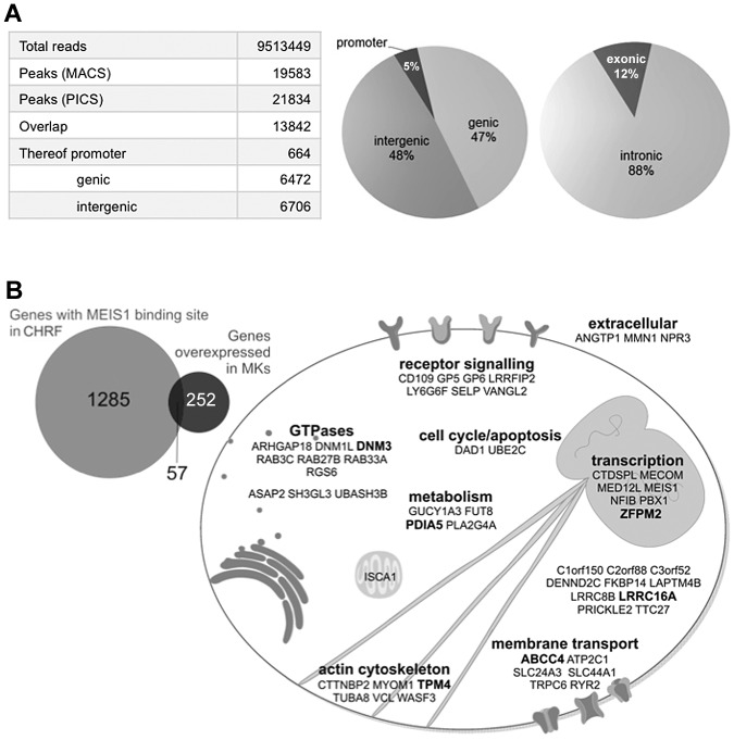
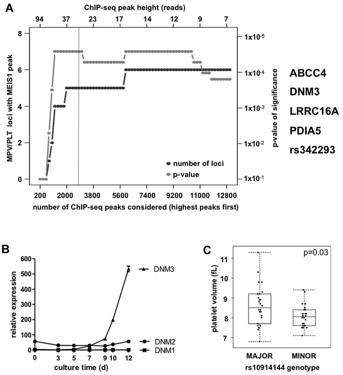
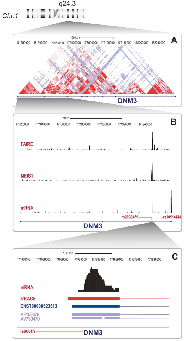
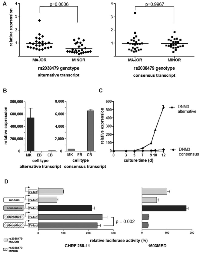
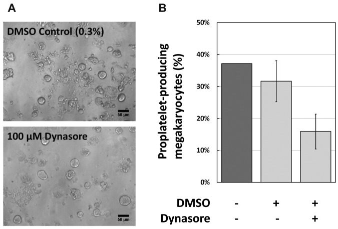
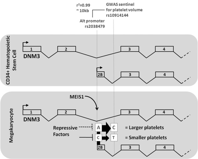
We recently identified 68 genomic loci where common sequence variants are associated with platelet count and volume. Platelets are formed in the bone marrow by megakaryocytes, which are derived from hematopoietic stem cells by a process mainly controlled by transcription factors. The homeobox transcription factor MEIS1 is uniquely transcribed in megakaryocytes and not in the other lineage-committed blood cells. By ChIP-seq, we show that 5 of the 68 loci pinpoint a MEIS1 binding event within a group of 252 MK-overexpressed genes. In one such locus in DNM3, regulating platelet volume, the MEIS1 binding site falls within a region acting as an alternative promoter that is solely used in megakaryocytes, where allelic variation dictates different levels of a shorter transcript. The importance of dynamin activity to the latter stages of thrombopoiesis was confirmed by the observation that the inhibitor Dynasore reduced murine proplatelet for-mation in vitro.


Figures

Figure 1
Figure 1
MEIS1 ChIP-seq identifies genes regulating MK and PLT biology. (A) MEIS1 ChIP-seq in CHRF 288-11 cells identifies mainly intronic and intergenic binding sites. (B) Cellular functions of 57 genes with MEIS1 binding events and overexpressed in MKs. Bold represents 6 loci containing sentinel SNPs associated with MPV or PLT count.
Figure 2
Figure 2
DNM3 harbors MEIS1 binding sites and is associated with differences in MPV. (A) Co-occurrence of MPV and PLT count association SNPs with MEIS1 ChIP-seq peaks. (B) Transcript levels of DNM genes during in vitro differentiation of MKs from human cord blood CD34+ cells, as measured by quantitative real-time RT-PCR (n = 3). Error bars represent SD. (C) MPV of the PLTs of persons according to genotype for sentinel SNP rs10914144 (n = 26 of 26). Mann-Whitney test: P = .03.
Figure 3
Figure 3
The DNM3 locus harbors a regulatory element and an alternative exon in annotated intron 2. (A) DNM3 gene locus showing CEU LOD plot (HapMap release 22). (B) Close-up of gene region, including exons 1-4 with regional plots from FAIRE-seq, MEIS1 ChIP-seq, and RNA-seq data. Also depicted are key SNPs rs10914144 and rs2038479. (C) Further close-up of the region of the MEIS1 peak in intron 2 showing RNA-seq peak, sequence location of the longest 5′RACE clone, alternative transcript ENST00000523513, and 2 EST clones as well as position of variant rs2038479.
Figure 4
Figure 4
Identification of functional SNP rs2038479 in a novel, lineage-specific promoter. (A) Relative DNM3 transcript levels in PLTs from persons homozygous for the major or minor allele of variant rs2038479. Quantitative real-time RT-PCR results with a probe specific for the novel exon 2B-exon 3 boundary (left) versus the annotated exon 2-exon 3 boundary (right) (n = 24/23, unpaired t test, including Welch correction for unequal variances). (B) Relative transcript abundance of tissues as measured by quantitative PCR. MK indicates MK; EB, erythroblast; and CB, cerebellum. n = 3. Error bars represent SD, relative to EB = 1. (C) Quantitative PCR comparison of 2 major DNM3 transcripts during MK maturation (n = 3). Error bars represent SD, relative to annotated transcript day 0 = 1. (D) In vitro dual Luciferase reporter assay of promoter activity of the region 5′ of alternative exon 2B, including variant rs2038479 compared with the consensus promoter 5′ of exon 1. Left: Megakaryocytic CHRF cells. Right: Medulloblastoma cells 1603MED, relative to empty firefly vector = 100%. Error bars represent SD. The significance of the effect of the variant at the alternative promoter (P = .002) was determined using a linear mixed model based on 3 independent experiments with 3 replicates per experiment.
Figure 5
Figure 5
Inhibition of dynamin in MK cultures. (A) Representative phase-contrast photomicrographs from the 2 treatments shown. DMSO was used at 0.3% final, DynaSore at 100μM final (taken with a 20× objective). Bar represents 50 μm. (B) The proportion of murine MK developing pro-PLTs after 4 days culture of fetal liver cells, quantified as stated in “Methods.”
Figure 6
Figure 6
From a PLT volume, GWAS variant at rs10914144 in the DNM3 locus to the discovery of the functional variant rs2038479 at a MEIS1/RUNX1 binding site. A schema depicting the transcription of the DNM3 gene at 1q24.3 in CD34+ hematopoietic stem cells (top panel) and in MKs (bottom panel), respectively. A GWAS identified variant rs10914144 in intron 2 as being associated with differences in MPV. The MPV of persons homozygous for the major (C) allele of the variant is higher compared with the values observed in persons who are homozygous for the minor (T) allele. Functional annotation of the genome of megakaryocytic cells identified binding sites for the transcription factor MEIS1 in intron 2 at a position of an alternative transcription start site, which is uniquely used in MKs. The same element harbors SNP rs2038479, which is in high linkage disequilibrium with the GWAS sentinel rs10914144. DNM3 transcript levels (represented by the sizes of arrows at the consensus and alternative promoters) in PLTs from persons homozygous major (T) for rs2038479 were significantly lower than in PLTs from homozygous minor ones (C).

Comment in

References

    1. Gieger C, Radhakrishnan A, Cvejic A, et al. New gene functions in megakaryopoiesis and platelet formation. Nature. 2011;480(7376):201–208. - PMC - PubMed
    1. Paul DS, Nisbet JP, Yang T-P, et al. Maps of open chromatin guide the functional follow-up of genome-wide association signals: application to hematological traits. PLoS Genet. 2011;7(6):e1002139. - PMC - PubMed
    1. Moskow JJ, Bullrich F, Huebner K, Daar IO, Buchberg AM. Meis1, a PBX1-related homeobox gene involved in myeloid leukemia in BXH-2 mice. Mol Cell Biol. 1995;15(10):5434–5443. - PMC - PubMed
    1. Thorsteinsdottir U, Kroon E, Jerome L, Blasi F, Sauvageau G. Defining roles for HOX and MEIS1 genes in induction of acute myeloid leukemia. Mol Cell Biol. 2001;21(1):224–234. - PMC - PubMed
    1. Pineault N, Buske C, Feuring-Buske M, et al. Induction of acute myeloid leukemia in mice by the human leukemia-specific fusion gene NUP98-HOXD13 in concert with Meis1. Blood. 2003;101(11):4529–4538. - PubMed

Publication types

MeSH terms

Substances