. 2012 Nov 1;120(18):3741-9.
doi: 10.1182/blood-2012-06-435362. Epub 2012 Sep 12.

Signaling through cannabinoid receptor 2 suppresses murine dendritic cell migration by inhibiting matrix metalloproteinase 9 expression

Affiliations

Signaling through cannabinoid receptor 2 suppresses murine dendritic cell migration by inhibiting matrix metalloproteinase 9 expression

Sabina Adhikary et al. Blood. .

Abstract

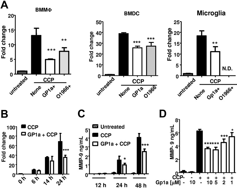
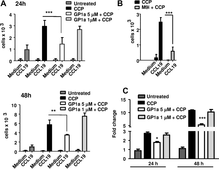
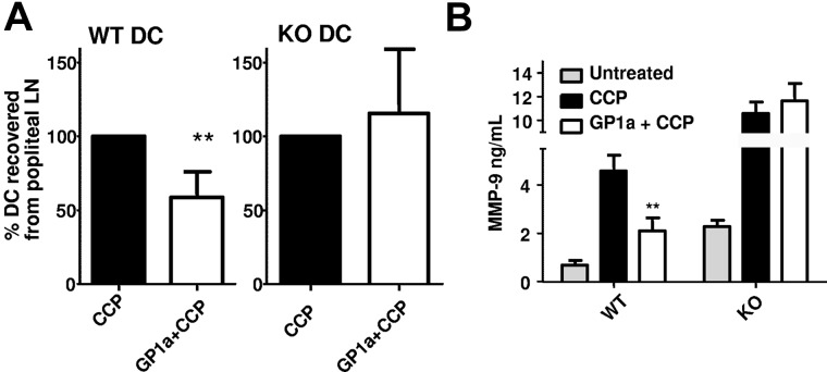
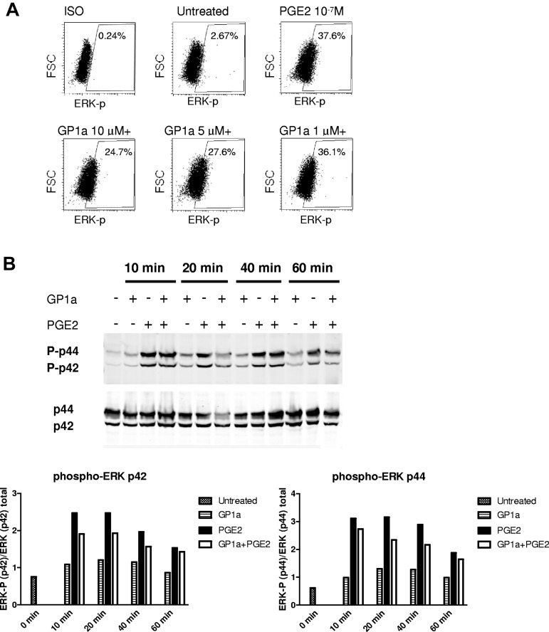
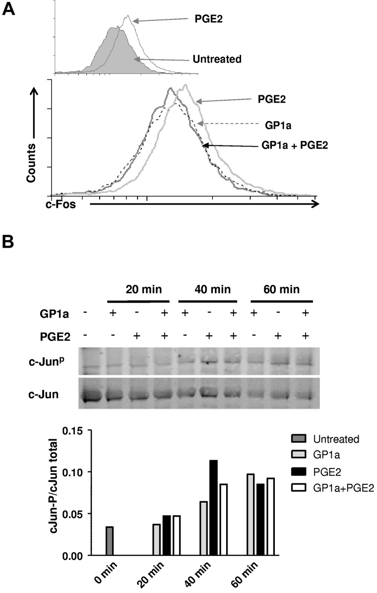
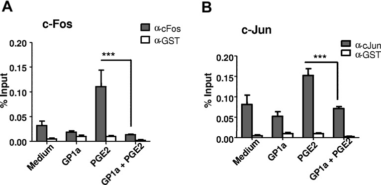
Administration of cannabinoid receptor 2 (CB2R) agonists in inflammatory and autoimmune disease and CNS injury models results in significant attenuation of clinical disease, and reduction of inflammatory mediators. Previous studies reported that CB2R signaling also reduces leukocyte migration. Migration of dendritic cells (DCs) to various sites is required for their activation and for the initiation of adaptive immune responses. Here, we report for the first time that CB2R signaling affects DC migration in vitro and in vivo, primarily through the inhibition of matrix metalloproteinase 9 (MMP-9) expression. Reduced MMP-9 production by DCs results in decreased migration to draining lymph nodes in vivo and in vitro in the matrigel migration assay. The effect on Mmp-9 expression is mediated through CB2R, resulting in reduction in cAMP levels, subsequent decrease in ERK activation, and reduced binding of c-Fos and c-Jun to Mmp-9 promoter activator protein 1 sites. We postulate that, by dampening production of MMP-9 and subsequent MMP-9-dependent DC migration, cannabinoids contribute to resolve acute inflammation and to reestablish homeostasis. Selective CB2R agonists might be valuable future therapeutic agents for the treatment of chronic inflammatory conditions by targeting activated immune cells, including DCs.


Figures

Figure 1
Figure 1
GP1a inhibits Mmp9 expression induced by CCP in myeloid immune cells. (A) BMMΦs, BMDCs, or primary microglia were treated with CCP with or without CB2R agonists GP1a (5μM) or O-1966 (5μM) for 24 hours. RNA was extracted and subjected to qRT-PCR for Mmp9. N.D. indicates not determined. (B) BMDCs were treated with CCP with or without GP1a (5μM), and RNA was extracted at different time points and subjected to qRT-PCR for Mmp9. (C) BMDCs were treated as described in panel B, and supernatants collected at various time points were subjected to MMP-9 ELISA. (D) BMDCs were treated with different concentrations of GP1a for 48 hours, and the resulting supernatants were subjected to MMP-9 ELISA. Data in panels A and B are normalized to housekeeping gene β-actin and presented as fold change compared with untreated samples. *P < .05, **P < .01, *** P < .001 compared with CCP. Data are representative of 2 (A-B) and 4 (C-D) independent experiments.
Figure 2
Figure 2
GP1a prevents DC matrigel migration. DC were treated with CCP with or without GP1a (5 μM or 1 μM) for 24 or 48h (A) or MMP-9 inhibitor (M9i) for 48h (B) and 105 cells were placed in upper Transwell chambers coated wtih Matrigel (100 μg). The bottom chambers were filled with serum free medium with or without CCL19 (100 ng/ml). 3h later migrated cells were collected from the lower chambers and counted by FACS. (C) DC were treated as in A and Mmp9 mRNA expression was determined by qRT-PCR. Data are representative of 3 independent experiments.
Figure 3
Figure 3
GP1a-treated DCs exhibit lower migratory capacity in vivo. (A) DCs generated from Cnr2+/+ (wt) or Cnr2−/− (ko) mice were treated with CCP with or without GP1a (5μM) for 48 hours and labeled with PKH-26. Then, 1 × 10⁶ labeled DCs were injected subcutaneously in the footpads of wt C57BL/6 mice preinjected with 40 ng of TNF-α s.c in the footpads 24 hours earlier. Recipient mice received CCP-treated DC (control) in the right footpad and CCP + GP1a–treated DCs in the left footpad. Forty-eight hours later, cells were collected from popliteal lymph nodes, and PKH-labeled cells were analyzed by FACS. Data from 3 different experiments are normalized by plotting the number of labeled cells from the control leg as 100%. (B) Supernatants collected from DC cultures before injection were analyzed for MMP-9 via ELISA.
Figure 4
Figure 4
GP1a reduces PGE2-induced MMP-9 through inhibition of cAMP induction. (A) DCs were treated with cytokine cocktail without PGE2 (CC) or cytokine cocktail + PGE2 (CCP). Culture supernatants collected at different time points were analyzed for MMP-9 by ELISA. (B) DCs were treated with PGE2 (0.1μM) with or without GP1a (5μM). Supernatant collected at different time points were analyzed for MMP-9 by ELISA. (C) DCs (1 × 10⁵ cells) were treated as described in panel B. Cell lysates collected at different time points were analyzed for total cAMP by ELISA. (D) DCs were treated with PGE2 or dbcAMP (10μM) with or without GP1a for 24 hours. Supernatants were analyzed for MMP-9 by ELISA. Data are representative of 3 independent experiments.
Figure 5
Figure 5
GP1a treatment reduces PGE2 induced ERK phosphorylation. (A) DCs were treated with PGE2 with or without different concentrations of GP1a for 20 minutes. Cells were fixed, permeabilized, stained intracellular with phospho-ERK antibody, and analyzed by FACS. (B) DCs were treated with PGE2 with or without GP1a for various time periods. Cells were lysed and analyzed for phosphorylation of ERK by Western blot. Densitometric analyses are plotted in graphs normalizing phospho-ERK to total ERK. Data are representative of 2 (B) and 3 (A) independent experiments.
Figure 6
Figure 6
GP1a treatment reduces both c-Fos induction and c-Jun phosphorylation. (A) DCs treated with PGE2 with or without GP1a for 40 minutes were fixed, permeabilized, stained intracellular with c-Fos antibody, and analyzed by FACS. (Top) Shaded area represents untreated cells. (Bottom) Black line represents GP1a + PGE2, gray line represents PGE2, and broken black line represents GP1a. (B) DCs were treated with PGE2 with or without GP1a for various time periods and analyzed for phospho-c-Jun by Western blot. Densitometric analyses are plotted in graphs normalizing phospho-c-Jun to total c-Jun. Data are representative of 2 (B) and 3 (A) independent experiments.
Figure 7
Figure 7
GP1a treatment leads to decreased binding of c-Fos and c-Jun to the Mmp9 AP-1 sites. DCs were treated with PGE2 with or without GP1a for 1 hour. Cells were fixed, sonicated, and subjected to ChIP analysis using antibodies to c-Fos (A), c-Jun (B), or control IgG. Precipitated DNA was isolated and evaluated by PCR using specific primers for the AP-1 sites in the Mmp9 promoter. ***P < .001 compared with PGE2. Data are representative of 2 independent experiments.

References

    1. Pertwee RG, Howlett AC, Abood ME, et al. International Union of Basic and Clinical Pharmacology. LXXIX. Cannabinoid receptors and their ligands: beyond CB(1) and CB(2). Pharmacol Rev. 2010;62(4):588–631. - PMC - PubMed
    1. Console-Bram L, Marcu J, Abood ME. Cannabinoid receptors: nomenclature and pharmacological principles. Prog Neuropsychopharmacol Biol Psychiatry. 2012;38(1):4–15. - PMC - PubMed
    1. Kubajewska I, Constantinescu CS. Cannabinoids and experimental models of multiple sclerosis. Immunobiology. 2010;215(8):647–657. - PubMed
    1. Basu S, Dittel BN. Unraveling the complexities of cannabinoid receptor 2 (CB2) immune regulation in health and disease. Immunol Res. 2011;51(1):26–38. - PMC - PubMed
    1. Zhang M, Martin BR, Adler MW, Razdan RK, Jallo JI, Tuma RF. Cannabinoid CB(2) receptor activation decreases cerebral infarction in a mouse focal ischemia/reperfusion model. J Cereb Blood Flow Metab. 2007;27(7):1387–1396. - PMC - PubMed

Publication types

MeSH terms

Substances