. 2012 Nov 1;120(18):3803-11.
doi: 10.1182/blood-2012-03-419663. Epub 2012 Sep 12.

Excess F-actin mechanically impedes mitosis leading to cytokinesis failure in X-linked neutropenia by exceeding Aurora B kinase error correction capacity

Affiliations

Excess F-actin mechanically impedes mitosis leading to cytokinesis failure in X-linked neutropenia by exceeding Aurora B kinase error correction capacity

Dale A Moulding et al. Blood. .

Abstract

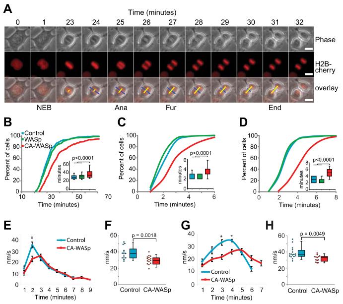
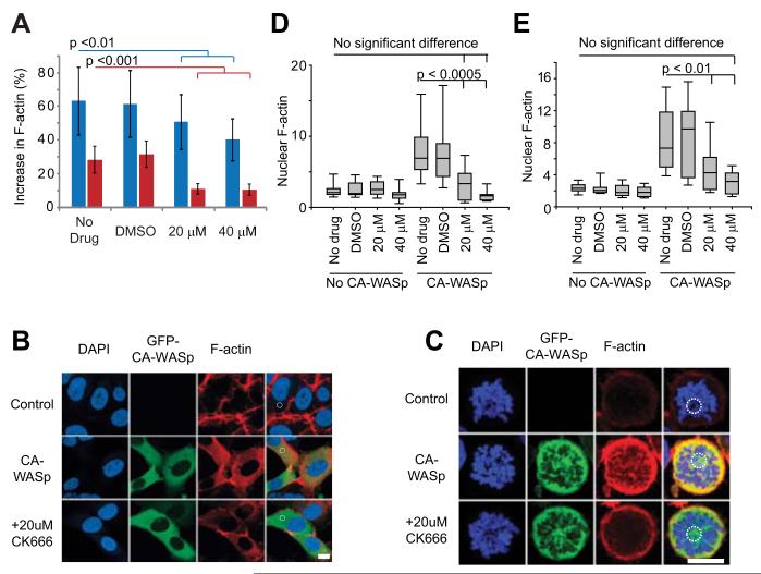
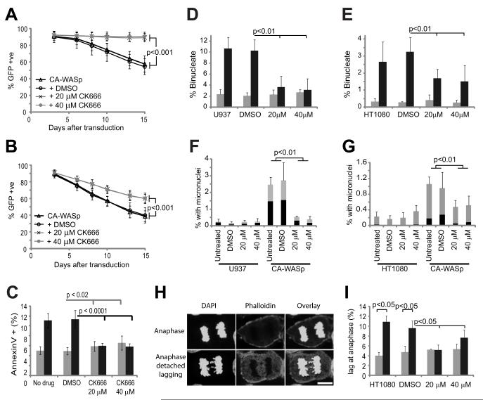
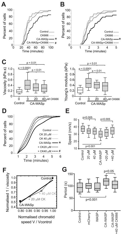
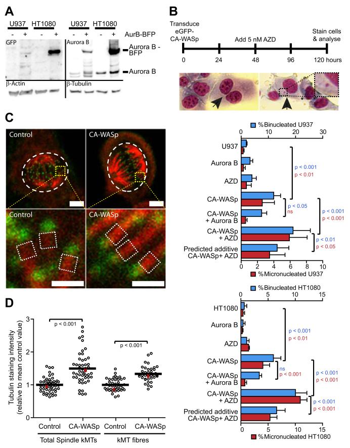
The constitutively active mutant of the Wiskott-Aldrich Syndrome protein (CA-WASp) is the cause of X-linked neutropenia and is linked with genomic instability and myelodysplasia. CA-WASp generates abnormally high levels of cytoplasmic F-actin through dysregulated activation of the Arp2/3 complex leading to defects in cell division. As WASp has no reported role in cell division, we hypothesized that alteration of cell mechanics because of increased F-actin may indirectly disrupt dynamic events during mitosis. Inhibition of the Arp2/3 complex revealed that excess cytoplasmic F-actin caused increased cellular viscosity, slowed all phases of mitosis, and perturbed mitotic mechanics. Comparison of chromosome velocity to the cytoplasmic viscosity revealed that cells compensated for increased viscosity by up-regulating force applied to chromosomes and increased the density of microtubules at kinetochores. Mitotic abnormalities were because of overload of the aurora signaling pathway as subcritical inhibition of Aurora in CA-WASp cells caused increased cytokinesis failure, while overexpression reduced defects. These findings demonstrate that changes in cell mechanics can cause significant mitotic abnormalities leading to genomic instability, and highlight the importance of mechanical sensors such as Aurora B in maintaining the fidelity of hematopoietic cell division.


Figures

Fig. 1
Fig. 1. All stages of mitosis are impeded by CA-WASp expression
(A) Time-lapse images of HT1080 cells expressing fluorescent Histone 2B showing nuclear envelope breakdown (NEB), anaphase onset (Ana) and the start (Fur) and completion (End) of furrowing. Bar = 10 μm. (B) Timing of NEB to anaphase, (C) anaphase to furrow initiation and (D) furrow duration, n > 200 cells over at least 4 independent experiments. (E) Chromatid speed during anaphase (mean +/− SEM, n = 14 control and n = 15 CA-WASp). (F) Peak chromatid speeds. (G) Furrow closure speed (mean +/− SEM n = 16 for control and CA-WASp). (H) Peak furrow closure speeds. Imaging used a Zeiss Axiovert 135 microscope fitted with an environmental chamber at 37°C with 5% CO2. Cells were cultured in phenol red free DMEM with 10% FCS. Fluorochromes were mCherry-H2B and eGFP-WASp. A Hamamatsu ORCA-ER CCD camera was used, and acquisition was controlled with Volocity software. Image analysis used imageJ.
Fig. 2
Fig. 2. CA-WASp requires Arp2/3 activity to increase F-actin production
(A) Percentage increase in total cellular F-actin due to CA-WASp, measured by flow cytometry in HT1080 cells (blue) and U937 cells (red) cultured with DMSO, 20 μM or 40 μM CK666. n = 3 mean +/− SD. (B,C) Confocal images of (B) interphase HT1080 and (C) prometaphase U937 cells showing DNA (DAP1, blue), GFP-CA-WASp (green), and F-actin (red). Dashed white circles show individual ~ 10 μm2 areas used to measure F-actin density. Bar = 10 μm. (D, E) Nuclear F-actin density in prometaphase (D) HT1080 and (E) U937 cells with and without CA-WASp expression cultured in the conditions shown, n > 10 for each condition. Confocal microscopy was performed with a Zeiss LSM 710 inverted confocal microscope with a 40x C-Apochromat NA 1.2 WD 280 mm objective. Image analysis used imageJ software. Fluorochromes were DAP1, eGFP-WASp and Alexafluor-647-phalloidin.
Fig. 3
Fig. 3. Arp2/3 complex inhibition rescues the proliferative and nuclear defects caused by CA-WASp
(A, B) Percentage of (A) U937 and (B) HT1080 cells expressing GFP-CA-WASp 3, 6, 8, 10,13 and 15 days after transduction cultured with DMSO, 20 μM or 40 μM CK666, n = 3 mean +/− SD. (C) Apoptosis measured by flow cytometry of annexinV stained U937 cells after culture for 3-8 days in the conditions shown, with (black) and without (grey) CA-WASp expression, n = 8. Percentage of binucleated (D) U937 and (E) HT1080 cells after culture for 3-10 days in the conditions shown, n = 8. Control (grey bars), CA-WASp (black bars). (F, G) Percentage of micronucleated (F) U937 and (G) HT1080 cells after culture for 3-10 days in the conditions shown, n = 6. Black represents cells with micronuclei alone, grey represents binucleated cells with micronuclei. (H) Lagging chromosomes at anaphase in HT1080 cells. Bar = 10 μm. (I) Percent of HT1080 cells with lagging anaphase chromosomes n = 3 with at least 300 cells analyzed per condition. Confocal microscopy was performed as in Figure 2.
Fig. 4
Fig. 4. Changes in cytoplasmic viscosity correlate with altered mitotic kinetics
(A) Time for control and CA-WASp U937 cells to progress from NEB to anaphase when cultured with DMSO or 20 μM CK666. n > 35 cells per condition over 4 independent experiments. (B) Time for U937 cells from panel A to progress from anaphase to the start of furrowing. (C) Apparent cytoplasmic viscosity and cellular elasticity in HT1080 cells. (D) Time for HT1080 cells to progress from anaphase to the start of furrowing when cultured with DMSO, 20 μM or 40 nM CK666. n > 33 cells per condition over 4 independent experiments. (E) Peak anaphase chromatid velocity of HT1080 cells shown in panel D. (F) Normalized anaphase chromatid velocity as a function of the inverse normalized apparent cytoplasmic viscosity in HT1080 cells. (G) Period of oscillation of prometaphase kinetochore pairs.* significant difference (p<0.01) from control, # significant difference (p<0.01) from CA-WASp.
Fig. 5
Fig. 5. CA-WASp mitotic abnormalities are sensitive to the Aurora B pathway and involve increased kinetochore microtubule intensity
(A) Western blot showing Aurora B-BFP overexpression in HT1080 and U937 cells. (B) Percentage binucleated and micronucleated U937 and HT1080 cells after 4 days expression of GFP-CA-WASp in cells overexpressing Aurora B or inhibition of Aurora B with 5 nM AZD1152 for 48 hours. Images show examples of binucleated and micronucleated HT1080 cells. (C) Confocal images of HT1080 cells blocked in metaphase by proteasome inhibition followed by 10 minutes in ice cold media stained for α-tubulin (red) and Hec1 (green, a kinetochore protein) to show kinetochore microtubule fibres. Bar = 5 μm (top panels) and 1 μm (bottom panels). (D) Quantification of kMT intensity from cells prepared as in (C). Values are relative to the mean control value from each experimental repeat (n≥3). kMT intensity was measured from the fluorescence intensity of the entire spindle (top panels, white dashed areas) and also as an average value of 0.25 μm2 sections of kMT fibres at 10 individual kinetochores per cell (lower panels, white dashed areas). Each point on the chart represents the kMT intensity of an individual cell, with the values from the cells shown in (C) highlighted as red points. Confocal microscopy performed with a Zeiss LSM 710 inverted confocal microscope with a 63x P-Apochromat NA 1.4 oil immersion objective, with fluorochromes eGFP-WASp, Alexafluor 568 and Alexafluor 647.

References

    1. Thrasher AJ, Burns SO. WASP: a key immunological multitasker. Nat Rev Immunol. 2010;10(3):182–192. - PubMed
    1. Moulding DA, Blundell MP, Spiller DG, et al. Unregulated actin polymerization by WASp causes defects of mitosis and cytokinesis in X-linked neutropenia. JExpMed. 2007;204(9):2213–2224. - PMC - PubMed
    1. Devriendt K, Kim AS, Mathijs G, et al. Constitutively activating mutation in WASP causes X-linked severe congenital neutropenia. NatGenet. 2001;27(3):313–317. - PubMed
    1. Ancliff PJ, Blundell MP, Cory GO, et al. Two novel activating mutations in the Wiskott-Aldrich syndrome protein result in congenital neutropenia. Blood. 2006;108(7):2182–2189. - PubMed
    1. Beel K, Cotter MM, Blatny J, et al. A large kindred with X-linked neutropenia with an I294T mutation of the Wiskott-Aldrich syndrome gene. Br J Haematol. 2009;144(1):120–126. - PMC - PubMed

Publication types

MeSH terms

Supplementary concepts