. 2012 Sep 12;32(37):12735-43.
doi: 10.1523/JNEUROSCI.0130-12.2012.

Peroxisome proliferator-activated receptor α mediates acute effects of palmitoylethanolamide on sensory neurons

Affiliations

Peroxisome proliferator-activated receptor α mediates acute effects of palmitoylethanolamide on sensory neurons

Iryna A Khasabova et al. J Neurosci. .

Abstract

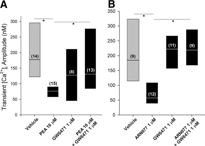
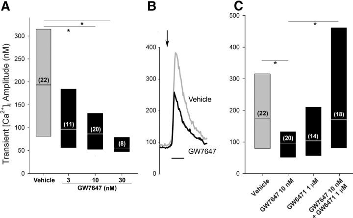
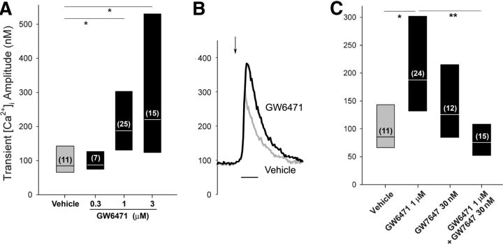
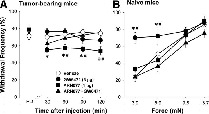
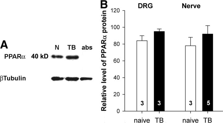
The amplitude of the depolarization-evoked Ca2+ transient is larger in dorsal root ganglion (DRG) neurons from tumor-bearing mice compared with that of neurons from naive mice, and the change is mimicked by coculturing DRG neurons with the fibrosarcoma cells used to generate the tumors (Khasabova et al., 2007). The effect of palmitoylethanolamide (PEA), a ligand for the peroxisome proliferator-activated receptor α (PPARα), was determined on the evoked-Ca2+ transient in the coculture condition. The level of PEA was reduced in DRG cells from tumor-bearing mice as well as those cocultured with fibrosarcoma cells. Pretreatment with PEA, a synthetic PPARα agonist (GW7647), or ARN077, an inhibitor of the enzyme that hydrolyzes PEA, acutely decreased the amplitude of the evoked Ca2+ transient in small DRG neurons cocultured with fibrosarcoma cells. The PPARα antagonist GW6471 blocked the effect of each. In contrast, the PPARα agonist was without effect in the control condition, but the antagonist increased the amplitude of the Ca2+ transient, suggesting that PPARα receptors are saturated by endogenous ligand under basal conditions. Effects of drugs on mechanical sensitivity in vivo paralleled their effects on DRG neurons in vitro. Local injection of ARN077 decreased mechanical hyperalgesia in tumor-bearing mice, and the effect was blocked by GW6471. These data support the conclusion that the activity of DRG neurons is rapidly modulated by PEA through a PPARα-dependent mechanism. Moreover, agents that increase the activity of PPARα may provide a therapeutic strategy to reduce tumor-evoked pain.


Figures

Figure 1.
Figure 1.
Pretreatment with PEA decreased the amplitude of the depolarization-evoked Ca2+ transient in DRG neurons cocultured with fibrosarcoma cells. A, The effect of exogenous PEA (10 μm) was blocked by pretreatment with the PPARα antagonist GW7647. B, An inhibitor of NAAA, the enzyme that degrades PEA, mimicked the effect of PEA. The label for the y-axis is the same as for A. Results are reported as the median and the 25th to 75th percentile range; the numbers in the parentheses indicate the number of neurons that responded to 30 mm KCl. *Different at p < 0.05 (Kruskal–Wallis one-way ANOVA on ranks followed by Dunn's test).
Figure 2.
Figure 2.
A synthetic PPARα agonist decreased the amplitude of the evoked Ca2+ transient in DRG neurons cocultured with fibrosarcoma cells. A, Pretreatment with GW7647 (5 min) decreased the amplitude of the Ca2+ transient in a concentration-dependent manner. The label of the y-axis is the same for B and C. B, Examples of Ca2+ transients evoked in small neurons maintained in the coculture condition during superfusion with vehicle (gray) or 10 nm GW7647 (black). The arrow indicates the time of superfusion with 30 mm KCl. Calibration: 30 s. C, GW6471 blocked the effect of GW7647 on the Ca2+ transient. Results are reported as the median and the 25th to 75th percentile range; the numbers in the parentheses indicate the number of neurons that responded to 30 mm KCl. *Different at p < 0.05 (Kruskal–Wallis one-way ANOVA on ranks followed by Dunn's test).
Figure 3.
Figure 3.
The PPARα antagonist increased the evoked Ca2+ transient in the small DRG neurons in the control condition. A, Pretreatment with GW6471 increased the amplitude of the Ca2+ transient in a concentration-dependent manner in small DRG neurons maintained in the control condition. The label of the y-axis is the same for B and C. B, Examples of Ca2+ transients evoked in small neurons maintained in the control condition during superfusion with vehicle (gray) or 1 μm GW6471 (black). The arrow indicates the time of superfusion with 30 mm KCl. Calibration: 30 s. C, Cotreatment with the PPARα agonist GW7647 blocked the effect of GW6471 on the amplitude of the Ca2+ transient. Results are reported as the median and the 25th to 75th percentile range; the numbers in the parentheses indicate the number of neurons with a positive response to 30 mm KCl. *Different at p < 0.05 (Kruskal–Wallis one-way ANOVA on ranks followed by Dunn's test).
Figure 4.
Figure 4.
The NAAA inhibitor and the PPARα antagonist had differential effects on mechanical sensitivity in tumor-bearing and naive mice. A, Mechanical hyperalgesia occurred in tumor-bearing mice before drug injection [predrug (PD)]. The NAAA inhibitor ARN077 (1 μg, intraplantar, ipsilateral to the tumor) decreased mechanical hyperalgesia, and its effect was blocked by coadministration of the PPARα antagonist GW6471 (3 μg, intraplantar). n = 4–9 mice/treatment. B, In naive mice, mechanical hyperalgesia was measured ipsilateral to intraplantar injection of the PPARα antagonist GW6471 (3 μg) at 1 h after drug injection. The mechanical hyperalgesia was blocked by coinjection of GW6471 with ARN077 (1 μg). The label for the y-axis is the same as for A. Results are expressed as the mean ± SEM. n = 3–11 mice/treatment. *Significantly different from vehicle and #different from GW6471 plus ARN077 at p < 0.02 (two-way ANOVA with Bonferroni's t test).
Figure 5.
Figure 5.
The tumor condition did not change the expression of PPARα protein in DRGs or tibial nerve. A, Representative images of PPARα- and β-Tubulin-immunoreactive bands from a Western blot prepared of tibial nerves from naive (N) and tumor-bearing (TB) mice. B, Quantification of PPARα protein in DRGs and tibial nerves from naive and TB mice. The optical density of PPARα band was divided by the optical density of β-tubulin band and multiplied by 100. Data represent the mean ± SEM. The numbers inside the bars represent the sample size.

References

    1. Agarwal N, Pacher P, Tegeder I, Amaya F, Constantin CE, Brenner GJ, Rubino T, Michalski CW, Marsicano G, Monory K, Mackie K, Marian C, Batkai S, Parolaro D, Fischer MJ, Reeh P, Kunos G, Kress M, Lutz B, Woolf CJ, et al. Cannabinoids mediate analgesia largely via peripheral type I cannabinoid receptors in nociceptors. Nat Neurosci. 2007;10:870–879. - PMC - PubMed
    1. Armirotti A, Romeo E, Ponzano S, Mengatto L, Dionisi M, Karacsonyi C, Bertozzi F, Garau G, Tarozzo G, Reggiani A, Bandiera T, Tarzia G, Mor M, Piomelli D. β-Lactones inhibit N-acylethanolamine acid amidase by S-acylation of the catalytic N-terminal cysteine. ACS Med Chem Lett. 2012;3:422–426. - PMC - PubMed
    1. Berridge MJ, Lipp P, Bootman MD. The versatility and universality of calcium signaling. Nat Rev Mol Cell Biol. 2000;1:11–21. - PubMed
    1. Blanquart C, Barbier O, Fruchart JC, Staels B, Glineur C. Peroxisome proliferator-activated receptor alpha (PPARalpha) turnover by the ubiquitin-proteasome system controls the ligand-induced expression level of its target genes. J Biol Chem. 2002;277:37254–37259. - PubMed
    1. Brown PJ, Stuart LW, Hurley KP, Lewis MC, Winegar DA, Wilson JG, Wilkison WO, Ittoop OR, Willson TM. Identification of a subtype selective human PPARalpha agonist through parallel-array synthesis. Bioorg Med Chem Lett. 2001;11:1225–1227. - PubMed

Publication types