. 2012 Sep 12;32(37):12854-61.
doi: 10.1523/JNEUROSCI.2024-12.2012.

Integrin dynamics produce a delayed stage of long-term potentiation and memory consolidation

Affiliations

Integrin dynamics produce a delayed stage of long-term potentiation and memory consolidation

Alex H Babayan et al. J Neurosci. .

Abstract

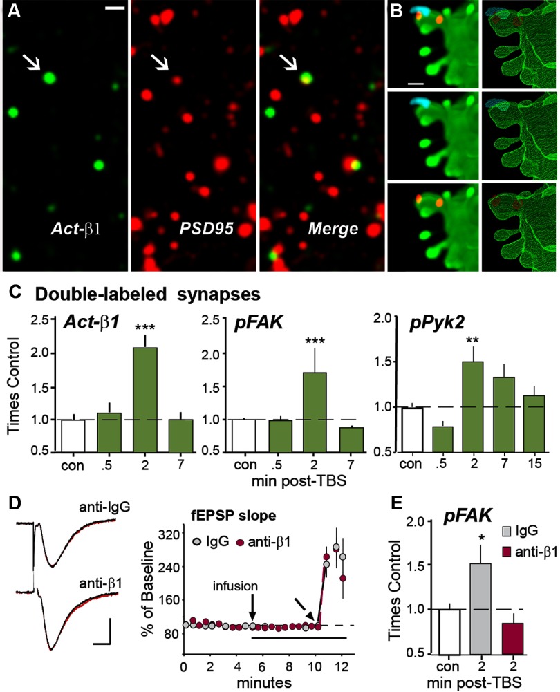
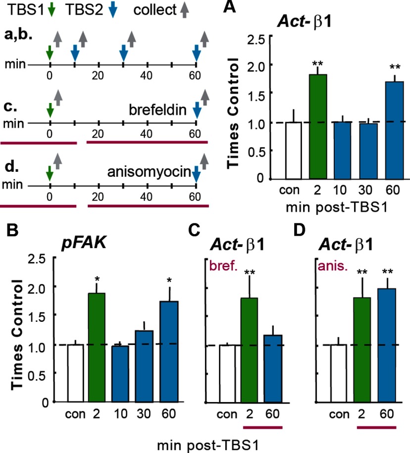
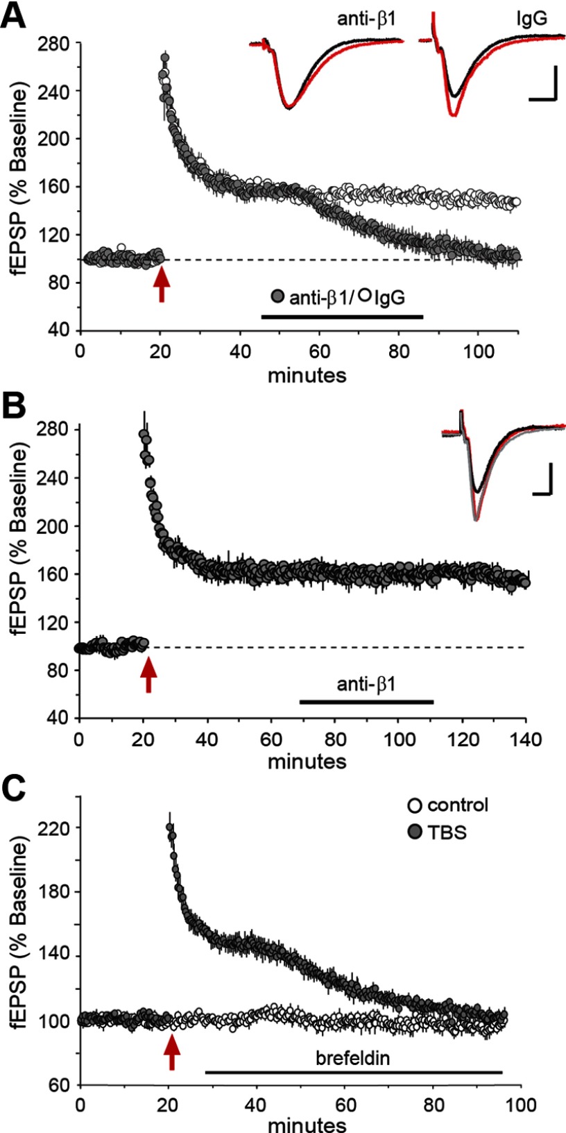
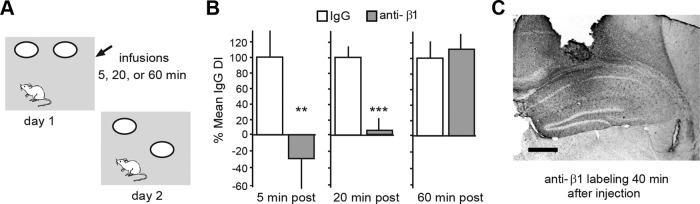
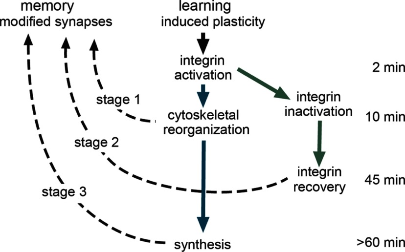
Memory consolidation theory posits that newly acquired information passes through a series of stabilization steps before being firmly encoded. We report here that in rat and mouse, hippocampus cell adhesion receptors belonging to the β1-integrin family exhibit dynamic properties in adult synapses and that these contribute importantly to a previously unidentified stage of consolidation. Quantitative dual immunofluorescence microscopy showed that induction of long-term potentiation (LTP) by theta burst stimulation (TBS) activates β1 integrins, and integrin-signaling kinases, at spine synapses in adult hippocampal slices. Neutralizing antisera selective for β1 integrins blocked these effects. TBS-induced integrin activation was brief (<7 min) and followed by an ∼45 min period during which the adhesion receptors did not respond to a second application of TBS. Brefeldin A, which blocks integrin trafficking to the plasma membrane, prevented the delayed recovery of integrin responses to TBS. β1 integrin-neutralizing antisera erased LTP when applied during, but not after, the return of integrin responsivity. Similarly, infusions of anti-β1 into rostral mouse hippocampus blocked formation of long-term, object location memory when started 20 min after learning but not 40 min later. The finding that β1 integrin neutralization was effective in the same time window for slice and behavioral experiments strongly suggests that integrin recovery triggers a temporally discrete, previously undetected second stage of consolidation for both LTP and memory.


Figures

Figure 1.
Figure 1.
TBS activates synaptic β1 integrins. A, Deconvolved images show localization of immunoreactivities for activated (Act)-β1 and PSD-95 alone, and merged, in field CA1b str. radiatum (bar = 1 μm); arrows indicate a double-labeled synapse. B, Immunolocalization of Act-β1 (blue) and pFAK (red) on a span of GFP-labeled dendrite from field CA1 of a mouse slice. The maximum intensity projection (left) and 3D reconstruction from confocal image Z-stack (right) shows localization of Act-β1 and its signaling kinase on the head of an individual dendritic spine. C, Graphs show numbers of PSD-95-immunopositive (+) synapses double-labeled for Act-β1, pFAK Tyr397 or pPyk2 Tyr402 (normalized to mean control-slice values) are transiently increased after TBS in the CA1b sample field and peak at the 2 min time point (Act-β1 and pFAK ANOVA: p < 0.0001; ***p < 0.001 vs con; pPyk2 ANOVA: p < 0.0003; **p < 0.01 vs con; n = 6–12 slices/group). D, Acute, topical infusions of neutralizing anti-β1, or control IgG, have no effect on baseline fEPSPs or initial potentiation following TBS. Left, Representative traces collected during baseline recordings (black) and 5 min after infusion of IgG or anti-β1 (red; bars: 1 mV, 5 ms). Right, Plot of fEPSP slopes collected over time (TBS applied at angled arrow). E, The same anti-β1 treatment completely suppressed TBS-induced increases in synaptic pFAK (*p < 0.05 vs con; p > 0.05 for anti-β1 group vs con, Student's t test; n = 12 slices/group).
Figure 2.
Figure 2.
Loss and recovery of integrin signaling. Time lines at top left summarize the experimental designs (time lines and associated results are denoted with lower and upper case letters, respectively). A, B, TBS was delivered twice (TBS1, TBS2) with different between-train intervals; slices were collected 2 min after the last TBS and processed for immunofluorescence. A, A single TBS train (green bar) increased numbers of PSD-95+ synapses containing immunoreactivity for Act-β1 integrin relative to counts from yoked control (con) slices given low-frequency (baseline) stimulation only. TBS2 had no effect on numbers of Act-β1+ synapses when delayed by 10 or 30 min after TBS1 but caused the normal increase in Act-β1+ synapses at 60 min (blue bars). B, Similar effects were obtained for TBS-induced increases in synaptic pFAK. C, Groups of slices were perfused (horizontal red bars in c) for 40 min with brefeldin before TBS1 or TBS2. Brefeldin had no effect on integrin activation by TBS1 but blocked that produced by TBS2. D, The design was the same as in C except that slices were treated with anisomycin. The anisomycin infusion had no effect on β1 activation by TBS1 or TBS2. ANOVA: p < 0.001 for all graphs; *p < 0.05 and **p < 0.01 vs con; ≥6 slices/group.
Figure 3.
Figure 3.
Integrin recovery is necessary for LTP consolidation. Hippocampal slices received stimulation of Schaffer-commissural projections and fEPSPs were recorded from CA1b str. radiatum; at different times following TBS (red arrow), neutralizing antisera to β1 integrin (anti-β1) or brefeldin A were infused into the slice. A, Plot of fEPSPs shows that neutralizing anti-β1 (closed circles), but not control IgG (open circles), destabilized LTP when applied to hippocampal slices during the recovery of integrin signaling (30–60 min post-TBS). Inset traces are fEPSPs recorded before (black) and 90 min after (red) TBS; waveforms after antibody-induced LTP reversal are the same as those for baseline responses (calibration: 1 mV, 5 ms). B, Anti-β1 had no effect on LTP when infused after integrin recovery was complete (starting 60 min post-TBS). Traces show fEPSPs collected before TBS (black), 55 min after TBS (gray), and 120 min after TBS (red); those collected 55 and 120 min post-TBS are not detectably different (calibration: 1 mV, 5 ms). C, Post-TBS infusion of brefeldin A (at horizontal bar, initiated 10 min after stimulation) caused a delayed decay of potentiation, similar to effects of anti-β1 shown in A.
Figure 4.
Figure 4.
β1 integrins contribute to the stabilization of object location memory. A, Object location paradigm for studying long-term memory. Panel shows the open field apparatus with two open circles indicating the placement of identical objects; the location of one object is changed on day 2. B, Bar graphs show the discrimination indices of mice treated with neutralizing antisera to β1-integrin (gray bars) expressed as a percentage of measures from paired IgG-treated controls (con; white bars); there were 8–10 mice in each group. As shown, anti-β1 infusion initiated 5 and 20 min after training reduced the time spent with the novel-location object on day 2, whereas anti-β1 infused 60 min after training had no effect (**p < 0.01; ***p < 0.0001 vs same time point IgG con, one-tailed t test). C, Immunostaining for rabbit IgG shows the spread of the anti-β1 in hippocampus for one case killed 40 min after infusion (using techniques used in behavioral studies); the antisera had clearly diffused throughout rostral hippocampus at this time point (scale bar, 0.5 mm).
Figure 5.
Figure 5.
Model for multistage consolidation of synaptic plasticity and memory. Schematic showing the proposed serial stages in LTP and memory consolidation. Prior studies have shown that learning and LTP undergo rapid stabilization (∼10 min) involving integrin-driven reorganization of the subsynaptic cytoskeleton (stage 1). The same events initiate local translation resulting in the later appearance of proteins needed for full stabilization of synaptic changes (stage 3). This constitutes a serial (early and late stages) consolidation pathway (blue arrows). The initial integrin activation is followed by inactivation and then, ∼45 min later, “recovery” of responsivity. The latter event is also necessary for stabilization of synaptic changes, thus producing a parallel path (green arrows) leading to an intermediate stage of consolidation (stage 2).

Similar articles

Cited by

References

    1. Barrett RM, Malvaez M, Kramar E, Matheos DP, Arrizon A, Cabrera SM, Lynch G, Greene RW, Wood MA. Hippocampal focal knockout of CBP affects specific histone modifications, long-term potentiation, and long-term memory. Neuropsychopharmacology. 2011;36:1545–1556. - PMC - PubMed
    1. Baudry M, Bi X, Gall C, Lynch G. The biochemistry of memory: the 26 year journey of a ‘new and specific hypothesis’. Neurobiol Learn Mem. 2011;95:125–133. - PMC - PubMed
    1. Bernard-Trifilo JA, Kramár EA, Torp R, Lin CY, Pineda EA, Lynch G, Gall CM. Integrin signaling cascades are operational in adult hippocampal synapses and modulate NMDA receptor physiology. J Neurochem. 2005;93:834–849. - PubMed
    1. Bourne JN, Harris KM. Nanoscale analysis of structural synaptic plasticity. Curr Opin Neurobiol. 2012;22:372–382. - PMC - PubMed
    1. Bozdagi O, Nagy V, Kwei KT, Huntley GW. In vivo roles for matrix metalloproteinase-9 in mature hippocampal synaptic physiology and plasticity. J Neurophysiol. 2007;98:334–344. - PMC - PubMed

Publication types

Substances