Rapid transepithelial transport of prions following inhalation
- PMID: 22973025
- PMCID: PMC3497678
- DOI: 10.1128/JVI.01930-12
Rapid transepithelial transport of prions following inhalation
Abstract
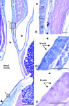
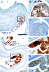
Prion infection and pathogenesis are dependent on the agent crossing an epithelial barrier to gain access to the recipient nervous system. Several routes of infection have been identified, but the mechanism(s) and timing of in vivo prion transport across an epithelium have not been determined. The hamster model of nasal cavity infection was used to determine the temporal and spatial parameters of prion-infected brain homogenate uptake following inhalation and to test the hypothesis that prions cross the nasal mucosa via M cells. A small drop of infected or uninfected brain homogenate was placed below each nostril, where it was immediately inhaled into the nasal cavity. Regularly spaced tissue sections through the entire extent of the nasal cavity were processed immunohistochemically to identify brain homogenate and the disease-associated isoform of the prion protein (PrP(d)). Infected or uninfected brain homogenate was identified adhering to M cells, passing between cells of the nasal mucosa, and within lymphatic vessels of the nasal cavity at all time points examined. PrP(d) was identified within a limited number of M cells 15 to 180 min following inoculation, but not in the adjacent nasal mucosa-associated lymphoid tissue (NALT). While these results support M cell transport of prions, larger amounts of infected brain homogenate were transported paracellularly across the respiratory, olfactory, and follicle-associated epithelia of the nasal cavity. These results indicate that prions can immediately cross the nasal mucosa via multiple routes and quickly enter lymphatics, where they can spread systemically via lymph draining the nasal cavity.
Figures


Similar articles
-
Specificity, Size, and Frequency of Spaces That Characterize the Mechanism of Bulk Transepithelial Transport of Prions in the Nasal Cavities of Hamsters and Mice.J Virol. 2016 Aug 26;90(18):8293-301. doi: 10.1128/JVI.01103-16. Print 2016 Sep 15. J Virol. 2016. PMID: 27384659 Free PMC article.
-
The nasal cavity is a route for prion infection in hamsters.J Virol. 2007 May;81(9):4482-91. doi: 10.1128/JVI.02649-06. Epub 2007 Feb 14. J Virol. 2007. PMID: 17301140 Free PMC article.
-
Direct prion neuroinvasion following inhalation into the nasal cavity.mSphere. 2024 Dec 19;9(12):e0086324. doi: 10.1128/msphere.00863-24. Epub 2024 Nov 29. mSphere. 2024. PMID: 39611853 Free PMC article.
-
Prion protein transgenes and the neuropathology in prion diseases.Brain Pathol. 1995 Jan;5(1):77-89. doi: 10.1111/j.1750-3639.1995.tb00579.x. Brain Pathol. 1995. PMID: 7767493 Review.
-
Pathogenesis of prion diseases.Acta Neuropathol. 2005 Jan;109(1):32-48. doi: 10.1007/s00401-004-0953-9. Epub 2005 Jan 12. Acta Neuropathol. 2005. PMID: 15645262 Review.
Cited by
-
Mucosal transmission and pathogenesis of chronic wasting disease in ferrets.J Gen Virol. 2013 Feb;94(Pt 2):432-442. doi: 10.1099/vir.0.046110-0. Epub 2012 Oct 24. J Gen Virol. 2013. PMID: 23100363 Free PMC article.
-
Intranasal inoculation of white-tailed deer (Odocoileus virginianus) with lyophilized chronic wasting disease prion particulate complexed to montmorillonite clay.PLoS One. 2013 May 9;8(5):e62455. doi: 10.1371/journal.pone.0062455. Print 2013. PLoS One. 2013. PMID: 23671598 Free PMC article.
-
Intra-host mathematical model of chronic wasting disease dynamics in deer (Odocoileus).Prion. 2016 Sep 2;10(5):377-390. doi: 10.1080/19336896.2016.1189054. Epub 2016 Aug 18. Prion. 2016. PMID: 27537196 Free PMC article.
-
A test for Creutzfeldt-Jakob disease using nasal brushings.N Engl J Med. 2014 Aug 7;371(6):519-29. doi: 10.1056/NEJMoa1315200. N Engl J Med. 2014. PMID: 25099576 Free PMC article.
-
The immunobiology of prion diseases.Nat Rev Immunol. 2013 Dec;13(12):888-902. doi: 10.1038/nri3553. Epub 2013 Nov 5. Nat Rev Immunol. 2013. PMID: 24189576 Review.
References
-
- Adams DR, McFarland LZ. 1972. Morphology of the nasal fossae and associated structures of the hamster (Mesocricetus auratus). J. Morphol. 137:161–180 - PubMed
-
- Åkesson CP, et al. 2011. Exosome-producing follicle associated epithelium is not involved in uptake of PrP from the gut of sheep (Ovis aries): an ultrastructural study. PLoS One 6:e22180 doi:10.1371/journal.pone.0022180 - DOI - PMC - PubMed
-
- Andréoletti O, et al. 2002. PrPsc accumulation in placentas of ewes exposed to natural scrapie: influence of foetal PrP genotype and effect on ewe-to-lamb transmission. J. Gen. Virol. 83:2607–2616 - PubMed
-
- Berk DA, Swartz MA, Leu AJ, Jain RK. 1996. Transport in lymphatic capillaries. II. Microscopic velocity measurement with fluorescence photobleaching. Am. J. Physiol. 270(1 Pt 2):H330–H337 - PubMed
Publication types
MeSH terms
Substances
Grants and funding
LinkOut - more resources
Full Text Sources
Research Materials

