. 2012 Dec;86(23):12695-707.
doi: 10.1128/JVI.00647-12. Epub 2012 Sep 12.

The NS1 protein of influenza A virus interacts with cellular processing bodies and stress granules through RNA-associated protein 55 (RAP55) during virus infection

Affiliations

The NS1 protein of influenza A virus interacts with cellular processing bodies and stress granules through RNA-associated protein 55 (RAP55) during virus infection

Bobo Wing-Yee Mok et al. J Virol. 2012 Dec.

Abstract

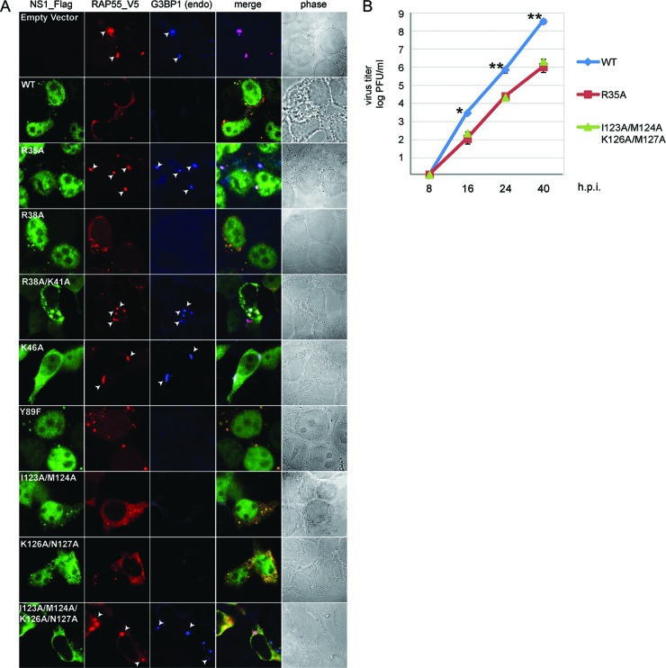
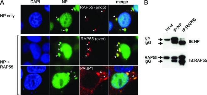
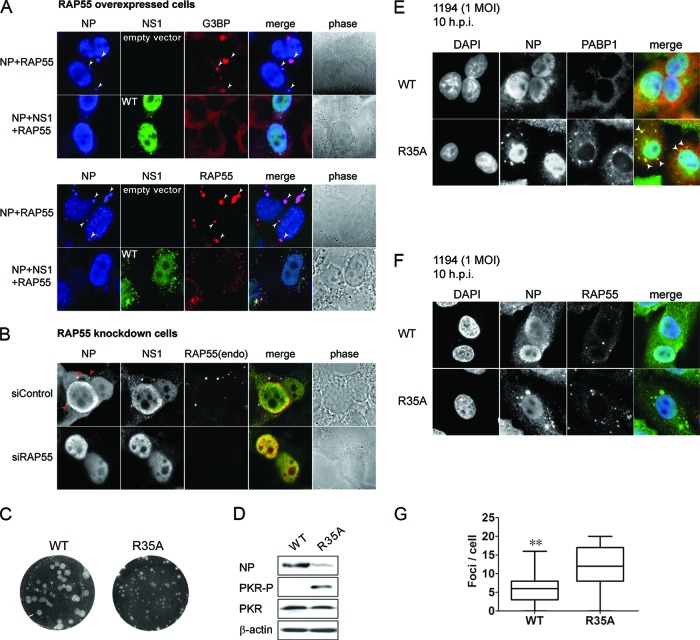
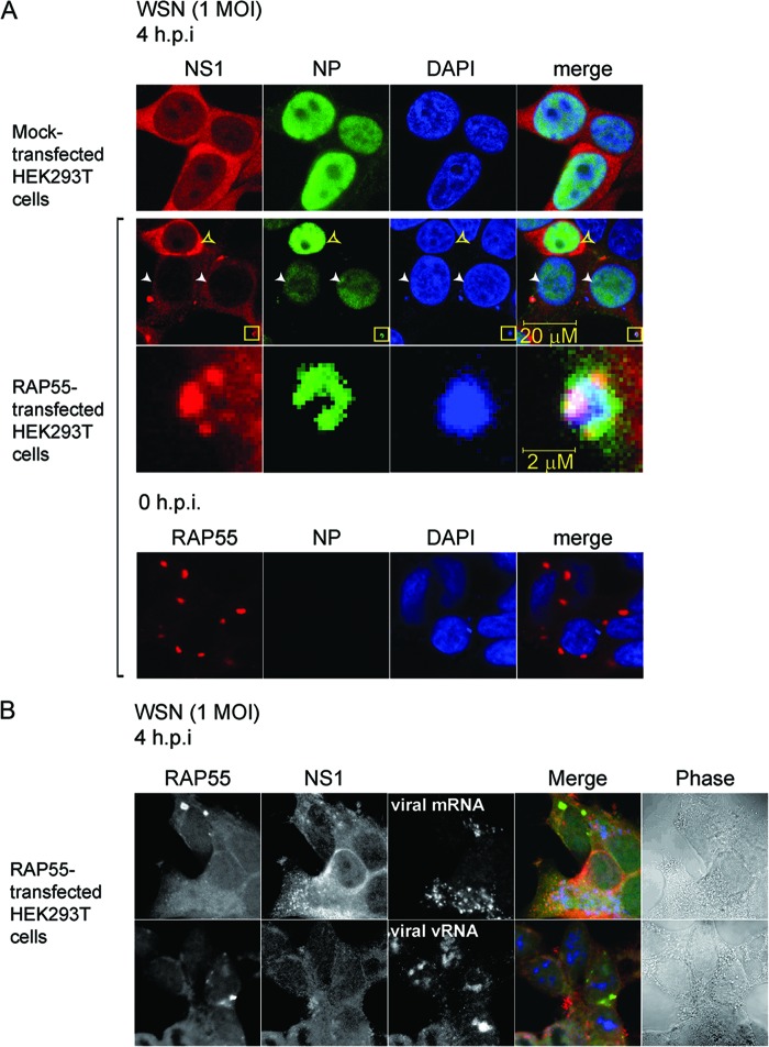
The nonstructural protein (NS1) of influenza A virus performs multiple functions in the virus life cycle. Proteomic screening for cellular proteins which interact with NS1 identified the cellular protein RAP55, which is one of the components of cellular processing bodies (P-bodies) and stress granules. To verify whether NS1 interacts with cellular P-bodies, interactions between NS1, RAP55, and other P-body-associated proteins (Ago1, Ago2, and DCP1a) were confirmed using coimmunoprecipitation and cellular colocalization assays. Overexpression of RAP55 induced RAP55-associated stress granule formation and suppressed virus replication. Knockdown of RAP55 with small interfering RNA (siRNA) or expression of a dominant-negative mutant RAP55 protein with defective interaction with P-bodies blocked NS1 colocalization to P-bodies in cells. Expression of NS1 inhibited RAP55 expression and formation of RAP55-associated P-bodies/stress granules. The viral nucleoprotein (NP) was found to be targeted to stress granules in the absence of NS1 but localized to P-bodies when NS1 was coexpressed. Restriction of virus replication via P-bodies occurred in the early phases of infection, as the number of RAP55-associated P-bodies in cells diminished over the course of virus infection. NS1 interaction with RAP55-associated P-bodies/stress granules was associated with RNA binding and mediated via a protein kinase R (PKR)-interacting viral element. Mutations introduced into either RNA binding sites (R38 and K41) or PKR interaction sites (I123, M124, K126, and N127) caused NS1 proteins to lose the ability to interact with RAP55 and to inhibit stress granules. These results reveal an interplay between virus and host during virus replication in which NP is targeted to P-bodies/stress granules while NS1 counteracts this host restriction mechanism.


Figures

Fig 1
Fig 1
Interaction of NS1 with RNA-associated protein 55 (RAP55). (A) Identification of association of RAP55 and other host factors with influenza virus NS1 by MS analysis. HEK 293T cells were transfected with an expression vector encoding NS1-Flag from A/Vietnam/1194/2004 (H5N1) and then cultured for 48 h. Cell lysates were precipitated with anti-Flag M2 (Sigma) monoclonal antibody, and precipitates were then separated by SDS-PAGE and visualized by silver staining. Each of the separated protein bands was individually excised and analyzed by MS. Only host factors reproducibly identified to associate with NS1 in repeated experiments are shown. (B) (Left) Coimmunoprecipitation of NS1 and RAP55 proteins. V5-tagged RAP55 and Flag-tagged wild-type (WT) and mutant (R35A, R38A/K41A, R46A, Y89F, I123A/M124A, K126A/M127A, and I123A/M124A/K126A/M127A) NS1 expression vectors were cotransfected into HEK 293T cells. RAP55 was used as bait to pull down the NS1 proteins, as described in Materials and Methods. For RNase treatment, 200 μg/ml of RNase A was added to cell lysates, which were then incubated on ice for 30 min prior to addition of antibody. Proteins coprecipitated with mouse anti-V5 antibody (IP) were probed with anti-NS1 rabbit polyclonal and anti-V5 mouse monoclonal antibodies (IB). IP, immunoprecipitation; IB, immunoblotting. (Right) HEK 293T cells were transfected with an expression vector encoding NS1-Flag derived from WSN virus. Cell lysates were collected at 48 h posttransfection and incubated with poly(I-C) immobilized on cyanogen bromide-activated Sepharose beads. Poly(I-C)-bound proteins were isolated and analyzed by Western blotting (IB) using antibodies against NS1. (C) Colocalization of endogenous RAP55 (red) and wild-type or R35A mutant NS1 (green) was assessed by cotransfection of RAP55-V5 and NS1-Flag expression vectors into HEK 293T cells. Expression of RAP55 and NS1 was examined by double immunostaining of cells with anti-RAP55 and anti-Flag mouse monoclonal antibodies (for NS1) at 24 h posttransfection. Signals were visualized using a confocal microscope. (D) Association of NS1 with RAP55 and other P-body-associated proteins. HEK 293T cells were cotransfected with NS1-Flag and Ago1-GFP, Ago2-GFP, or DCP1a-GFP expression vector. Colocalization of NS1 with these P-body components was examined by double immunostaining, using anti-Flag for NS1, anti-RAP55 for endogenous RAP55, and anti-GFP for Ago1, Ago2, and DCP1a. (E) NS1 association with P-bodies in RAP55 knockdown and control HEK 293T cells. Flag-tagged NS1 was expressed in control knockdown (siControl) and RAP55 knockdown (siRAP55) HEK 293T cells, and cells were examined with anti-Flag and anti-RAP55 antibodies. (F) NS1 fails to localize to P-bodies in cells which overexpress a dominant-negative RAP55 mutant (34). Flag-tagged NS1 and wild-type or mutated GFP-fused RAP55 expression vectors were transfected into HEK 293T cells, which were later examined with anti-Flag and anti-GFP antibodies.
Fig 2
Fig 2
Colocalization of wild-type or mutant NS1 proteins with overexpressed RAP55 and suppression of RAP55-associated stress granule formation by different versions of NS1. HEK 293T cells were cotransfected with V5-tagged RAP55 and wild-type and mutated Flag-tagged NS1 expression vectors (NS1 mutants are shown in Fig. 1B). Signals were examined by double immunostaining using anti-Flag and anti-V5 antibodies at 16 to 20 h posttransfection. Formation of stress granules was detected using an anti-mouse monoclonal antibody specific for endogenous G3BP1. (B) Growth kinetics of WSN wild-type and mutant (R35A and I123/A/M124A/K125A/M127A) viruses. A549 cells were infected with WSN WT, R35A mutant, or I123/A/M124A/K125A/M127A mutant virus at an MOI of 0.05. Viral titers were determined by plaque assay at the indicated time points. Values represent the means for three independent experiments, and error bars show standard deviations. *, P < 0.05; **, P < 0.005.
Fig 3
Fig 3
RAP55 negatively regulates influenza A virus replication. (A) Effects of RAP55, Ago1, and Ago2 on influenza A virus polymerase complex activity. Plasmids expressing RNP complex components (PB1, PB2, PA, and NP), with or without an expression vector for NS1 derived from A/Vietnam/1194/2004, were cotransfected into HEK 293T cells along with a vector expressing one of the P-body components (RAP55, Ago1, or Ago2), plus a luciferase reporter, as described in Materials and Methods. Knockdown of RAP55 cells was also used to measure the effect of RAP55 on RNP polymerase activity in this experiment. A plasmid expressing Renilla luciferase was cotransfected as an internal control for data normalization. (Top) Luciferase activities were estimated at 20 h posttransfection. Results shown are averages for three independent experiments, and error bars indicate standard deviations. *, P < 0.05; ***, P < 0.001. (Bottom) Whole-cell lysates were analyzed by immunoblotting with antibodies specific for phospho-PKR, total PKR, and NS1, as indicated. (B) RAP55 inhibits influenza virus replication. Immunostaining of NP expression is shown for vector-transfected (upper panels) or RAP55-overexpressing (middle and lower panels) HEK 293T cells infected with the A/Wisconsin/1933 (WSN) strain at an MOI of 0.005. The average percentage of infected cells was calculated from five randomly selected fields (magnification, ×200). NP expression was not detected in cells where RAP55 granules were observed, as indicated by white arrows in the high-power images (lower panels). h.p.i, hours postinfection. (C) Degradation of RAP55 granules during the course of virus infection. Visualization of RAP55 granule dynamics is shown for A549 cells infected with WSN virus at an MOI of 1. Cells were fixed at 0, 3, and 7 h postinfection, as indicated, and then stained with anti-NP and anti-RAP55 mouse monoclonal antibodies. To enhance the image contrast, single staining of NP (green), RAP55 (red), and DAPI (blue) was adjusted to a monochrome presentation in this panel of images. (D) The number of RAP55-associated granules per cell was quantitated for 100 randomly selected WSN-infected A549 cells at each indicated time point. Data observed are shown with a box-and-whisker plot. *, P < 0.05; ***, P < 0.001. (E) Effects of WT and R35A mutant virus infection on expression of RAP55 and other P-body and stress granule components. Cell lysates collected at different time points were subjected to Western blot analysis with various antibodies. Beta-tubulin was used a loading control. (F) Mock-transfected or RAP55-transfected HEK 293T cells were infected with WSN virus at an MOI of 1, and the replication efficiency was analyzed by plaque assay. The average number of PFU was quantified for three individual sets of experiments. **, P < 0.01. (G) Expression of viral NS1 protein in WSN-infected and mock- or RAP55-transfected HEK 293T cells was analyzed by Western blot analysis at the indicated hours postinfection (h.p.i.). Numbers below the α-NS1 panel indicate the relative expression levels of NS1 at 24 h.p.i. compared to the levels in mock-transfected cells. β-Actin was used as a loading control. Quantification of band intensities was performed using Image J software (developed at the National Institutes of Health).
Fig 4
Fig 4
Interaction of NP with RAP55. (A) Colocalization patterns of NP and endogenous and plasmid-expressed RAP55 were examined in HEK 293T cells transfected with expression vectors for V5-tagged RAP55 and Flag-tagged NP. NP and RAP55 complexes were detected by double staining with anti-Flag antibody for NP and anti-RAP55 antibody for endogenous RAP55 (upper panels) or anti-V5 for overexpressed RAP55 protein (middle panels). Formation of NP-associated stress granules was detected by double staining with anti-Flag antibody for NP and anti-PABP1 antibody (lower panels). (B) Coimmunoprecipitation of RAP55 and NP proteins. V5-tagged RAP55 and Flag-tagged NP proteins were coexpressed in HEK 293T cells by plasmid transfection. NP was used as bait to pull down plasmid-expressed RAP55, and vice versa, as described in Materials and Methods.
Fig 5
Fig 5
NS1 inhibits formation of RAP55-associated stress granules. (A and B) NP and RAP55 expression vectors were cotransfected with an NS1 expression plasmid or control vector into HEK 293T cells. Formation of stress granules was examined using antibodies specific for G3BP and RAP55. (A) (Top) Formation of stress granules was revealed by immunostaining for endogenous G3BP protein, as shown by white arrows. (Bottom) Formation of RAP55-associated granules was examined by immunostaining of RAP55, as indicated by white arrows. (B) Localization of NP to P-bodies in the presence of NS1 and RAP55. NP and NS1 expression vectors were cotransfected into RAP55 knockdown and control siRNA-treated HEK 293T cells. Red arrows indicate the localization of NP to cytoplasmic P-bodies. (C) Plaque phenotypes of wild-type NS1 and R35A mutant NS1 viruses. (D) Western blot analysis of A549 cells infected with WT or R35A mutant NS1 virus, using antibodies specific for NP, phospho-PKR, total PKR, and β-actin, as indicated. (E and F) A549 cells were infected with wild-type NS1 or the R35A NS1 mutant of the A/Vietnam/1194/2004 strain at an MOI of 1. At 10 h postinfection, NP and RAP55 colocalization patterns were examined by double immunostaining analysis. Formation of stress granules and P-bodies was detected using anti-PABP1 (E) and anti-RAP55 (F), respectively. Stress granules are indicated by white arrows in the bottom row of panel E. (G) Numbers of RAP55-associated granules were estimated by examining 100 randomly selected cells infected with wild-type or R35A mutant A/Vietnam/1194/2004 virus at 10 h postinfection. Data observed are shown with a box-and-whisker plot. **, P < 0.005.
Fig 6
Fig 6
Detection of cytoplasmic granules composed of NP, NP-bound nucleic acids, and NS1 in RAP55-overexpressing cells. (A) HEK 293T cells were first transfected with a RAP55 expression vector and then infected with WSN virus at an MOI of 1 at 24 h posttransfection. NS1 and NP expression was examined by immunostaining WSN-infected HEK 293T cells in the presence or absence of RAP55 overexpression. White arrowheads indicate cells with low levels of NP expression in RAP55-overexpressing cell cultures. Yellow boxes indicate a complex where colocalization of NS1, NP, and NP-bound nucleic acids occurs; this area is shown in an enlarged view in the third row. Overexpression of RAP55 in HEK 293T cells was examined prior to virus infection (0 h) to ensure that most of the cells had been transfected successfully (bottom row). (B) HEK 293T cells were transfected with RAP55 expression vector and then infected with WSN virus at an MOI of 1. At 4 h postinfection, the NS1 and RAP55 proteins were detected by costaining with rabbit anti-NS1 polyclonal antibodies (red) and mouse anti-RAP55 monoclonal antibodies (green). Viral mRNA and vRNA were detected by FISH as described in Materials and Methods (blue). To enhance the image contrast, single-color panels were adjusted to a monochrome presentation, while the merge panels are shown in color.
Fig 7
Fig 7
Working model for the interplay between NS1 and NP-induced stress granules (SG) and P-bodies (PB) during influenza A virus infection. Influenza virus mRNA is synthesized by a messenger ribonucleoprotein (mRNP) complex in the nucleus and exported to the cytoplasm for protein translation. Expression of NP during virus infection induces assembly of P-bodies and stress granules, which may trap NP and NP-bound viral RNA and thereby stall viral mRNA translation. Expression of NS1 dissociates (or inhibits formation of) NP-associated P-bodies/stress granules and releases NP and NP-bound mRNA, allowing translation of viral proteins.

References

    1. Abdel-Ghafar AN, et al. 2008. Update on avian influenza A (H5N1) virus infection in humans. N. Engl. J. Med. 358:261–273 - PubMed
    1. Albrecht M, Lengauer T. 2004. Novel Sm-like proteins with long C-terminal tails and associated methyltransferases. FEBS Lett. 569:18–26 - PubMed
    1. Aragon T, et al. 2000. Eukaryotic translation initiation factor 4GI is a cellular target for NS1 protein, a translational activator of influenza virus. Mol. Cell. Biol. 20:6259–6268 - PMC - PubMed
    1. Arribere JA, Doudna JA, Gilbert WV. 2011. Reconsidering movement of eukaryotic mRNAs between polysomes and P bodies. Mol. Cell 44:745–758 - PMC - PubMed
    1. Basler CF, et al. 2001. Sequence of the 1918 pandemic influenza virus nonstructural gene (NS) segment and characterization of recombinant viruses bearing the 1918 NS genes. Proc. Natl. Acad. Sci. U. S. A. 98:2746–2751 - PMC - PubMed

Publication types

MeSH terms

LinkOut - more resources