Comparative Study
. 2012 Dec;86(23):12503-11.
doi: 10.1128/JVI.01350-12. Epub 2012 Sep 12.

Herpes B virus, macacine herpesvirus 1, breaks simplex virus tradition via major histocompatibility complex class I expression in cells from human and macaque hosts

Affiliations
Comparative Study

Herpes B virus, macacine herpesvirus 1, breaks simplex virus tradition via major histocompatibility complex class I expression in cells from human and macaque hosts

Mugdha Vasireddi et al. J Virol. 2012 Dec.

Abstract

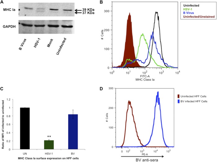
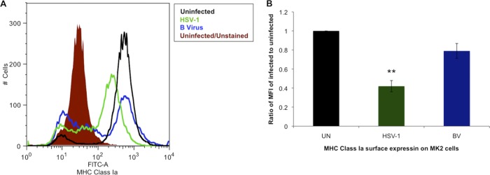
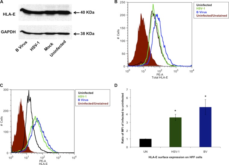
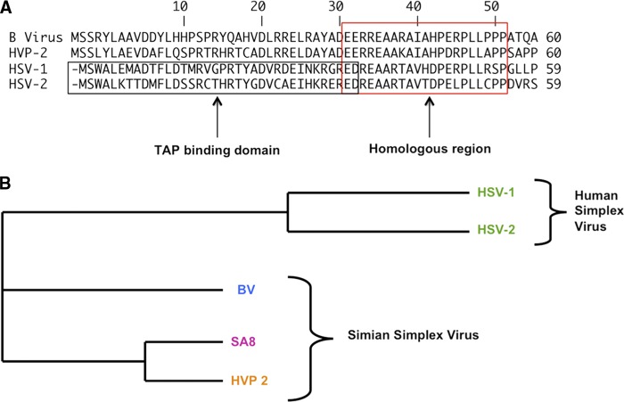
B virus of the family Herpesviridae is endemic to rhesus macaques but results in 80% fatality in untreated humans who are zoonotically infected. Downregulation of major histocompatibility complex (MHC) class I in order to evade CD8(+) T-cell activation is characteristic of most herpesviruses. Here we examined the cell surface presence and total protein expression of MHC class I molecules in B virus-infected human foreskin fibroblast cells and macaque kidney epithelial cells in culture, which are representative of foreign and natural host initial target cells of B virus. Our results show <20% downregulation of surface MHC class I molecules in either type of host cells infected with B virus, which is statistically insignificantly different from that observed in uninfected cells. We also examined the surface expression of MHC class Ib molecules, HLA-E and HLA-G, involved in NK cell inhibition. Our results showed significant upregulation of HLA-E and HLA-G in host cells infected with B virus relative to the amounts observed in other herpesvirus-infected cells. These results suggest that B virus-infected cell surfaces maintain normal levels of MHC class Ia molecules, a finding unique among simplex viruses. This is a unique divergence in immune evasion for B virus, which, unlike human simplex viruses, does not inhibit the transport of peptides for loading onto MHC class Ia molecules because B virus ICP47 lacks a transporter-associated protein binding domain. The fact that MHC class Ib molecules were significantly upregulated has additional implications for host-pathogen interactions.


Figures

Fig 1
Fig 1
MHC class Ia regulation in B virus-infected HFF cells. (A) HFF cells were infected for 18 h with B virus or HSV-1 at an MOI of 10, and the amounts of total MHC class Ia (HLA-A, -B, and -C) antigen in the cell lysates were quantified by using anti-human MHC class I clone W6/32. The band corresponding to a 39-kDa molecular mass represents normal MHC class Ia, while the 37-KDa band represents nonglycosylated MHC class Ia due to incomplete processing of MHC class Ia. (B) Uninfected HFF cells and HSV-1- or B virus-infected (MOI, 10) HFF cells were formaldehyde fixed and stained for detection of surface MHC class Ia by using FITC-conjugated anti-human MHC class Ia W6/32 antibody (FITC-A). The shaded histogram represents uninfected cells unstained for autofluorescence, the black histogram represents uninfected stained cells, the blue histogram represents B virus-infected stained cells, and the green histogram represents HSV-1-infected stained cells. (C) MFIs of B virus (BV)- and HSV-1-infected HFF cells relative to the MFI of uninfected (UN) HFF cells. **, P ≤ 0.01. (D) The blue histogram represents B virus-infected HFF cells fixed, labeled with BV antiserum, and stained with anti-human IgG (PE-A)-conjugated antibody.
Fig 2
Fig 2
MHC class Ia is not downregulated in B virus-infected macaque epithelial cells. (A) MK2 cells were infected with B virus or HSV-1 at an MOI of 10, and formaldehyde-fixed cells were stained for surface MHC class Ia using FITC-conjugated anti-human MHC class I clone W6/32 (FITC-A). The shaded histogram represents uninfected cells unstained for autofluorescence, the black histogram represents uninfected stained cells, the blue histogram represents B virus-infected stained cells, and the green histogram represents HSV-1-infected stained cells. (B) MFIs of B virus (BV)- and HSV-1-infected MK2 cells relative to the MFI of uninfected (UN) HFF cells. **, P ≤ 0.01.
Fig 3
Fig 3
Regulation of HLA-E in HFF cells infected with B virus. (A) Western blot analysis of HFF cells infected with B virus or HSV-1 and probed with anti-HLA-E polyclonal antibody. (B) HFF cells were infected with B virus or HSV-1 at an MOI of 10, and total HLA-E expression was determined by using flow cytometry. The shaded histogram represents uninfected cells unstained for autofluorescence, the black histogram represents uninfected cells stained with PE-conjugated anti-HLA-E antibody clone 3D12 (PE-A), the blue histogram represents B virus-infected stained cells, and the green histogram represents HSV-1-infected stained cells. (C) HLA-E surface expression determined by using flow cytometry. (D) MFIs of B virus (BV)- and HSV-1-infected HFF cells relative to the MFI of uninfected (UN) HFF cells. *, P ≤ 05.
Fig 4
Fig 4
Differential regulation of HLA-G in HFF cells infected with B virus. (A) Western blot analysis of HFF cells infected with B virus or HSV-1 using anti-HLA-G antibody (clone MEM G/1) to detect all forms of HLA-G. (B) HFF cells were infected with B virus or HSV-1 at an MOI of 10, and surface HLA-G1 and -expression was determined by using flow cytometry. The shaded histogram represents uninfected cells unstained for autofluorescence, the black histogram represents uninfected cells stained with PE-conjugated anti-HLA-G clone 87G (PE-A), the blue histogram represents B virus-infected stained cells, and the green histogram represents HSV-1-infected stained cells. (C) MFIs of B virus (BV)- and HSV-1-infected HFF cells relative to that of uninfected (UN) HFF cells. *, P ≤ 02; **, P ≤ 0.005. (D) HLA-G total expression determined by using flow cytometry.
Fig 5
Fig 5
ICP47 sequence comparison between human herpesviruses (HSV-1 and HSV-2) and simian herpesviruses (B virus, HVP-2, and SA8). (A) Sequence homology among HSV-1, HSV-2, B virus, and HVP-2 ICP47 in amino acids 1 to 60. The N-terminal region of HSV ICP47 is the TAP binding domain, which is not conserved in B virus ICP47 or in HVP-2 ICP47. The C-terminal region of ICP47, however, is the most homologous region in all four viruses. (B) Phylogenic tree indicating the divergence of ICP47 between human and simian HSVs. This phylogenic tree was generated using ClustalW program. BV, B virus.

Similar articles

Cited by

References

    1. Abendroth A, Lin I, Slobedman B, Ploegh H, Arvin AM. 2001. Varicella-zoster virus retains major histocompatibility complex class I proteins in the Golgi compartment of infected cells. J. Virol. 75:4878–4888 - PMC - PubMed
    1. Ambagala AP, Gopinath RS, Srikumaran S. 2004. Peptide transport activity of the transporter associated with antigen processing (TAP) is inhibited by an early protein of equine herpesvirus-1. J. Gen. Virol. 85:349–353 - PubMed
    1. Ambagala AP, Hinkley S, Srikumaran S. 2000. An early pseudorabies virus protein down-regulates porcine MHC class I expression by inhibition of transporter associated with antigen processing (TAP). J. Immunol. 164:93–99 - PubMed
    1. Artenstein AW, Hicks CB, Goodwin BS, Jr, Hilliard JK. 1991. Human infection with B virus following a needlestick injury. Rev. Infect. Dis. 13:288–291 - PubMed
    1. Benson PM, Malane SL, Banks R, Hicks CB, Hilliard JK. 1989. B virus (Herpesvirus simiae) and human infection. Arch. Dermatol. 125:1247–1248 - PubMed

Publication types

MeSH terms

Substances