. 2012 Nov;26(11):1896-906.
doi: 10.1210/me.2012-1188. Epub 2012 Sep 12.

The different ligand-binding modes of relaxin family peptide receptors RXFP1 and RXFP2

Affiliations

The different ligand-binding modes of relaxin family peptide receptors RXFP1 and RXFP2

Daniel J Scott et al. Mol Endocrinol. 2012 Nov.

Abstract

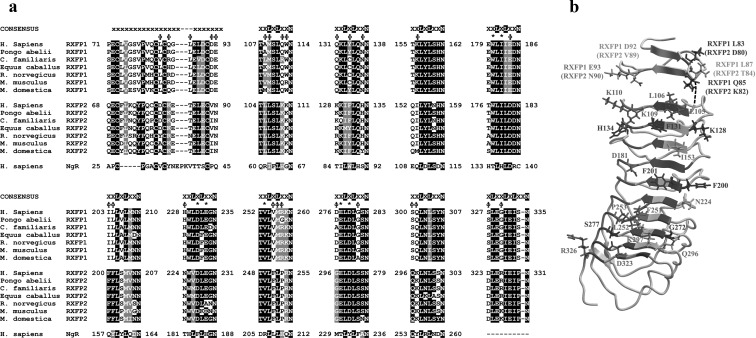
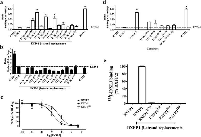
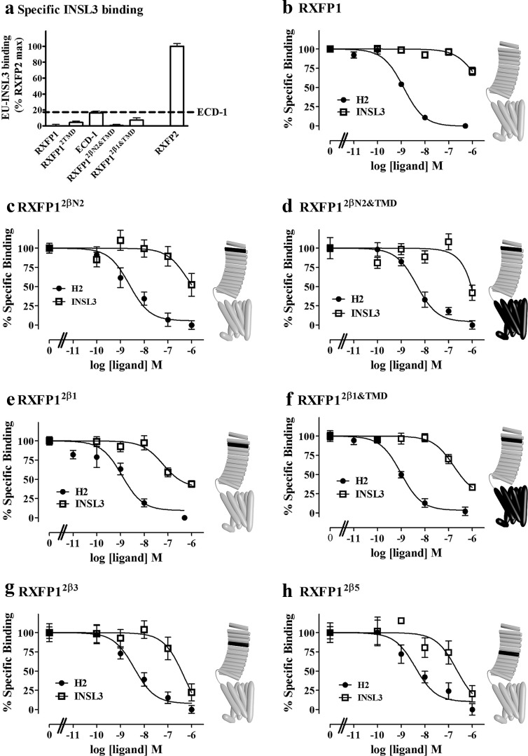
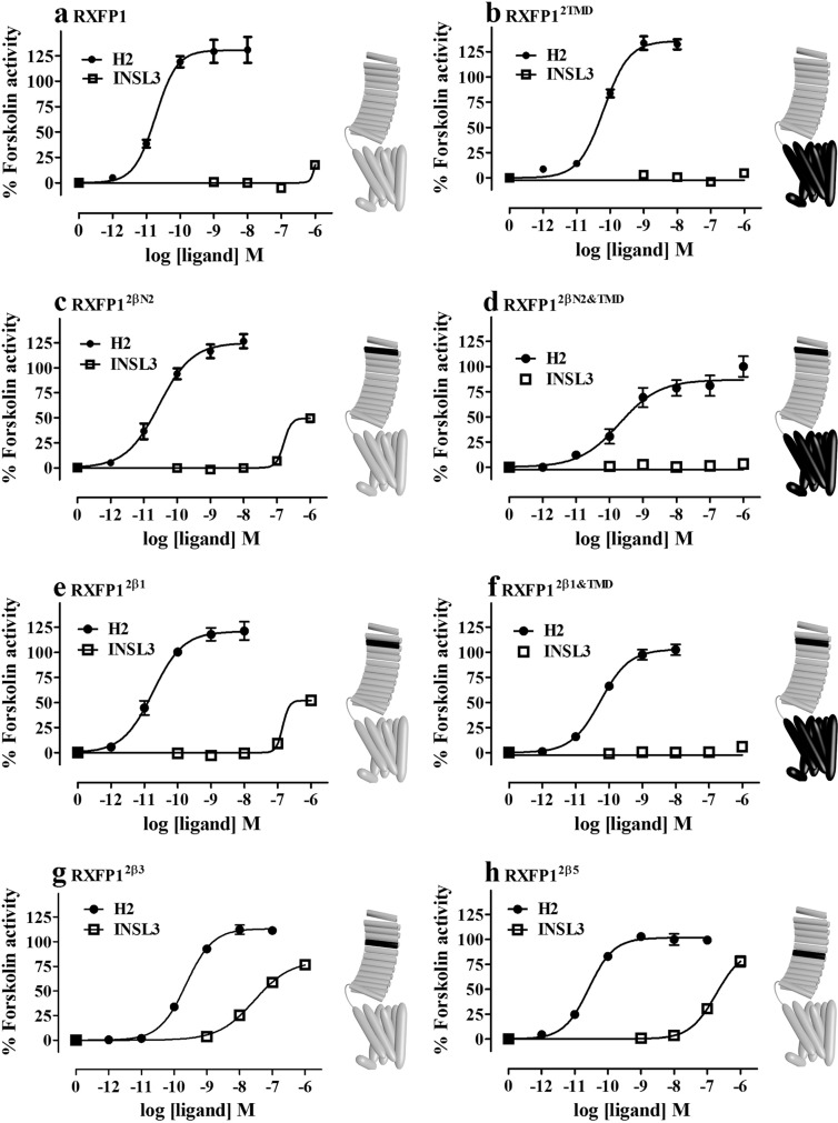
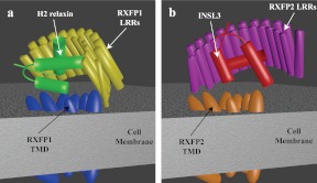
Relaxin and insulin-like peptide 3 (INSL3) are peptide hormones with a number of important physiological roles in reproduction, regulation of extracellular matrix turnover, and cardiovascular function. Relaxin and INSL3 mediate their actions through the closely related G-protein coupled receptors, relaxin family peptide receptors 1 and 2 (RXFP1 and RXFP2), respectively. These receptors have large extracellular domains (ECD) that contain high-affinity ligand-binding sites within their 10 leucine-rich repeat (LRR)-containing modules. Although relaxin can bind and activate both RXFP1 and RXFP2, INSL3 can only bind and activate RXFP2. To investigate whether this difference is related to the nature of the high-affinity ECD binding site or to differences in secondary binding sites involving the receptor transmembrane (TM) domain, we created a suite of constructs with RXFP1/2 chimeric ECD attached to single TM helices. We show that by changing as little as one LRR, representing four amino acid substitutions, we were able to engineer a high-affinity INSL3-binding site into the ECD of RXFP1. Molecular modeling of the INSL3-RXFP2 interaction based on extensive experimental data highlights the differences in the binding mechanisms of relaxin and INSL3 to the ECD of their cognate receptors. Interestingly, when the engineered RXFP1/2 ECD were introduced into full-length RXFP1 constructs, INSL3 exhibited only low affinity and efficacy on these receptors. These results highlight critical differences both in the ECD binding and in the coordination of the ECD-binding site with the TM domain, and provide new mechanistic insights into the binding and activation events of RXFP1 and RXFP2 by their native hormone ligands.


Figures

Fig. 1.
Fig. 1.
A, Multiple sequence alignment of the inner faces of the LRR of RXFP1 and RXFP2 from human (H. sapiens), orangutan (P. abelii), dog (C. familiaris), horse (E. caballus), rat (R. norvegicus), mouse (M. musculus), and opossum (M. domestica) and compared with the human Nogo receptor (NgR), which was used for the homology models. Residues involved with INSL3 binding to RXFP2 and conserved in RXFP1 are indicated (*), whereas RXFP2-specific residues that were investigated experimentally are labeled with φ. B, Homology model of the LRR of RXFP2 with receptor-specific residues identified in the multiple sequence alignment highlighted as previously described (7).
Fig. 2.
Fig. 2.
A, INSL3 binding to chimeric ECD-only receptors expressed on the surface of cells. Data are expressed as a ratio of specific Eu-INSL3 binding to the cell surface expression of each construct and are normalized to RXFP2. *, P < 0.01 vs. ECD-1. B, Eu-H2 relaxin binding to chimeric ECD constructs. C, Eu-INSL3 competition binding curves on RXFP2 (solid squares), ECD-1 (black crosses), and ECD-12β1 (open circles). D, INSL3 binding to ECD-1 mutants containing the individual substitutions that constitute ECD-12β1. *, P < 0.01 vs. ECD-1. E, Specific [125I]INSL3 binding to full-length chimeric receptors (with TMD).
Fig. 3.
Fig. 3.
A, Eu-INSL3 binding to full-length chimeric receptors, normalized to that of RXFP2. Eu-H2 relaxin competition binding curves on (B) RXFP1, (C) RXFP12βN2, (D) RXFP12βN2&TMD, (E) RXFP12β1, (F) RXFP12β1&TMD, (G) RXFP12β3, and (H) RXFP12β5 expressed on the surface of cells. Eu-H2 relaxin was competed with either H2 relaxin (solid circles) or INSL3 (open squares). Schematic representations of the receptor chimeras are displayed to the right of relevant graphs. Gray regions indicate RXFP1 sequence with black regions representing the insertion of RXFP2 sequences.
Fig. 4.
Fig. 4.
Dose-response cAMP activity curves were measured from cells expressing (A) RXFP1, (B) RXFP2TMD, (C) RXFP12βN2, (D) RXFP12βN2&TMD, (E) RXFP12β1, (F) RXFP12β1&TMD, (G) RXFP12β3, and (H) RXFP12β5. Cells were stimulated with increasing concentrations of either H2 relaxin (solid circles) or INSL3 (open squares). Schematic representations of the receptor chimeras are displayed to the right of relevant graphs. Gray regions indicate RXFP1 sequence with black regions representing the insertion of RXFP2 sequences.
Fig. 5.
Fig. 5.
Haddock model of INSL3 binding to RXFP2 ECD. RXFP2 ECD is shown as a transparent surface with side chains from key residues involved in the interaction shown as sticks. INSL3 is shown in ribbon representation, highlighting the positioning of the B-chain helix along the concave surface of the ECD. Interacting residues are labeled with single-letter amino acid codes and residue numbers, and the N- and C-termini of the INSL3 A and B chain are labeled with residue numbers for clarity. The figure was generated in PyMol.
Fig. 6.
Fig. 6.
Schematic representation of how RXFP1 and RXFP2 may bind H2 relaxin and INSL3. A, H2 relaxin binds to RXFP1 with the relaxin B chain α-helix binding across the face of the LRR at an angle of 45°. This positions the A chain N-terminal α-helix across the extracellular loops of the TMD of RXFP1. Interactions between the LRR and the TMD of RXFP1 hold the receptor in the conformation needed to coordinate the sites and facilitate H2 relaxin binding. These interactions, however, hinder the binding mode of INSL3 to the ECD, which requires a 90° binding angle between the LRR β-strands and the B chain α-helix (B).

References

    1. Bathgate RAD, Hsueh AJ, Sherwood OD. 2006. Physiology and molecular biology of the relaxin peptide family. In: (Secondary Bathgate RAD, Hsueh AJ, Sherwood OD.) (Neill JD, ed. Physiology of reproduction. 3rd ed San Diego: Elsevier; 679–770
    1. Bathgate RA, Ivell R, Sanborn BM, Sherwood OD, Summers RJ. 2006. International Union of Pharmacology: recommendations for the nomenclature of receptors for relaxin family peptides. Pharmacol Rev 58:7–31 - PubMed
    1. Hsu SY, Liang SG, Hsueh AJ. 1998. Characterization of two LGR genes homologous to gonadotropin and thyrotropin receptors with extracellular leucine-rich repeats and a G protein-coupled, seven-transmembrane region. Mol Endocrinol 12:1830–1845 - PubMed
    1. Scott DJ, Layfield S, Yan Y, Sudo S, Hsueh AJ, Tregear GW, Bathgate RA. 2006. Characterization of novel splice variants of LGR7 and LGR8 reveals that receptor signaling is mediated by their unique LDLa modules. J Biol Chem 281:34942–34954 - PubMed
    1. Hopkins EJ, Layfield S, Ferraro T, Bathgate RA, Gooley PR. 2007. The NMR solution structure of the relaxin (RXFP1) receptor lipoprotein receptor class A module and identification of key residues in the N-terminal region of the module that mediate receptor activation. J Biol Chem 282:4172–4184 - PubMed

Publication types

MeSH terms