. 2012 Nov;26(11):1868-82.
doi: 10.1210/me.2012-1159. Epub 2012 Sep 12.

Identification of a dynamic mitochondrial protein complex driving cholesterol import, trafficking, and metabolism to steroid hormones

Affiliations

Identification of a dynamic mitochondrial protein complex driving cholesterol import, trafficking, and metabolism to steroid hormones

Malena B Rone et al. Mol Endocrinol. 2012 Nov.

Abstract

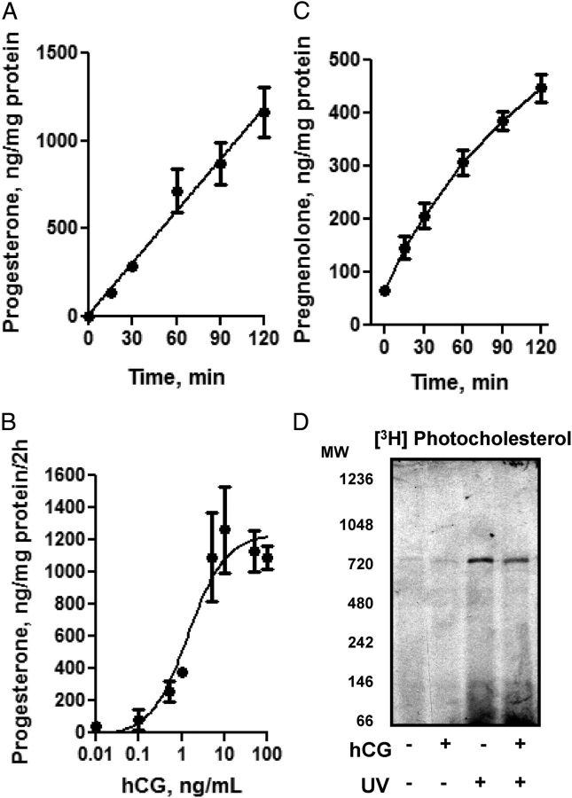
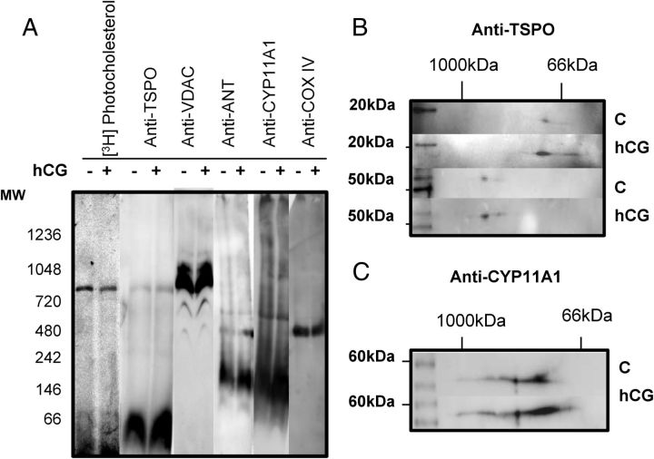
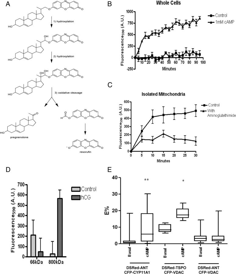
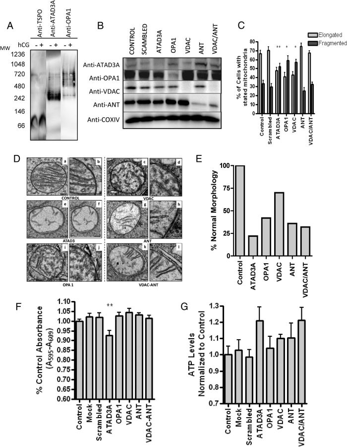
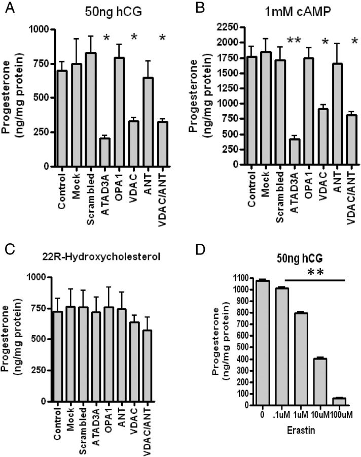
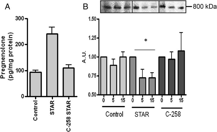
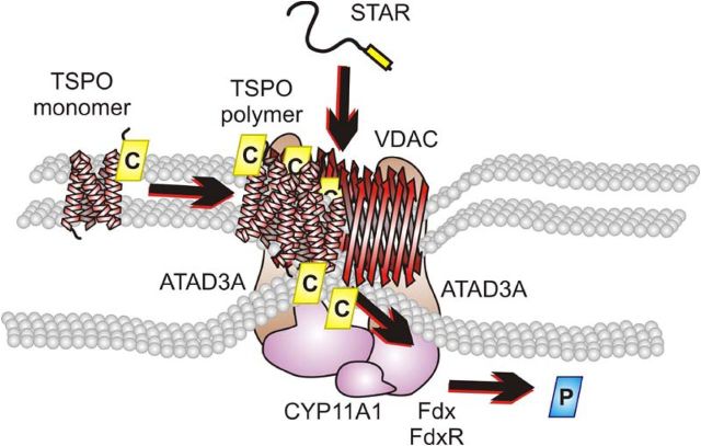
Steroid hormones are critical for organismal development and health. The rate-limiting step in steroidogenesis is the transport of cholesterol from the outer mitochondrial membrane (OMM) to the cytochrome P450 enzyme CYP11A1 in the inner mitochondrial membrane (IMM). Cholesterol transfer occurs through a complex termed the "transduceosome," in which cytosolic steroidogenic acute regulatory protein interacts with OMM proteins translocator protein and voltage-dependent anion channel (VDAC) to assist with the transfer of cholesterol to OMM. It has been proposed that cholesterol transfer from OMM to IMM occurs at specialized contact sites bridging the two membranes composed of VDAC and IMM adenine nucleotide translocase (ANT). Blue native PAGE of Leydig cell mitochondria identified two protein complexes that were able to bind cholesterol at 66- and 800-kDa. Immunoblot and mass spectrometry analyses revealed that the 800-kDa complex contained the OMM translocator protein (18-kDa) and VDAC along with IMM CYP11A1, ATPase family AAA domain-containing protein 3A (ATAD3A), and optic atrophy type 1 proteins, but not ANT. Knockdown of ATAD3A, but not ANT or optic atrophy type 1, in Leydig cells resulted in a significant decrease in hormone-induced, but not 22R-hydroxycholesterol-supported, steroid production. Using a 22-phenoxazonoxy-5-cholene-3-beta-ol CYP11A1-specific probe, we further demonstrated that the 800-kDa complex offers the microenvironment needed for CYP11A1 activity. Addition of steroidogenic acute regulatory protein to the complex mobilized the cholesterol bound at the 800-kDa complex, leading to increased steroid formation. These results identify a bioactive, multimeric protein complex spanning the OMM and IMM unit that is responsible for the hormone-induced import, segregation, targeting, and metabolism of cholesterol.


Figures

Fig. 1.
Fig. 1.
hCG-induced mitochondrial steroid formation. A, Time-dependent effect of 50 ng/ml hCG incubation on MA-10 Leydig cell progesterone formation. B, Dose-dependent production of progesterone after a 2-h incubation of stated concentration of hCG on MA-10 Leydig cells. Results shown in A and B are means ± sem (n = 2). C, Cultured MA-10 cells were washed with serum-free medium and treated with hCG (1 nm) and aminoglutethimide (0.76 mm). At the indicated time points mitochondrial were prepared, and the rate of pregnenolone formation was measured, as described in Material and Methods. The results shown are the means ± sem (n = 3). D. Mitochondria were isolated from control or hCG-treated MA-10 cells and incubated with [3H]Photocholesterol for 30 min. Upon completion, half of the sample was cross-linked with a UV light, solubilized with 1% digitonin buffer, and run on a BN-PAGE gel. The gel was transferred to polyvinylidene difluoride and exposed to film, and the [3H]Photocholesterol was identified at 66-kDa and 800-kDa molecular masses. MW, Molecular weight (molecular mass).
Fig. 2.
Fig. 2.
MS Analysis of BN-PAGE gel identifies protein complex at 800 kDa. Mitochondria from control and hCG-treated cells were isolated, and protein complexes were isolated by BN-PAGE. The gel was divided into 18 slices (3 mm in width) and subjected to MS analysis. The resulting protein identification was classified according to gel slice/molecular mass of the BN-PAGE and the average samples identified of the three gel slices from control and hCG treatment. VDAC isoforms (panel A), ANT isoforms (panel B), CYP11A1 (panel C), hexokinase isoforms (panel D), Cox IV (panel E), and electron transport chain supracomplexes (panel F) were each identified at the stated molecular mass with VDAC and CYP11A1 present at 800 kDa.
Fig. 3.
Fig. 3.
A, Identification of cholesterol-binding mitochondrial protein complexes. Mitochondria were isolated and treated as previously stated. Immunoblot analysis of BN-PAGE identified TSPO at 66 and 800 kDa, same as the [3H]Photocholesterol, CYP11A1 at 146 kDa and higher molecular masses, VDAC laddering at 440, 600, and 800 kDa and higher complexes, and CoxIV was located at 440 kDa. B, 2D-SDS-PAGE was performed on BN-PAGE gel strip, identifying TSPO as an 18-kDa monomer at the 66-kDa complex and a 54-kDa polymer at 800-kDa complex. C, 2D SDS-PAGE gel identified CYP11A1 forming a band at 55 kDa. MW, Molecular weight (molecular mass).
Fig. 4.
Fig. 4.
Cholesterol-Resorufin probe detects CYP11A1 activity. A, The enzymatic activity of CYP11A1 on the cholesterol-resorufin probe results in the production of fluorescent resorufin and pregnenolone. B, MA-10 cells incubated with 5 μm of the cholesterol resorufin probe for 24 h, after which the addition of cAMP results in a time-dependent increase in fluorescence; measurement was at 530ex/595em at the stated time points. C, Isolated mitochondria were incubated with 5 μm cholesterol-resorufin probe or cholesterol-resorufin probe and CYP11A1 inhibitor aminoglutethimide (AG). Measurement at 530ex/595em at stated time points demonstrate an increase in fluorescence in mitochondria not incubated with AG. D, Cholesterol-resorufin probe (50 μm) was incubated in control and hCG-stimulated gel slices. After addition of isocitrate to stimulate steroid production, CYP11A1 activity was measured at 530ex/595em, demonstrating increased activity at the 800-kDa complexes upon hCG treatment. E, Transiently transfected MA-10 cells were imaged 48 h after transfection of DsRed-ANT, CFP-CYP11A1 pair; DsRed-ANT, CFP-VDAC pair; or DsRed-TSPO, CFP-VDAC pair. Cells were treated for 2 h with dbcAMP and imaged. Mann-Whitney U test was performed to determine statistical differences of the E% between samples obtained before and after cAMP treatment. Results shown are means ± sem (n = 3); *, P < 0.05 **, P < 0.01.
Fig. 5.
Fig. 5.
Effect of ATAD3A, VDAC, OPA1, and ANT knockdown on MA-10 mitochondrial morphology. A, BN-PAGE immunoblot identifies TSPO, OPA1, and ATAD3A at the 800-kDa molecular mass complex. B, MA-10 cells were transfected with the siRNA targeted for the indicated gene products. After 3 d of treatment, specific protein expression was assessed by immunoblot analysis. Immunoblots shown are representative of three independent experiments. C, The percent elongated vs. tubular mitochondria of the mitochondria treated with stated siRNA as indicated in panel B. One-way ANOVA with Dunnett's Multiple Comparison Test on the difference in percent elongated was performed. Results shown are means ± sem (n = 2; 200 cells scored each); *, P < 0.05, **, P < 0.01. D, Transmission EM images of MA-10 cells treated with or without mitochondrial protein-specific siRNAs. a, c, e, g, I, and k are low magnification images (200 nm) whereas b, d, f, h, j, and i are higher magnification images (100 nm) of representative mitochondria from cells treated as indicated under each panel. E, Quantitative evaluation of altered mitochondrial morphology in response to the various siRNA treatments obtained as described under Materials and Methods. Values are reported as percentage of normal mitochondria relative to control; control, scrambled, and mock have the same percentage of transfection. F, MTT analysis compared with control (%) and (G) ATP levels normalized to control in siRNA-treated cells. Samples are ± sem (n = 2; each point in triplicate); **, P < 0.01.
Fig. 6.
Fig. 6.
Role of ATAD3A, VDAC, OPA1, and ANT in MA-10 steroidogenesis. MA-10 cells treated with various siRNA were stimulated with 50 ng/ml hCG (A) 1 mm dbcAMP (B) or 20 μm 22R-hydroxycholesterol (C). One-way ANOVA with Dunnett's Multiple Comparison Test was performed with the results shown as means ± sem (n = 3); *, P < 0.05, **, P < 0.01. D, Effect of the VDAC permeability inhibitor erastin on MA-10 cell steroid formation. Cells were incubated with the indicated concentrations of erastin and hCG for 2 h before harvesting. Results shown are means ± sem (n = 2; each point in triplicate); **, P < 0.01.
Fig. 7.
Fig. 7.
STAR mobilizes cholesterol bound at the 800-kDa complex. A, Steroid production with isolated mitochondria incubated with in vitro transcription/translation-generated WT or mutant C258 STAR demonstrated that mutant STAR and control samples are unable to stimulate steroid production as compared with WT. B, Isolated mitochondria preloaded with [3H]Photocholesterol were incubated with WT and mutant STAR. Aliquots were removed after 5 and 15 min, cross-linked, and run on BN-PAGE. Decrease in cholesterol binding was observed at 800 kDa in WT STAR but not control or mutant STAR. Results shown are means ± sem (n = 3) or representative blots from at least three experiments. *, P < 0.05.
Fig. 8.
Fig. 8.
Schematic representation of the hormone-responsive steroidogenic metabolon (hormonad). The TSPO monomer was demonstrated to bind cholesterol at 66 kDa. Upon hormonal stimulation, TSPO undergoes polymerization and associates with the 800-kDa complex where it is associated with VDAC. The pooling of cholesterol bound to TSPO is acted upon by STAR where it is able to be translocated to the IMM through the formation of a contact site by VDAC and the mitochondrial matrix protein ATAD3A present in the 800-kDa complex. Cholesterol is then metabolized by CYP11A1, also present in the 800-kDa complex, supported by the electron transfer proteins, ferredoxin and ferredoxin reductase, to form pregnenolone. The proteins function together to form a hormone-dependent mitochondrial metabolon, the hormonad.

References

    1. Srere PA. 1987. Complexes of sequential metabolic enzymes. Annu Rev Biochem 56:89–124 - PubMed
    1. Rone MB , Fan J , Papadopoulos V. 2009. Cholesterol transport in steroid biosynthesis: role of protein-protein interactions and implications in disease states. Biochim Biophys Acta 1791:646–658 - PMC - PubMed
    1. Liu J , Rone MB , Papadopoulos V. 2006. Protein-protein interactions mediate mitochondrial cholesterol transport and steroid biosynthesis. J Biol Chem 281:38879–38893 - PubMed
    1. Hauet T , Yao ZX , Bose HS , Wall CT , Han Z , Li W , Hales DB , Miller WL , Culty M , Papadopoulos V. 2005. Peripheral-type benzodiazepine receptor-mediated action of steroidogenic acute regulatory protein on cholesterol entry into Leydig cell mitochondria. Mol Endocrinol 19:540–554 - PubMed
    1. Bose HS , Lingappa VR , Miller WL. 2002. Rapid regulation of steroidogenesis by mitochondrial protein import. Nature 417:87–91 - PubMed

Publication types

MeSH terms