. 2012 Nov;23(21):4188-202.
doi: 10.1091/mbc.E12-06-0424. Epub 2012 Sep 12.

Proinflammatory cytokine secretion is suppressed by TMEM16A or CFTR channel activity in human cystic fibrosis bronchial epithelia

Affiliations

Proinflammatory cytokine secretion is suppressed by TMEM16A or CFTR channel activity in human cystic fibrosis bronchial epithelia

Guido Veit et al. Mol Biol Cell. 2012 Nov.

Abstract

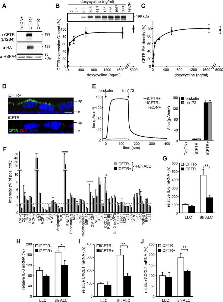
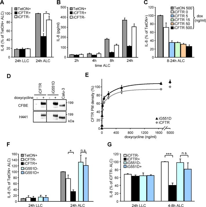
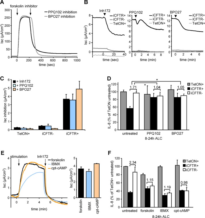
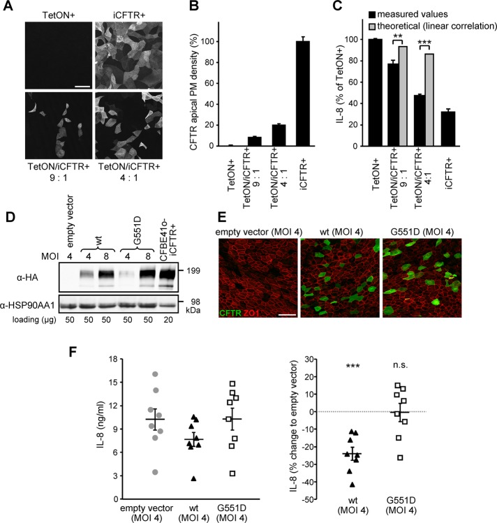
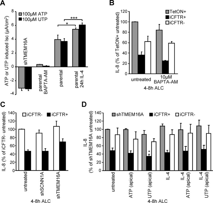
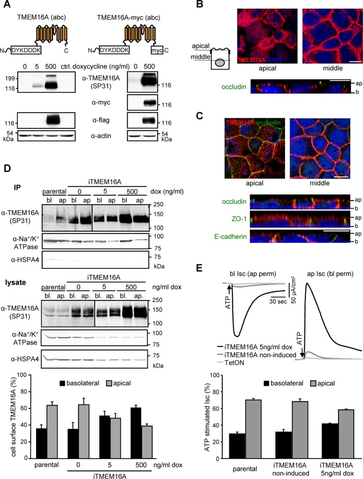
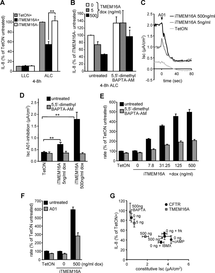
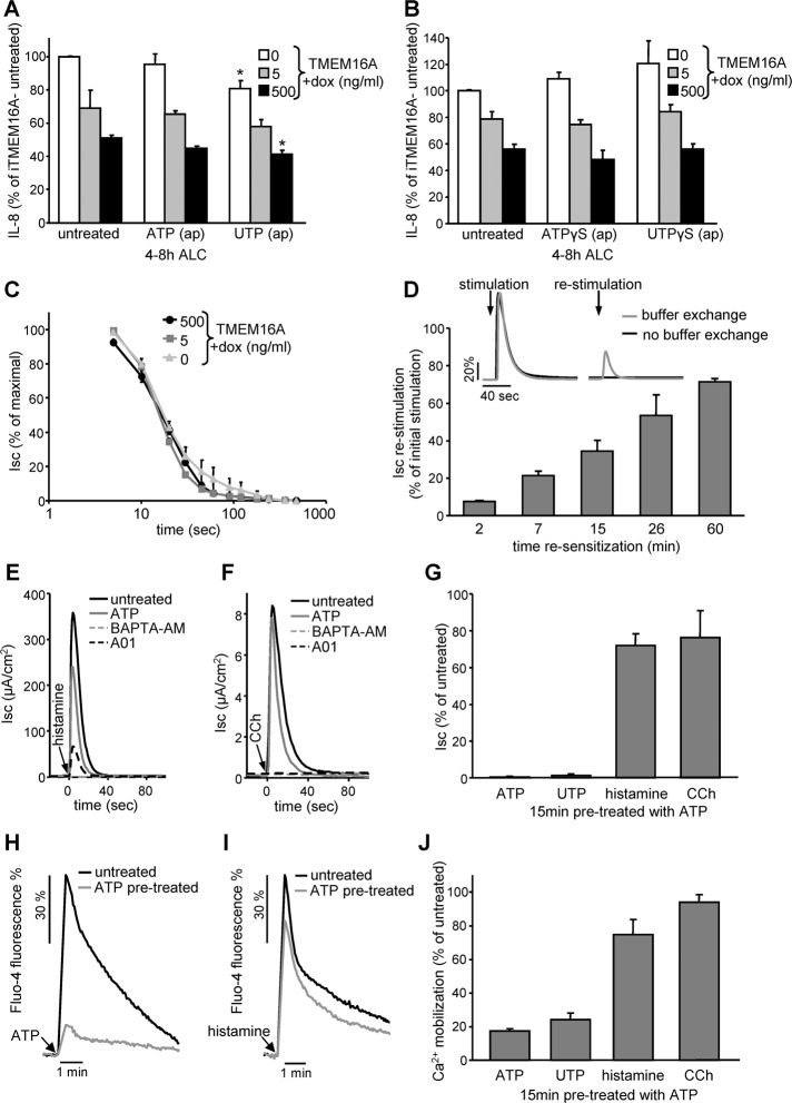
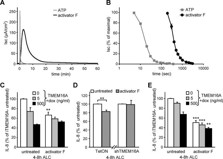
Cystic fibrosis (CF) is caused by the functional expression defect of the CF transmembrane conductance regulator (CFTR) chloride channel at the apical plasma membrane. Impaired bacterial clearance and hyperactive innate immune response are hallmarks of the CF lung disease, yet the existence of and mechanism accounting for the innate immune defect that occurs before infection remain controversial. Inducible expression of either CFTR or the calcium-activated chloride channel TMEM16A attenuated the proinflammatory cytokines interleukin-6 (IL-6), IL-8, and CXCL1/2 in two human respiratory epithelial models under air-liquid but not liquid-liquid interface culture. Expression of wild-type but not the inactive G551D-CFTR indicates that secretion of the chemoattractant IL-8 is inversely proportional to CFTR channel activity in cftr(∆F508/∆F508) immortalized and primary human bronchial epithelia. Similarly, direct but not P2Y receptor-mediated activation of TMEM16A attenuates IL-8 secretion in respiratory epithelia. Thus augmented proinflammatory cytokine secretion caused by defective anion transport at the apical membrane may contribute to the excessive and persistent lung inflammation in CF and perhaps in other respiratory diseases associated with documented down-regulation of CFTR (e.g., chronic obstructive pulmonary disease). Direct pharmacological activation of TMEM16A offers a potential therapeutic strategy to reduce the inflammation of CF airway epithelia.


Figures

FIGURE 1:
FIGURE 1:
Inducible CFTR expression in CF human bronchial epithelia (CFBE) attenuates the proinflammatory cytokines IL-8, IL-6, and CXCL1/2. (A) IB of CFBE transduced with transactivator (TetON) or in combination with inducible CFTR carrying an extracellular 3HA tag (iCFTR) with (+) or without (–) Dox (500 ng/ml)–induced expression for 3 d. (B, C) Dox concentration dependence of CFTR cellular expression determined by IB (B) or by cell surface ELISA (C). Densitometric analysis of the complex-glycosylated CFTR expression is expressed as percentage of the maximum (B, bottom). (D) Apical localization of CFTR (green) in polarized CFBE cells by confocal microscopy. ZO1 was used as tight junction marker (red), and nuclei were stained with DAPI (blue). Ap, apical; b, basal. Bar, 5 μm. (E) Maximal CFTR current (Isc) was measured through stimulation with 10 μM forskolin, followed by inhibition with 20 μM Inh172 (quantification depicted in bar chart) after basolateral permeabilization with amphotericin B (100 μM). (F) Antibody array comparing the basolateral cytokine secretion of polarized CFBE with or without induced CFTR expression. The basolateral media were conditioned for the time period 4–8 h after switch to ALC; the dotted line represents twofold background, and the prespotted positive control was used to normalize between arrays. (G–J) CFTR expression–dependent mRNA levels of IL-8 (G), IL-6 (H), CXCL1 (I), and CXCL2 (J) in CFBE cells kept under LLC or switched for 8 h to ALC, determined by quantitative PCR as described in Materials and Methods. Values show means ± SEM from three independent experiments (B, E, G–J), means ± SD of two independent experiments (F), or means ± SD of one representative experiment (C). *p < 0.05; **p < 0.01; ***p < 0.001.
FIGURE 2:
FIGURE 2:
Functional CFTR expression attenuates IL-8 secretion in human bronchial epithelia under ALC. (A) CFTR expression–dependent basolateral IL-8 secretion of polarized CFBE cells subjected for 24 h to ALC or kept under LLC. (B) Time dependence of the basolateral IL-8 secretion after the switch from LLC to ALC. (C) CFTR expression level dependence of IL-8 secretion of polarized CFBE epithelia kept under ALC. (D) IB of CFBE or H441 cells transduced with inducible wild-type (iCFTR) or G551D (iG551D) CFTR with (+) or without (–) Dox in comparison to the endogenous CFTR expression in Calu3 cells. (E) The dependence of wt and G551D CFTR PM densities on Dox concentration as determined by cell-surface ELISA. (F, G) wt or G551D expression–dependent basolateral IL-8 secretion of polarized (F) CFBE or (G) H441 cells switched to ALC or kept under LLC for the indicated times. IL-8 levels were determined by ELISA. Values represent means ± SEM from three (A, D, F, G) or two (C) independent experiments or means ± SD of one representative experiment (B, E). n.s., not significant; *p < 0.05; ***p < 0.001.
FIGURE 3:
FIGURE 3:
CFTR function is required to suppress IL-8 secretion. (A) PPQ102 (25 μM) or BPO27 (25 μM) inhibits CFTR activity as measured by Isc after stimulation with 10 μM forskolin. (B) The constitutive activity of CFTR in iCFTR+ CFBE14o- cells was unmasked by CFTR inhibitor 172 (Inh172, 20 μM), PPQ102 (25 μM), or BPO27 (25 μM). Measurements were performed in intact monolayers. (C) Quantification of the maximal inhibition as determined in B. (D) Basolateral IL-8 secretion of polarized CFBE cells after channel inhibition with PPQ102 or BPO27. Ratios between basolateral IL-8 secretion with or without induced CFTR expression are depicted, and significance was tested using the ratios derived from three independent experiments. (E) CFTR channel activation in CFBE epithelia with leaked expression of CFTR with forskolin (1 μM), IBMX (0.5 mM), or cpt-cAMP (0.5 mM) determined by Isc. (F) Basolateral IL-8 secretion of polarized CFBE cells under ALC after CFTR channel activation with 1 μM forskolin, 0.5 mM IBMX, or 0.5 mM cpt-cAMP. Numbers indicate the ratio between cells with or without induced CFTR expression. Values show means ± SEM from three (C–E) or two (F) independent experiments. *p < 0.05.
FIGURE 4:
FIGURE 4:
A limited number of wt CFTR–expressing cells suppresses the overall IL-8 secretion of CFBE epithelia and primary HBE. (A, B) CFTR expression monitored by confocal microscopy (A) or CFTR PM density (B) in polarized CFBE TetON or iCFTR cells, as well as in 9:1 or 4:1 mixtures of both, after 5 d induction with Dox. (C) Basolateral IL-8 secretion of polarized CFBE TetON, iCFTR cells, or mixtures of both. The theoretical values estimating a linear correlation between the number of CFTR-expressing cells and IL-8 secretion are depicted for comparison and were used for significance testing. (D, E) IB (D) and confocal microscopy pictures (E) of primary CF HBE transduced with lentiviral particles transferring wt, G551D CFTR (green), or empty vector cDNA at a MOI of 4 or 8. ZO1 was used as tight junction marker (red). (F) Basolateral IL-8 secretion from CF HBE (n = 8) transduced at a MOI of 4 and grown on filter supports for 15 d. Left, absolute IL-8 values; right, the percentage change in comparison to empty vector. Values indicate means ± SEM from two (B), three (C), or eight (F) independent experiments. n.s., not significant; **p < 0.01; ***p < 0.001. Bar, 50 μm.
FIGURE 5:
FIGURE 5:
Altering the endogenous TMEM16A activity in CFBE epithelia by shRNA, inhibitor, or transcriptional activation cannot modulate IL-8 secretion. (A) ATP or UTP (each 100 μM) stimulated peak current quantification in CFBE cells in combination with TMEM16A shRNA expression, BAPTA-AM (10 μM, 30 min), or IL-4 (10 ng/ml, 24 h) pretreatment. (B, C) CFTR expression–dependent basolateral IL-8 secretion of polarized CFBE cells under ALC in combination with (B) 10 μM BAPTA-AM treatment or (C) shRNA expression targeting TMEM16A or SCNN1A. (D) IL-8 secretion in CFBE epithelia with or without induced CFTR expression or with shRNA silencing of TMEM16A in combination with IL-4 treatment to increase endogenous TMEM16A expression and channel activation with 100 μM ATP or UTP. Significance tested in comparison to untreated cells. Values indicate means ± SEM from three to five independent experiments. *p <0.05; ***p < 0.001.
FIGURE 6:
FIGURE 6:
TMEM16A is localized at the apical and lateral PM of polarized epithelia. (A) Schematic depiction of the TMEM16A proteins and IB of inducible expression in CFBE cells. (B, C) Apical and lateral localization of TMEM16A (red) in (B) polarized CFBE or (C) MDCK cells by confocal microscopy. Occludin or ZO1 was used as tight junction marker (green), lateral membranes were labeled with E-cadherin (green), and nuclei were stained with DAPI (blue). Ap, apical; b, basal. Bars, 10 μm. (D) Expression of TMEM16A in the apical and basolateral membranes of polarized CFBE probed by cell surface biotinylation as described in Materials and Methods. (E) ATP-stimulated change (100 μM) in apical or basolateral chloride currents after permeabilization of the basolateral or apical membranes with nystatin (100 μM) in presence of an outward-directed chloride gradient. Values represent means ± SEM from three to five independent experiments. IP, immunoprecipitation.
FIGURE 7:
FIGURE 7:
The constitutive activity of overexpressed TMEM16A suppresses IL-8 secretion in CFBE epithelia. (A) CFBE cells transduced with transactivator alone (TetON) or in combination with inducible TMEM16A with (+) or without (–) Dox (500 ng/ml)–induced expression were subjected to ALC or kept under LLC. Basolateral IL-8 secretion was determined by ELISA. (B) IL-8 secretion of polarized CFBE cells at different TMEM16A expression level (0, 5, or 500 ng/ml Dox) or after lowering of cytosolic Ca2+ with 5,5′-dimethyl BAPTA-AM (10 μM). (C) Constitutive TMEM16A activity was determined by Isc measurements in intact CFBE monolayers with (5 or 500 ng/ml Dox) or without TMEM16A expression after the addition of the A01 inhibitor (100 μM). (D) Bar graph summarizes the constitutive TMEM16A activity with or without pretreatment with 10 μM 5,5′-dimethyl BAPTA-AM. (E, F) Maximal fluorescence quenching rate after addition of iodide buffer to CFBE cells expressing the halide-sensitive YFP and TMEM16A induced with the indicated Dox concentrations. As controls, TMEM16A activity was reduced by 10 μM 5,5′-dimethyl BAPTA-AM (E) or using A01 (F). (G) Correlation between IL-8 secretion and constitutive Isc of CFBE epithelia expressing different levels of CFTR or TMEM16A in combination with channel activation or inhibition. Values show means ± SEM from three or four independent experiments. BAPTA, 5,5′-dimethyl BAPTA-AM; frk, forskolin; cAMP, cpt-cAMP. *p < 0.05; **p < 0.01.
FIGURE 8:
FIGURE 8:
P2YR activation cannot potentiate the suppressive effect of TMEM16A on IL-8 secretion. (A, B) TMEM16A expression level–dependent IL-8 secretion under ALC in polarized CFBE in combination with channel activation with (A) 100 μM ATP or UTP or (B) 100 μM stable nucleoside triphosphate analogue ATPγS or UTPγS. (C) Time-dependent desensitization of 100 μM ATP–stimulated peak Isc measured in polarized CFBE epithelia expressing different levels of TMEM16A. (D) Resensitization of ATP-stimulated TMEM16A activity. After initial stimulation (100 μM ATP, 5 min) ATP was removed and the cells were allowed to resensitize for the indicated period before restimulation with 100 μM ATP. Inset, representative traces of one experiment. (E, F) Activation of TMEM16A by stimulation with histamine receptor agonist (100 μM histamine, E) or muscarinic receptor agonist (100 μM carbachol [CCh], F) in the presence or absence of 100 μM ATP (15-min pretreatment), 10 μM BAPTA-AM (1-h pretreatment), or 10 μM A01 (5-min pretreatment) determined by Isc measurement. (G) Quantification of the peak Isc after 15-min pretreatment with ATP. (H, I) Cytoplasmic calcium mobilization measured by Fluo-4 fluorescence. CFBE cells with or without ATP pretreatment (100 μM, 15 min) were stimulated with 100 μM ATP (H) or 100 μM histamine (I). (J) Quantification of peak Fluo-4 fluorescence after 15-min pretreatment with ATP. Bar charts depict means ± SEM from three to five independent experiments as percentage of controls. *p < 0.05.
FIGURE 9:
FIGURE 9:
Activator F results in prolonged stimulation of TMEM16A and decreases IL-8 secretion in CFBE and H441 expressing low TMEM16A cannel density. (A, B) Sample traces (A) and inactivation kinetics (B) of TMEM16A in polarized CFBE stimulated with 100 μM ATP or 20 μM activator F. Isc was determined in intact epithelia. (C–E) IL-8 secretion in polarized CFBE (C, D) or H441 (E) with different levels of TMEM16A expression (induced with 0, 5, or 500 ng/ml Dox; C, E) or endogenous TMEM16A or shRNA silencing (D) with or without channel activation by activator F (20 μM, added at 0 h ALC). Values show means ± SEM from three independent experiments. **p < 0.01; ***p < 0.001.

Similar articles

Cited by

References

    1. Abu-El-Haija M, Sinkora M, Meyerholz DK, Welsh MJ, McCray PB Jr, Butler J, Uc A. An activated immune and inflammatory response targets the pancreas of newborn pigs with cystic fibrosis. Pancreatology. 2011;11:506–515. - PMC - PubMed
    1. Accurso FJ, Moss RB, Wilmott RW, Anbar RD, Schaberg AE, Durham TA, Ramsey BW. Denufosol tetrasodium in patients with cystic fibrosis and normal to mildly impaired lung function. Am J Respir Crit Care Med. 2011;183:627–634. - PubMed
    1. Adcock IM, Tsaprouni L, Bhavsar P, Ito K. Epigenetic regulation of airway inflammation. Curr Opin Immunol. 2007;19:694–700. - PubMed
    1. Anderson MP, Welsh MJ. Regulation by ATP and ADP of CFTR chloride channels that contain mutant nucleotide-binding domains. Science. 1992;257:1701–1704. - PubMed
    1. Armstrong DS, Hook SM, Jamsen KM, Nixon GM, Carzino R, Carlin JB, Robertson CF, Grimwood K. Lower airway inflammation in infants with cystic fibrosis detected by newborn screening. Pediatr Pulmonol. 2005;40:500–510. - PubMed

Publication types

MeSH terms