Review
. 2012:2012:194069.
doi: 10.1100/2012/194069. Epub 2012 Sep 2.

T2-weighted cardiac magnetic resonance imaging of edema in myocardial diseases

Affiliations
Review

T2-weighted cardiac magnetic resonance imaging of edema in myocardial diseases

Yasuo Amano et al. ScientificWorldJournal. 2012.

Abstract

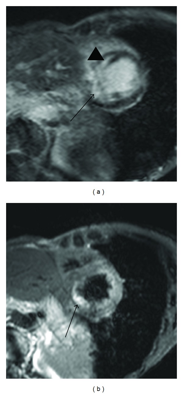
The purpose of this paper is to describe imaging techniques and findings of T2-weighted magnetic resonance imaging (MRI) of edema in myocardial diseases. T2-weighted cardiac MRI is acquired by combining acceleration techniques with motion and signal suppression techniques. The MRI findings should be interpreted based on coronary artery supply, intramural distribution, and comparison with delayed-enhancement MRI. In acute myocardial diseases, such as acute myocardial infarction and myocarditis, the edema is larger than myocardial scarring, whereas the edema can be smaller than the scarring in some types of nonischemic cardiomyopathy, including hypertrophic cardiomyopathy. T2-weighted MRI of edema identifies myocardial edema associated with ischemia, inflammation, vasculitis, or intervention in the myocardium and provides information complementary to delayed-enhancement MRI.


Figures

Figure 1
Figure 1
Myocardial T2 mapping generated from multiecho T2-weighted imaging.
Figure 2
Figure 2
63-year-old female with myocardial infarction. Short-axis delayed-enhancement MRI (a) shows myocardial infarction at the inferior myocardium (arrows) and the anterior myocardium (arrowhead). Short-axis T2-weighted cardiac MRI (b) shows only myocardial edema associated with acute myocardial infarction in the inferior myocardium and right ventricular myocardium (arrows) that are consistent with blood supply from the right coronary artery.
Figure 3
Figure 3
25-year-old male with acute myocarditis. T2-weighted MRI shows myocardial edema associated with acute myocarditis (arrows). The edema predominantly involves the epicardial or transmural myocardium in the lateral wall.
Figure 4
Figure 4
50-year-old female with Churg-Strauss syndrome. Short-axis T2-weighted MRI shows patchy myocardial edema of the papillary muscle (arrow) in the eosinophilic myocarditis associated with this syndrome.
Figure 5
Figure 5
73-year-old female with cardiac sarcoidosis presenting with lower ejection fraction and conduction disturbance. Short-axis delayed-enhancement MRI (a) shows abnormal enhancement consistent with the edema (arrow) as well as the septal mesocardial myocardial scarring (arrowhead). T2-weighted cardiac MRI (b) shows myocardial edema at the transmural myocardium of the inferior region (arrow).
Figure 6
Figure 6
41-year-old male with cardiac sarcoidosis presenting with premature ventricular contraction. Short-axis T2-weighted MRI (a) shows a small myocardial edema at the inferior septal myocardium (arrow). 18FDG-PET (b) shows the focal inflammation (arrow), consistent with the edema on MRI.
Figure 7
Figure 7
77-year-old female with hypertrophic cardiomyopathy presenting with chest pain. Delayed-enhancement MRI (a) shows patchy enhancement at the anterior and inferior septal myocardium (arrows). T2-weighted cardiac MRI (b) shows myocardial edema at the inferior and inferior septal myocardium (arrow).
Figure 8
Figure 8
Comparison between myocardial edema (white) and scarring (dotted) in the myocardium. (a) At the acute stage of ischemic or inflammatory cardiomyopathy, myocardial edema is associated with smaller myocardial scarring or its absence. (b) In some types of nonischemic cardiomyopathy, myocardial edema is smaller than the scarring, indicating relapsed ischemia.
Figure 9
Figure 9
55-year-old female with takotsubo cardiomyopathy. Long-axis delayed enhancement (a) shows no myocardial scarring of the apical myocardium, whereas the T2-weighted MRI (b) shows circumferential myocardial edema at the apical and midventricular regions (arrows). The T2-weighted MRI at its remission (c) shows no myocardial edema. The flow artifact is seen in the apical cavity (arrow), due to the insufficient blood signal suppression by black-blood prepulse.
Figure 10
Figure 10
66-year-old male who has undergone cryoablation for ventricular tachycardia associated with chronic myocardial infarction. Delayed-enhancement MRI (a) visualizes apical myocardial scarring with various transmurality (arrows). T2-weighted MRI (b) shows myocardial edema after ablation (arrow), where nontransmural scarring has been seen in the delayed-enhancement MRI.

Similar articles

Cited by

  • The ebb and flow of cardiac lymphatics: a tidal wave of new discoveries.
    Harris NR, Bálint L, Dy DM, Nielsen NR, Méndez HG, Aghajanian A, Caron KM. Harris NR, et al. Physiol Rev. 2023 Jan 1;103(1):391-432. doi: 10.1152/physrev.00052.2021. Epub 2022 Aug 11. Physiol Rev. 2023. PMID: 35953269 Free PMC article. Review.
  • Arrhythmias in Cardiac Sarcoidosis Bench to Bedside: A Case-Based Review.
    Rosenfeld LE, Chung MK, Harding CV, Spagnolo P, Grunewald J, Appelbaum J, Sauer WH, Culver DA, Joglar JA, Lin BA, Jellis CL, Dickfeld TM, Kwon DH, Miller EJ, Cremer PC, Bogun F, Kron J, Bock A, Mehta D, Leis P, Siontis KC, Kaufman ES, Crawford T, Zimetbaum P, Zishiri ET, Singh JP, Ellenbogen KA, Chrispin J, Quadri S, Vincent LL, Patton KK, Kalbfleish S, Callahan TD, Murgatroyd F, Judson MA, Birnie D, Okada DR, Maulion C, Bhat P, Bellumkonda L, Blankstein R, Cheng RK, Farr MA, Estep JD. Rosenfeld LE, et al. Circ Arrhythm Electrophysiol. 2021 Feb;14(2):e009203. doi: 10.1161/CIRCEP.120.009203. Epub 2021 Feb 16. Circ Arrhythm Electrophysiol. 2021. PMID: 33591816 Free PMC article. Review.
  • Novel Imaging Technologies for Accurate Assessment of Cardiac Allograft Performance.
    Higuita ML, Jain R, Osho AA, Rabi SA, Pruett TL, Pierson RN, Iaizzo PA, Tessier SN. Higuita ML, et al. Curr Transplant Rep. 2023 Sep;10(3):100-109. doi: 10.1007/s40472-023-00400-w. Epub 2023 Jul 17. Curr Transplant Rep. 2023. PMID: 39015560 Free PMC article.
  • Texture analysis of T2-weighted cardiovascular magnetic resonance imaging to discriminate between cardiac amyloidosis and hypertrophic cardiomyopathy.
    Huang S, Shi K, Zhang Y, Yan WF, Guo YK, Li Y, Yang ZG. Huang S, et al. BMC Cardiovasc Disord. 2022 May 21;22(1):235. doi: 10.1186/s12872-022-02671-0. BMC Cardiovasc Disord. 2022. PMID: 35597906 Free PMC article.
  • Imaging of metabolic and overload disorders in tissues and organs.
    Bruno F, Albano D, Agostini A, Benenati M, Cannella R, Caruso D, Cellina M, Cozzi D, Danti G, De Muzio F, Gentili F, Giacobbe G, Gitto S, Grazzini G, Grazzini I, Messina C, Palmisano A, Palumbo P, Bruno A, Grassi F, Grassi R, Fusco R, Granata V, Giovagnoni A, Miele V, Barile A; Young SIRM Working Group. Bruno F, et al. Jpn J Radiol. 2023 Jun;41(6):571-595. doi: 10.1007/s11604-022-01379-7. Epub 2023 Jan 21. Jpn J Radiol. 2023. PMID: 36680702 Review.

References

    1. Mahrholdt H, Wagner A, Judd RM, Sechtem U, Kim RJ. Delayed enhancement cardiovascular magnetic resonance assessment of non-ischaemic cardiomyopathies. European Heart Journal. 2005;26(15):1461–1474. - PubMed
    1. Cummings KW, Bhalla S, Javidan-Nejad C, Bierhals AJ, Gutierrez FR, Woodard PK. A pattern-based approach to assessment of delayed enhancement in nonischemic cardiomyopathy at MR imaging. Radiographics. 2009;29(1):89–103. - PubMed
    1. Tadamura E, Yamamuro M, Kubo S, et al. Effectiveness of delayed enhanced MRI for identification of cardiac sarcoidosis: comparison with radionuclide imaging. American Journal of Roentgenology. 2005;185(1):110–115. - PubMed
    1. Tandri H, Saranathan M, Rodriguez ER, et al. Noninvasive detection of myocardial fibrosis in arrhythmogenic right ventricular cardiomyopathy using delayed-enhancement magnetic resonance imaging. Journal of the American College of Cardiology. 2005;45(1):98–103. - PubMed
    1. Lehrke S, Lossnitzer D, Schöb M, et al. Use of cardiovascular magnetic resonance for risk stratification in chronic heart failure: prognostic value of late gadolinium enhancement in patients with non-ischaemic dilated cardiomyopathy. Heart. 2011;97(9):727–732. - PubMed

MeSH terms