. 2012;10(9):e1001386.
doi: 10.1371/journal.pbio.1001386. Epub 2012 Sep 4.

Smooth muscle tension induces invasive remodeling of the zebrafish intestine

Affiliations

Smooth muscle tension induces invasive remodeling of the zebrafish intestine

Christoph Seiler et al. PLoS Biol. 2012.

Abstract

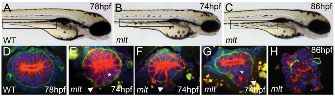
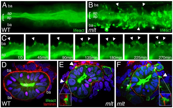
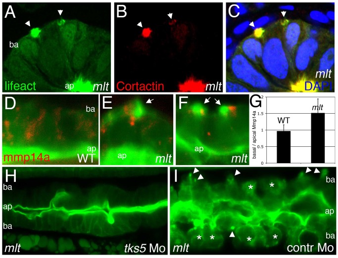
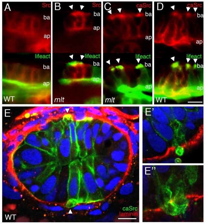
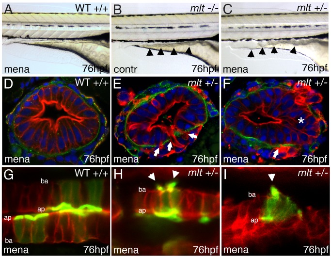
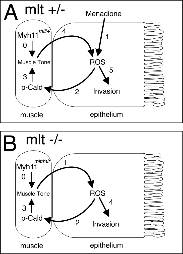
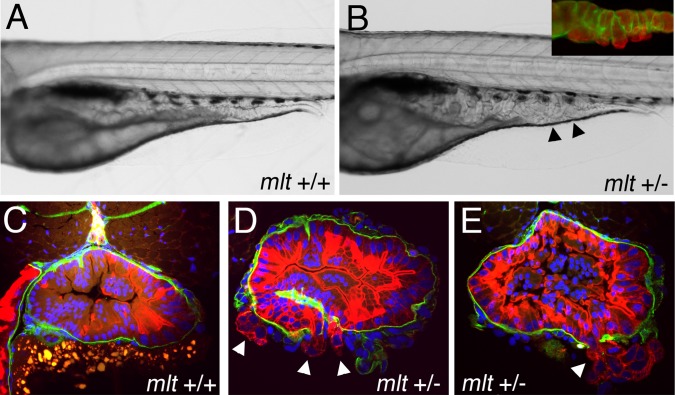
The signals that initiate cell invasion are not well understood, but there is increasing evidence that extracellular physical signals play an important role. Here we show that epithelial cell invasion in the intestine of zebrafish meltdown (mlt) mutants arises in response to unregulated contractile tone in the surrounding smooth muscle cell layer. Physical signaling in mlt drives formation of membrane protrusions within the epithelium that resemble invadopodia, matrix-degrading protrusions present in invasive cancer cells. Knockdown of Tks5, a Src substrate that is required for invadopodia formation in mammalian cells blocked formation of the protrusions and rescued invasion in mlt. Activation of Src-signaling induced invadopodia-like protrusions in wild type epithelial cells, however the cells did not migrate into the tissue stroma, thus indicating that the protrusions were required but not sufficient for invasion in this in vivo model. Transcriptional profiling experiments showed that genes responsive to reactive oxygen species (ROS) were upregulated in mlt larvae. ROS generators induced invadopodia-like protrusions and invasion in heterozygous mlt larvae but had no effect in wild type larvae. Co-activation of oncogenic Ras and Wnt signaling enhanced the responsiveness of mlt heterozygotes to the ROS generators. These findings present the first direct evidence that invadopodia play a role in tissue cell invasion in vivo. In addition, they identify an inducible physical signaling pathway sensitive to redox and oncogenic signaling that can drive this process.


Conflict of interest statement

The authors have declared that no competing interests exist.

Figures

Figure 1
Figure 1. Intestinal epithelial invasion in mlt larvae.
(A–C) Live images of wild type (WT) and mlt larvae. In WT (A) the posterior intestine forms a smooth cylindrical tube (box), whereas in mlt at 74 hpf the intestinal contour is irregular (B). Cystic expansion of the intestine is evident in 86 hpf mlt larvae (C). (D–H) Histological cross-sections through the posterior intestine of larvae immunostained for laminin (green) and cytokeratin (red). The WT intestine is comprised of a simple epithelial sheet consisting of a single layer of cells, whereas in mlt epithelial stratification (asterisks) and invasive cells that have breached the basement membrane are evident (E–G arrowheads). The initial invasive behavior is followed by expansive growth and loss of epithelial architecture (H).
Figure 2
Figure 2. Actin-rich protrusions in invasive epithelial cells of the mlt
intestine. (A, B) Full thickness 3-D rendering of sagittal confocal sections through the intestine of 78 hpf wild type (WT) (A) and mlt (B) larvae. Actin is labeled by transgenic Lifeact-GFP expression (green). (A) In WT, the majority of the label is present in the epithelial cell apical brush border (bracket). (B) In mlt, actin-rich invadopodia-like protrusions of the basal epithelial cell membrane are detected (arrows), in addition to brush border actin (bracket). (C) Time lapse analysis of protrusion development. Single sagittal confocal scans through the intestine of a mlt larva beginning at 74 hpf. Basal invadopodia-like protrusions (arrowheads) precede cell invasion, which is first detected at 135 min. Asterisks mark invasive cells at 270 min (see also B). (D–F) Histological cross-sections through the intestine of 74 hpf immunostained mlt larvae. Basement membrane is labeled red (laminin immunostain) and actin labeled green (GFP immunostain in Lifeact-GFP transgenics). Nuclei stained blue with DAPI. Actin rich protrusions in mlt co-localize with sites of basal lamina degradation (arrowheads and insets E, F). During progression of the phenotype epithelial cells invade the tissue stroma through degraded regions of the basal lamina (asterisks in F). ap, apical epithelial cell border; ba, basal cell epithelial cell border.
Figure 3
Figure 3. Actin rich protrusions in mlt are invadopodia homologs.
(A–C) Histological cross-sections through the posterior intestine of 74 hpf mlt mutant larvae immunostained with antibodies against GFP (green) (labeling Lifeact-GFP) and Cortactin (red). DAPI-stained nuclei, blue. Lifeact-GFP and Cortactin co-localize in actin rich basal membrane protrusions (arrowheads) and in the apical brush border (ap). (D–G) Sagittal confocal scans through the intestine of 74 hpf Lifeact-GFP transgenic wild type (WT) and mlt larvae. Lifeact-GFP binds actin in the apical brush border (ap) of WT (D) and mlt (E, F) epithelial cells, as well as basal (ba) invadopodia-like protrusions in mlt (E, F, arrows). In WT, the Mmp14a-mCherry fusion protein (red) is distributed throughout the epithelial cells. In mlt, Mmp14a-mCherry is preferentially localized to the basal region of the epithelial cells. (n = 33 WT and 33 mlt cells examined; 6 larvae each genotype). (G) Ratio of basal to apical Mmp14a-mCherry in WT versus mlt epithelial cells (error bars, standard deviation. * p<.001). (H–I) Sagittal confocal scans through the intestines of 84 hpf Lifeact-GFP transgenic mlt larvae. Invadopodia (arrowheads I) and invasive cells (asterisks I) are present in the mlt larvae injected with a control morpholino (I), but are not detected in the larvae injected with the tks5 morpholino (H).
Figure 4
Figure 4. Src induces formation of invadopodia-like protrusions in the intestine of wild type zebrafish larvae.
(A–D) Sagittal confocal scans through the intestine of 74 hpf wild type and mlt larvae that express Src-mCherry (red) and Lifeact-GFP (green) in the intestinal epithelium. (A) In WT, Src (red) is localized at the apical (ap) and lateral epithelial cell membrane. (B) In mlt, Src also localizes to sites of actin-rich (green) invadopodia-like protrusions (arrowheads B) arising from the basal epithelial cell membrane (ba). (C) Constitutively active Src (caSrc; red) localizes to invadopodia-like protrusions (green) in mlt (arrowheads). (D) caSrc induces formation of the protrusions in WT (arrowheads). (E) Histological cross-sections through the intestine of a 74 hpf wild type larva showing caSrc-rich protrusions (green) protruding through small degraded regions of the basal lamina (laminin immunostain, red). Additional examples are shown in high power images (E, E′, and E″).
Figure 5
Figure 5. Src inhibition rescues invasion but not formation of invadopodia-like protrusions in mlt.
(A–C) Lateral views of live 5 dpf WT (A) and mlt (B, C) larvae. Treatment with the Src-I1 inhibitor rescues invasion in mlt (C). The size of the intestinal epithelium in the treated mlt larva (C) is reduced compared with the untreated mlt larva (B). (D–G) Histological cross-sections of 4 dpf WT (D) and mlt (E–G) Tg(miR194:Lifeact-GFP) larvae immunostained with antibodies against laminin (red) and GFP (green). Nuclei stained blue with DAPI. Arrows point to invadopodia-like protrusions arising from the basal epithelial cell membrane of Src-I1 treated mlt larvae (F, G). Pronounced invasion with distortion of intestinal architecture is evident in the untreated mlt larva (E). Note that invasion is markedly reduced in the Src-I1 treated mlt larvae despite the presence of the invadopodia-like protrusions (white arrows in F, G).
Figure 6
Figure 6. Tonic intestinal smooth muscle contraction in mlt mutants.
Sagittal confocal images from time lapse movies (Movies S7 and S8) of transgenic WT (A–C) and mlt (D–H) larvae (anterior left) expressing fluorescent reporters. Intestinal epithelium (I) and pronephric duct (P) labeled red; intestinal smooth muscle labeled green. WT images beginning at 76 hpf show cycling of the contracted (A, C) and relaxed state (B); brackets depict intestinal diameter. (D–F) The mlt intestine at this stage shows progressive contraction that disrupts intestinal architecture. (G, H) Smooth muscle contraction is not detected in the mlt intestine at the onset of invasive remodeling (72 hpf). (I) Western blot showing premature phosphorylation of h-CaD (p-Cad) in 72 hpf mlt larvae, before the onset of smooth muscle contraction (ratio WT:mlt = 0.17/1, relative to Actin loading control; in four independent experiments, the ratio averaged 0.13/1; p = 0.002). Phospho-h-CaD is present at the onset of contraction in WT at 78 hpf and is comparable to mlt (ratio WT:mlt = 1.12/1, relative to Actin; no significant difference in four independent experiments). Levels of phospho-Myosin light chain (p-Mlc) are low at 72 hpf in both mlt and WT (ratio WT:mlt = 1.16/1, relative to Actin) and are increased when contractions occur at 78 hpf (ratio WT:mlt = 0.96/1, relative to Actin). There was no significant difference between WT and mlt at either time point in four independent experiments. Actin (beta-Actin), loading control.
Figure 7
Figure 7. Smooth muscle contraction drives epithelial invasion but does not alter tissue rigidity.
(A–C) Lateral views of live 5 dpf larvae injected with Sma or control morpholino. (A) Sma knockdown has no effect on WT intestinal morphology. (B) Control morpholino injection in mlt. (C) Sma knockdown rescues invasion in mlt. Residual invasive cells persist in this Sma deficient mlt larva (arrowhead). (D–I) Histological cross-sections through the posterior intestine of 74 hpf WT and mlt larvae immunostained with anti-keratin (red) and anti-laminin (green) antibodies. (D) WT. (E, F) mlt larvae injected with control (E) and Sma (D, F) morpholinos. Invasive cells in mlt (arrowheads, E) are rescued by Sma knockdown. (G, H) Injection of a morpholino targeting the high molecular weight isoform of Caldesmon (cald) has no effect on WT intestinal morphology but induces invasion (arrowhead) and stratification (asterisk) in an mlt heterozygote. (I) Treatment of an mlt heterozygote with L-NAME causes invasion (arrowhead) and epithelial stratification (asterisk). (J) Force displacement measurements show identical compliance of intestines dissected from mlt and WT larvae before the phenotype develops at 70 hpf, and a modest increase in compliance at the outer surface of the intestine (<4 micron indentation) when invasion is present at 74 hpf. (K) Compliance is indicated by Young's modulus, which is proportional to the slope of the Force versus Tissue indentation curve.
Figure 8
Figure 8. Heterozygous mlt larvae are sensitized to oxidative stress.
(A) Quantitative RT-PCR shows increased intestinal gpx expression in 74 hpf mlt homozygotes (mlt −/−) compared with WT (+/+) and mlt heterozygotes (mlt +/−) (** p<.01). (A′) Quantitative RT-PCR shows intestinal gpx expression is increased in menadione treated homozygous WT larvae (+/+ mena) compared with untreated WT (contr). A much stronger response to menadione is seen in mlt heterozygotes (+/− mena) (*** p<.001). (B) ROS production in intestinal epithelial cells of 76 hpf WT control larvae (contr) versus menadione treated homozygous WT and heterozygous mlt larvae (*** p<.001). Bar graphs in (A) and (A′) show mean and standard deviation of three independent experiments. Bar graph in (B) shows mean of six larvae for each genotype; 15–25 cells per larva. (C–F) Histological cross-sections of larvae processed for fluorescent RNA in situ hybridization. Menadione induced gpx expression (red) in the epithelium but not smooth muscle (green) of heterozygous mlt larvae (F). This is comparable to the gpx expression pattern in control homozygous mlt larvae (D). (G) Western blot showing premature h-CaD phosphorylation (pCad) in dissected intestines from menadione treated heterozygotes but not homozygous WT larvae (lane 4 versus lane 3; ratio phospho-h-CaD WT:mlt = 0.02/1, relative to total h-CaD; CaD; in six experiments the ratio averaged 0.016/1; p<.001). h-CaD is prematurely phosphorylated in intestines dissected from 74 hpf mlt homozygotes versus mlt heterozygotes (lane 1 versus lane 2). (H) Western blot showing h-CaD phosphorylation (pCad) in the menadione treated heterozygous intestines prior to dissociation (lane 2) but not after dissociation into free cell populations (lane 5). Phospho-h-CaD persists in dissociated cells from homozygous intestines (lane 3), but is not detected in control intestines dissected from mlt heterozygotes, before (lane 1) or after (lane 4) dissociation into free cell populations. No phopho-h-CaD was detected in any dissociated samples in three independent experiments. Loading control, phospho-Myosin light chain (pMlc).
Figure 9
Figure 9. Oxidative stress induces invasive remodeling in mlt heterozygous larvae.
(A–C) Lateral images of live WT (A), mlt homozygous (B), and mlt heterozygous larvae (C). The WT and mlt heterozygous larvae received 3 h of treatment with Menadione beginning at 73 hpf. Menadione treated heterozygote (C) larvae have an intestinal phenotype (arrowheads) resembling the untreated mlt homozygous larvae (B). (D–F) Corresponding histological cross-sections (representative of larvae in A, C) with intestinal epithelial cells labeled red (anti-keratin immunostain) and basement membrane in green (anti-laminin immunostain). Menadione causes epithelial cell invasion (arrows) and stratification (asterisks) in mlt heterozygous larvae (E, F) but does not affect epithelial architecture in the WT intestines (D). (G–I) Sagittal confocal scans through the intestine of WT and mlt heterozygotes treated with menadione. Both larvae express LifeAct-GFP in a subset of intestinal epithelial cells. Actin-rich invadopodia-like protrusions (green) are seen arising from the basal epithelial cell membrane of menadione treated heterozygous larvae (arrowheads, H, I). Actin is located nearly exclusively in the apical brush border of WT epithelial cells (G): Red -membrane mCherry; ba, basal epithelial cell border; ap, apical epithelial cell border.
Figure 10
Figure 10. An amplification feedback signaling loop controls smooth muscle contraction and invasion in mlt larvae.
(A) In mlt heterozygotes the expression of mutant Myh11 protein does not generate sufficient smooth muscle tension to induce epithelial invasion or stratification (0). Menadione treatment induces ROS production in the epithelium (1). Epithelial ROS signaling leads to premature phosphorylation of Caldesmon in heterozygous smooth muscle cells (2). The resulting increase in smooth muscle tone (3) leads to an amplified ROS response in the epithelium (4), establishing a feed forward loop causing additional h-CaD phosphorylation and increased smooth muscle tension. Together these stimuli induce invasive remodeling of the epithelium (5). (B) Endogenous smooth muscle tone in mlt homozygous larvae (0) induces epithelial ROS (1) and h-CaD phosphorylation via epithelial signaling (2, 3). Ultimately, this culminates in epithelial invasion (4), as in menadione treated mlt heterozygotes.
Figure 11
Figure 11. Activation of oncogenic signaling enhances sensitivity of mlt heterozygotes to oxidative stress.
(A, B) Lateral views of live, menadione treated 5 dpf axin mutant larvae that express mutant KRAS in the intestinal epithelium (Kras-axin). (A) Hypertrophy of the intestinal epithelium in a Kras-axin larva that is homozygous for the wildtype myh11 allele (mlt +/+) larvae is unchanged by treatment with menadione. (B) Menadione treatment causes pronounced cystic expansion of the posterior intestinal epithelium of the Kras-axin mlt heterozygote (arrowheads) that resembles the homozygous mlt phenotype. Inset, immunolabeling of the basal lamina (laminin, green) and epithelium (cytokeratin, red) shows epithelial cell invasion through the basement membrane. (C–E) Histological cross-sections through the intestine of immunostained larvae show invasive cells in menadione treated Kras-axin mlt heterozygotes (arrowheads; D, E). Invasion is not detected in menadione-treated Kras-axin mlt larvae that are homozygous for the wildtype myh11 allele (C).

Similar articles

Cited by

References

    1. de Rooij J, Kerstens A, Danuser G, Schwartz MA, Waterman-Storer CM (2005) Integrin-dependent actomyosin contraction regulates epithelial cell scattering. J Cell Biol 171: 153–164. - PMC - PubMed
    1. Yeung T, Georges PC, Flanagan LA, Marg B, Ortiz M, et al. (2005) Effects of substrate stiffness on cell morphology, cytoskeletal structure, and adhesion. Cell Motil Cytoskeleton 60: 24–34. - PubMed
    1. Paszek MJ, Zahir N, Johnson KR, Lakins JN, Rozenberg GI, et al. (2005) Tensional homeostasis and the malignant phenotype. Cancer Cell 8: 241–254. - PubMed
    1. Papusheva E, Heisenberg CP (2010) Spatial organization of adhesion: force-dependent regulation and function in tissue morphogenesis. EMBO J 29 (16) 2753–2768. - PMC - PubMed
    1. Lehoux S, Tedgui A (2003) Cellular mechanics and gene expression in blood vessels. J Biomech 36: 631–643. - PubMed

Publication types

MeSH terms