. 2012 Aug 28:6:56.
doi: 10.3389/fnbeh.2012.00056. eCollection 2012.

Perception of biological motion in visual agnosia

Affiliations

Perception of biological motion in visual agnosia

Elisabeth Huberle et al. Front Behav Neurosci. .

Abstract

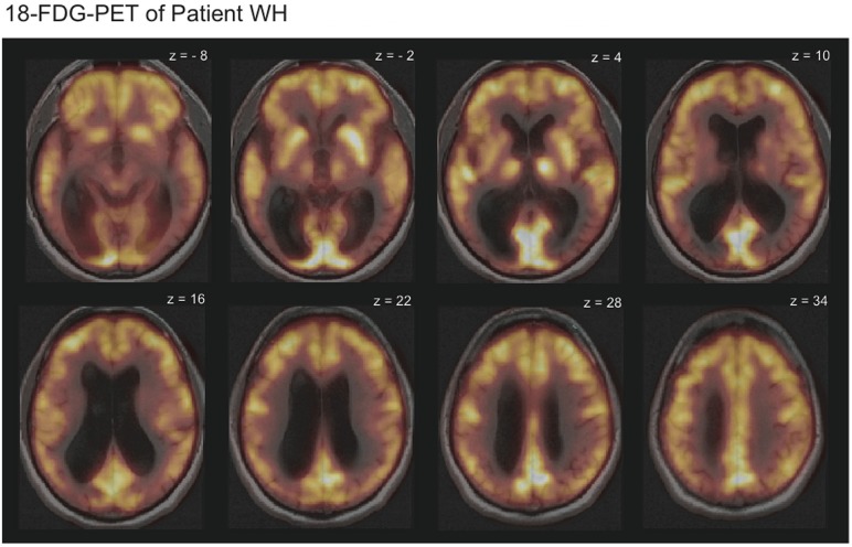
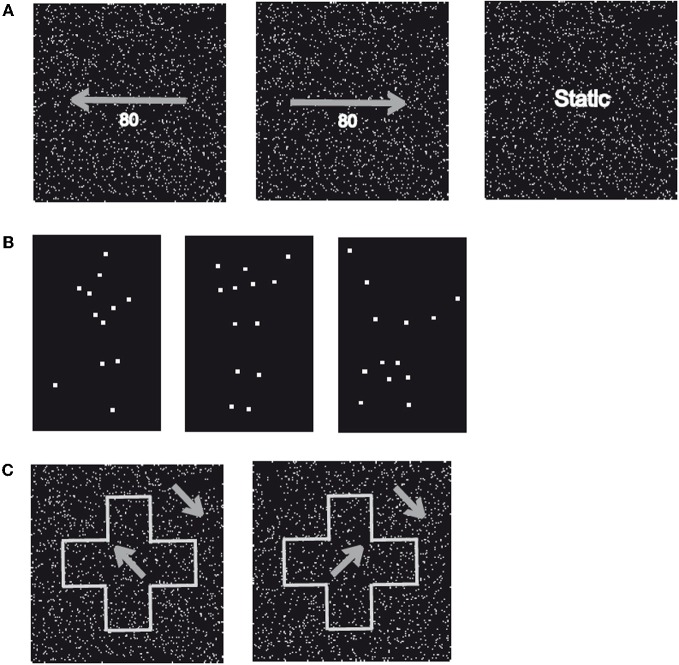
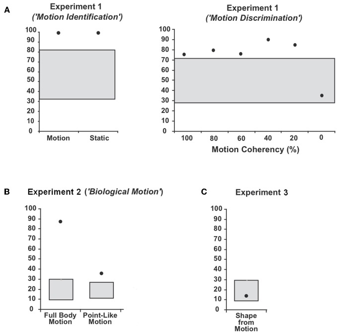
Over the past 25 years, visual processing has been discussed in the context of the dual stream hypothesis consisting of a ventral ("what") and a dorsal ("where") visual information processing pathway. Patients with brain damage of the ventral pathway typically present with signs of visual agnosia, the inability to identify and discriminate objects by visual exploration, but show normal perception of motion perception. A dissociation between the perception of biological motion and non-biological motion has been suggested: perception of biological motion might be impaired when "non-biological" motion perception is intact and vice versa. The impact of object recognition on the perception of biological motion remains unclear. We thus investigated this question in a patient with severe visual agnosia, who showed normal perception of non-biological motion. The data suggested that the patient's perception of biological motion remained largely intact. However, when tested with objects constructed of coherently moving dots ("Shape-from-Motion"), recognition was severely impaired. The results are discussed in the context of possible mechanisms of biological motion perception.

Keywords: agnosia; biological motion; perception; ventral stream; vision.


Figures

Figure 1
Figure 1
18-Fluorodeoxyglucose positron emission tomography (FDG-PET-CT) scans overlaid with the magnetic resonance imaging (MRI) scans revealed reduced metabolism in the temporo-occipital cortex bilaterally for patient WH. Displayed is HW's 18-FDG-uptake in comparison to the average of 20 healthy subjects. Bright yellow colors indicate high and dark red colors low 18-FDG-uptake.
Figure 2
Figure 2
Sample stimuli. (A) Displayed are two patterns of randomly moving dots with a motion coherency of 80% toward the left (left) or right (centre). In addition, static patterns of randomly assigned dots (right) with identical stimulus parameters were applied. (B) Displayed are three examples (“Running,” “Jumping,” and “Turning a Cartwheel”) of the human movements used in Experiment 2 (“Biological Motion”). Stimuli consisted of white dots placed at locations on (invisible) lines connecting the main joints of upper arm, forearm, upper leg, and lower leg and were presented on a black background (“Point-Light Motion”). (C) Displayed is a global shape (“Cross”) that consists of coherently moving dots with a direction of motion rotated 45° counter clockwise (left) or clockwise (right) from the vertical axis presented on a background of coherently moving dots rotated 45° counter clockwise from the vertical axis (“Shape-from Motion”).
Figure 3
Figure 3
Average percentage and 95%-confidence interval for the binomial distribution around chance level for patient WH in (A) Experiment 1 (“Motion Identification/ Discrimination”), (B) Experiment 2 (“Biological Motion”) and (C) Experiment 3 (“Shape-from Motion”). The black dots indicate the average percentage of correct responses (CR) and the grey boxes the 95%-confidence interval for the binomial distribution around chance level.

References

    1. Akiyama T., Kato M., Muramatsu T., Saito F., Umeda S., Kashima H. (2006). Gaze but not arrows: a dissociative impairment after right superior temporal gyrus damage. Neuropsychologia 44, 1804–1810 10.1016/j.neuropsychologia.2006.03.007 - DOI - PubMed
    1. Altmann C. F., Deubelius A., Kourtzi Z. (2004). Shape saliency modulates contextual processing in the human lateral occipital complex. J. Cogn. Neurosci. 16, 794–804 10.1162/089892904970825 - DOI - PubMed
    1. Battelli L., Cavanagh P., Thronton I. M. (2003). Perception of biological motion in parietal patients. Neuropsychologia 41, 1808–1816 10.1016/S0028-3932(03)00182-9 - DOI - PubMed
    1. Beintema J. A., Lappe M. (2002). Perception of biological motion without local image motion. Proc. Natl. Acad. Sci. U.S.A. 99, 5661–5663 10.1073/pnas.082483699 - DOI - PMC - PubMed
    1. Billino J., Braun D. I., Böhm K. D., Bremmer F., Gegenfurtner K. R. (2009). Cortical networks for motion processing: effects of focal brain lesions onf perception of different motion types. Neuropsychologia. 47, 2133–2144 10.1016/j.neuropsychologia.2009.04.005 - DOI - PubMed