. 2012 Aug 28:3:305.
doi: 10.3389/fmicb.2012.00305. eCollection 2012.

The Parasitic Dinoflagellates Blastodinium spp. Inhabiting the Gut of Marine, Planktonic Copepods: Morphology, Ecology, and Unrecognized Species Diversity

Affiliations

The Parasitic Dinoflagellates Blastodinium spp. Inhabiting the Gut of Marine, Planktonic Copepods: Morphology, Ecology, and Unrecognized Species Diversity

Alf Skovgaard et al. Front Microbiol. .

Abstract

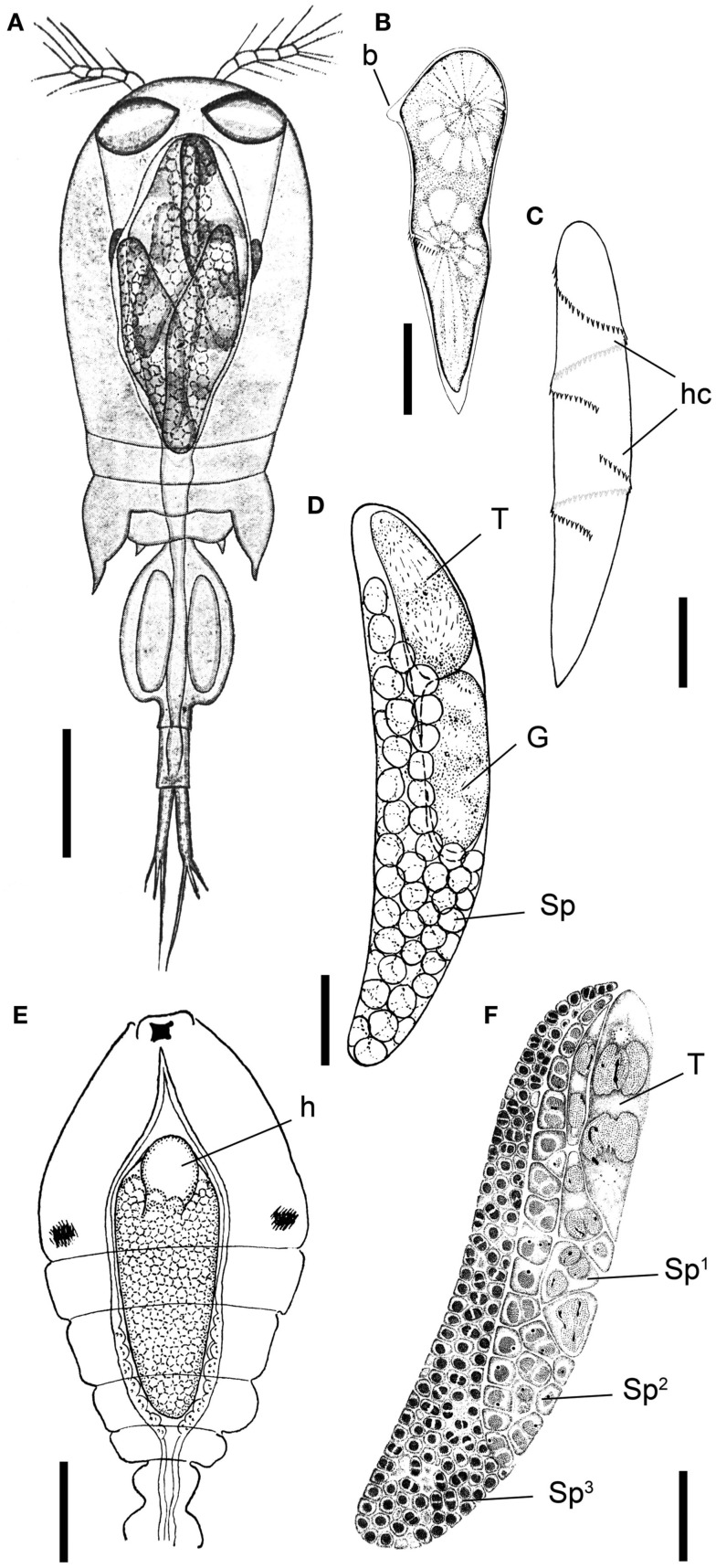
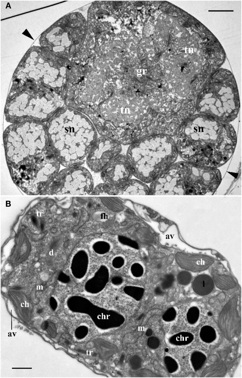
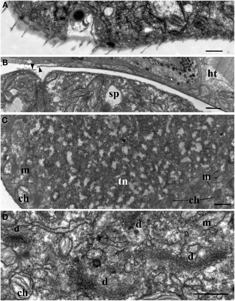
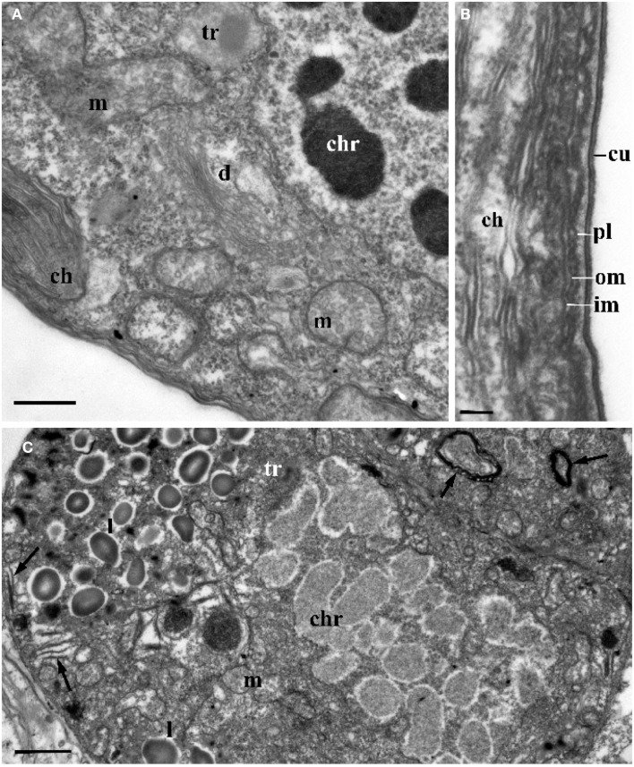
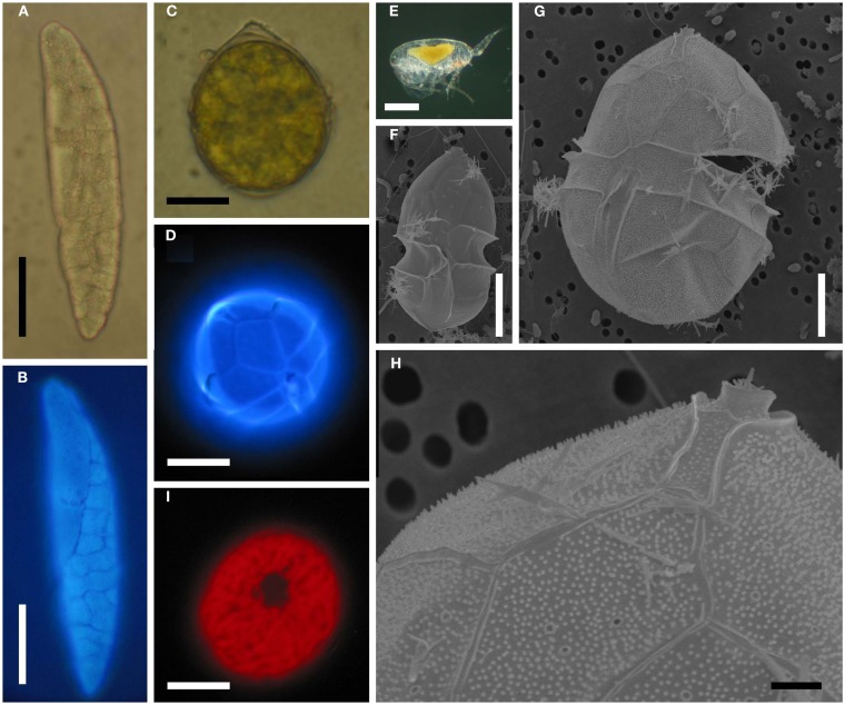
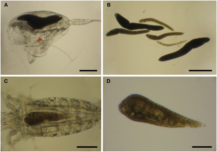
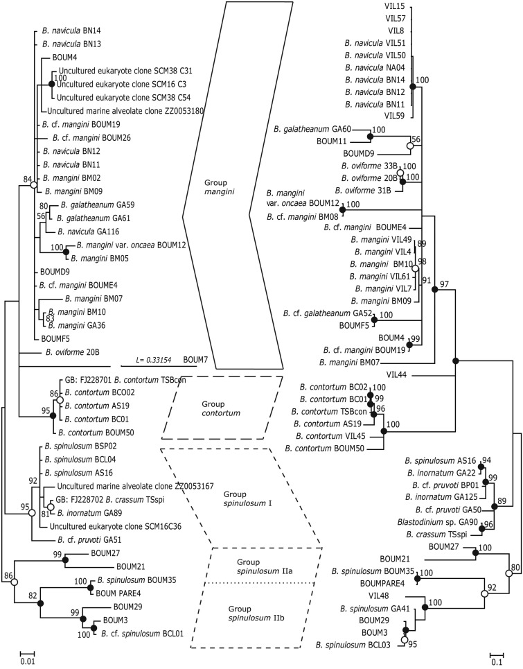
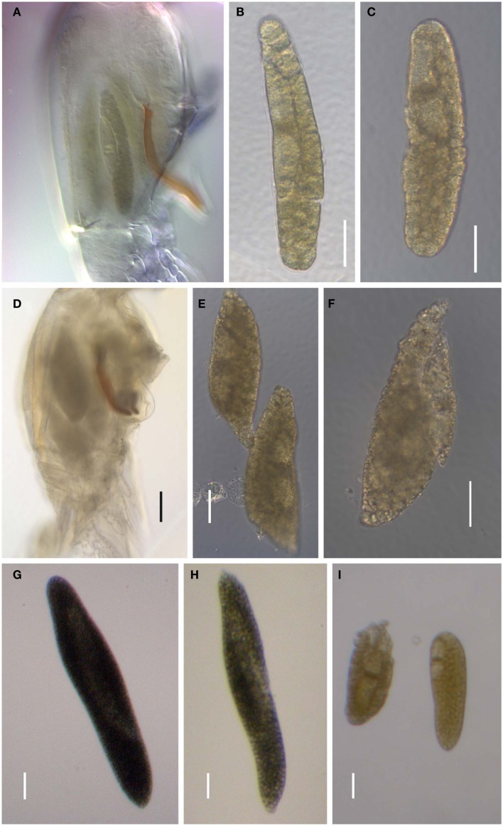
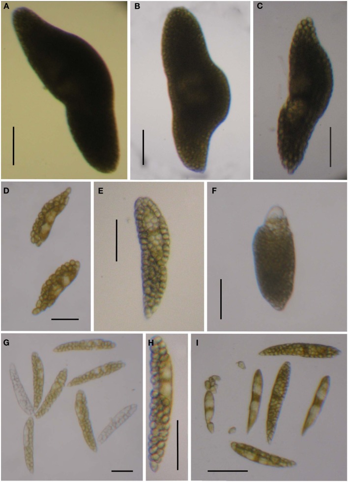
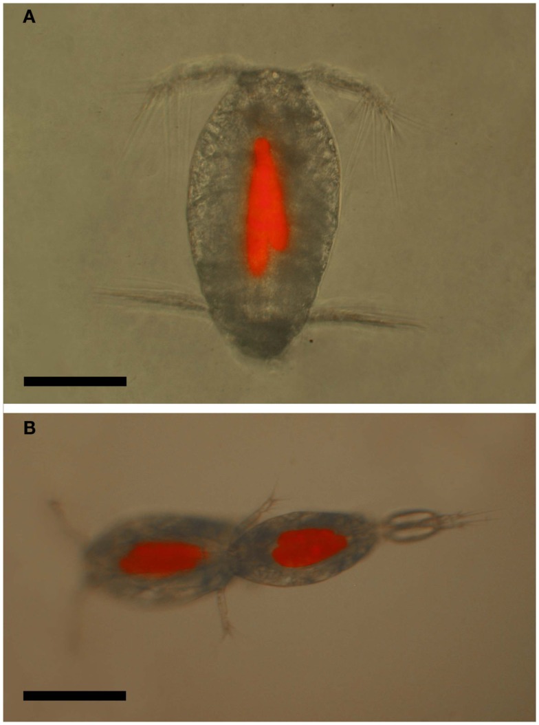
Blastodinium is a genus of dinoflagellates that live as parasites in the gut of marine, planktonic copepods in the World's oceans and coastal waters. The taxonomy, phylogeny, and physiology of the genus have only been explored to a limited degree and, based on recent investigations, we hypothesize that the morphological and genetic diversity within this genus may be considerably larger than presently recognized. To address these issues, we obtained 18S rDNA and ITS gene sequences for Blastodinium specimens of different geographical origins, including representatives of the type species. This genetic information was in some cases complemented with new morphological, ultrastructural, physiological, and ecological data. Because most current knowledge about Blastodinium and its effects on copepod hosts stem from publications more than half a century old, we here summarize and discuss the existing knowledge in relation to the new data generated. Most Blastodinium species possess functional chloroplasts, but the parasitic stage, the trophocyte, has etioplasts and probably a limited photosynthetic activity. Sporocytes and swarmer cells have well-developed plastids and plausibly acquire part of their organic carbon needs through photosynthesis. A few species are nearly colorless with no functional chloroplasts. The photosynthetic species are almost exclusively found in warm, oligotrophic waters, indicating a life strategy that may benefit from copepods as microhabitats for acquiring nutrients in a nutrient-limited environment. As reported in the literature, monophyly of the genus is moderately supported, but the three main groups proposed by Chatton in 1920 are consistent with molecular data. However, we demonstrate an important genetic diversity within the genus and provide evidences for new groups and the presence of cryptic species. Finally, we discuss the current knowledge on the occurrence of Blastodinium spp. and their potential impact on natural copepod populations.

Keywords: Blastodinium; copepod; parasite; phylogeny; plankton; symbiont; ultrastructure.


Figures

Figure 1
Figure 1
Blastodinium spp. (A) Several B. navicula inside the gut of the copepod Corycaeus giesbrechti. (B) A young trophocyte of B. pruvoti with anterior beak (b). (C) Early stage B. spinulosum with helicoidal crests (hc). (D) B. pruvoti showing trophycyte (T), gonocyte (G), and sporocytes (Sp). (E) B. oviforme with hilum (h) in Oithona nana. (F) B. pruvoti with three progressive layers of sporocytes (Sp1–Sp3). Adapted from Chatton (1920). (A,E) Scale bars = 100 μm. (B–F) Scale bars = 50 μm.
Figure 2
Figure 2
(A) Cross section through the central part of Blastodinium cf. mangini (from Farranula rostrata) showing a big trophocyte with several layers of sporocytes covered by common a cuticle (arrowheads). gr, golgi region; sn, nucleus of sporocyte; tn, nucleus of trophocyte. Scale bar = 5 μm. (B) Ultrastructure of mature Blastodinium cf. navicula sporocyte (from F. rostrata). av, amphiesmal vesicle (alveolus); ch, chloroplast; chr, chromosome; cu, cuticle; d, dictyosome; fh, vesicle with future flagellar hairs; l, lipid droplet; m, mitochondrion; tr, trichocyst. Scale bar = 1 μm.
Figure 3
Figure 3
Ultrastructure of Blastodinium. (A) Cell covering of Blastodinium cf. mangini (from Farranula rostrata) in crest region. (B) Cuticle of trophont attached tightly to the gut tissue of the host (ht); arrowheads show broken cuticle on the left. Blastodinium cf. mangini (from F. rostrata). (C) Portion of trophocyte of Blastodinium cf. navicula sporocyte (from F. rostrata) with huge nucleus (tn) containing decondensed chromosomes (light zones) and granular material (possibly ribosomal subunits), small chloroplasts (ch) and mitochondria (m). (D) Golgi region of trophocyte of B. cf. mangini (from F. rostrata) with prominent dictyosomes (d), chloroplasts (ch), and mitochondria (m). Scale bar = 1 μm.
Figure 4
Figure 4
The ultrastructure of Blastodinium sporocyte. (A) Portion of nucleus and cytoplasm of Blastodinium cf. navicula sporocyte (from F. rostrata). (B) Structure of coverings of Blastodinium cf. navicula sporocyte (from F. rostrata). (C) Colorless representative of Blastodinium (Blastodinium cf. hyalinum from Paracalanus parvus). ch, chloroplast; chr, chromosome; cu, cuticle; d, dictyosome; im, inner and outer (om) membrane of alveoli; l, lipid droplets; m, mitochondrion; pl, plasma membrane; tr, maturing trichocyst. Arrows show reduced presumed plastids. (A) Scale bar = 2.5 μm. (B) Scale bar = 0.1 μm. (C) Scale bar = 1 μm.
Figure 5
Figure 5
Blastodinium spp. (A) B. mangini from Oncaea sp. (B) Same as (A) but stained with Calcofluor White. (C) B. oviforme dinospore from O. similis. (D) Same as (C) but stained with Calcuflour White. (E) Clausocalanus sp. infected with Blastodinium sp. (F) B. contortum dinospore, SEM. (F) B. contortum dinospore, SEM. (G,H) Dinospore from the parasite in (E), SEM. (I) Blastodinium sp. dinospore showing autofluorescence of chloroplasts viewed in epifluorescent light. (A,B) Scale bar = 50 μm. (C,D,I) Scale bars = 10 μm. (E) Scale bar = 200 μm. (F,G) Scale bars = 5 μm. (H) Scale bar = 1 μm.
Figure 6
Figure 6
Blastodinium sp. in Euchaeta sp. from the Atlantic Ocean. (A,C) Parasites inside their hosts. (B) Six parasites of an unidentifiable species from the host in (A). (C) A single unidentifiable parasite from the host in (C). (A,B) Scale bars = 500 μm. (C) Scale bar = 200 μm. (D) Scale bar = 100 μm.
Figure 7
Figure 7
Phylogenetic trees based on Bayesian analyses of alignments comprising SSU rDNA sequences (left) and ITS1, 5.8S rDNA, and ITS2 sequences (right) of Blastodinium spp. Numbers above nodes are Maximum Likelihood bootstrap values; only values above 50% are shown. Filled circles at nodes denote that the clade had Bayesian posterior probabilities (PP) of 1.00; open circles denote PP of 0.95–0.99. PP < 95 are not shown. Labels at branches are species names and/or isolate names. Two sequences are identified by their GenBank accession number; these have the prefix “GB:” GenBank accession numbers for all sequences are given in Table A1 in Appendix.
Figure 8
Figure 8
Blastodinium spp. (A–C) Blastodinium mangini. (A) Three individuals inside Farranula rostrata (Villefranche-sur-mer, 2 of September 2009). Monoblastic stage (I-128). (B) Individual extracted from F. rostrata (Villefranche-sur-mer, 3 of September 2009, VIL49). Monoblastic stage (I-64). (C) Individual extracted from F. rostrata (Villefranche-sur-mer, 2 of September 2009, VIL52). Monoblastic stage (I-128). (D–F) B. navicula. (D) Four individuals inside F. rostrata (Villefranche-sur-mer, 2 of September 2009, VIL50). (E,F) Different individuals extracted from the precedent copepod host (VIL50). Monoblastic stage (I-128). (G) Blastodinium cf. galatheanum from Acartia negligens (North Atlantic, GA52). (H) B. galatheanum. from A. negligens (North Atlantic, GA60). Scale bar = 50 μm. I. B. mangini var. oncaea. Two individuals from Oncaea sp. (NW Mediterranean Sea, BM05). (B,C) Scale bars = 50μm. (D) Scale bar = 100 μm. (E–I) Scale bars = 50μm. (G)
Figure 9
Figure 9
Blastodinium spp. Sequences from samples are used for the phylogenetic analyses in Figure 7. (A–C) Solitary individual of B. inornatum Group spinulosum I from Clausocalanus spp. (A) GA22. (B) GA89. (C) GA125. (D–F) Gregarious individuals of Blastodinium spp. Group spinulosum I from Clausocalanus spp. (D) Blastodinium cf. pruvoti, BP01. (E) Blastodinium cf. pruvoti, GA50. (F) B. oviforme from Oithona similis. (G,H) Gregarious individuals of B. spinulosum Group spinulosum II. (G) Blastodinium cf. spinulosum, BCL01, from Clausocalanus cf. arcuicornis. Note two colorless individuals. (H) B. spinulosum, GA41, from Paracalanus parvus. (I) B. spinulosum, BCL04, Group spinulosum I from Clausocalanus sp. Scale bars = 100 μm.
Figure 10
Figure 10
Location of current reports of Blastodinium spp. in marine copepods. Red circles represent photosynthetic species; black circles are colorless species; and white circles are environmental DNA sequences with high similarity to Blastodinium.
Figure 11
Figure 11
Blastodinium spp. in calanoid copepods. (A) Calanus sp. from Greenlandic waters with B. hyalinum. (B) Parasite from (A). (C) Cosmocalanus vulgaris off the West coast of Africa with Blastodinium cf. chattoni. (D) Parasite from (C). (E) Temora stylifera from the Central Atlantic Ocean with Blastodinium cf. contortum. (F) Parasite from (E). (A,C,E) Scale bars = 500 μm; (B,D,F) scale bars = 200 μm.
Figure 12
Figure 12
Oncaea cf. scottodicarloi with Blastodinium mangini in epifluorescent illumination showing autofluorescence of parasite chloroplasts. (A) Female with two parasites. (B) Copulation male (right) and female (left), both infected with 2–3 parasites. Scale bars = 100 μm.

Similar articles

Cited by

References

    1. Alves-de-Souza C., Cornet C., Nowaczyk A., Gasparini S., Skovgaard A., Guillou L. (2011). Blastodinium spp. infect copepods in the ultra-oligotrophic marine waters of the Mediterranean Sea. Biogeosciences 8, 2125–2136
    1. Apstein B. (1911). Parasiten von Calanus finmarchicus. Wiss. Meeresunters. Abt. Kiel 19, 206–223
    1. Cattley J. G. (1948). Sex reversal in copepods. Nature 161, 937–93710.1038/161937a0 - DOI - PubMed
    1. Cavalier-Smith T. (1993). Kingdom Protozoa and its 18 phyla. Microbiol. Rev. 57, 953–994 - PMC - PubMed
    1. Chatton É. (1906). Les Blastodinides, ordre nouveuax de Dinoflagellés parasites. C. R. Acad. Sci. 144, 981–983

LinkOut - more resources