

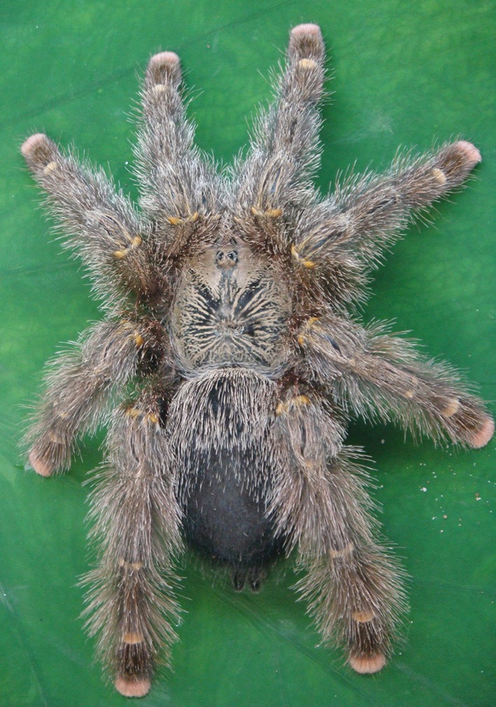
Juruin: an antifungal peptide from the venom of the Amazonian Pink Toe spider, Avicularia juruensis, which contains the inhibitory cystine knot motif
- PMID: 22973266
- PMCID: PMC3437525
- DOI: 10.3389/fmicb.2012.00324
Juruin: an antifungal peptide from the venom of the Amazonian Pink Toe spider, Avicularia juruensis, which contains the inhibitory cystine knot motif
Abstract
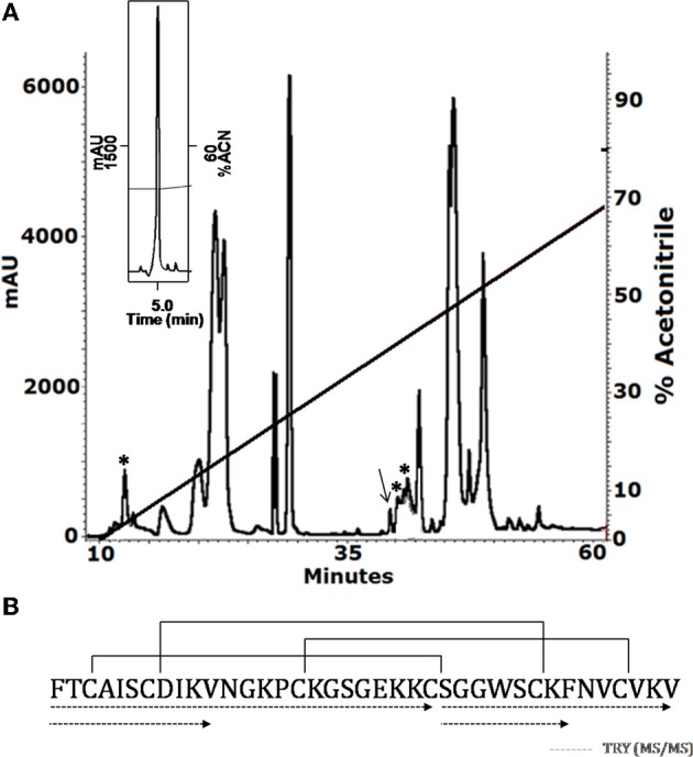
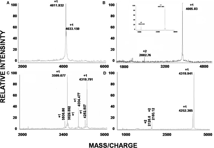
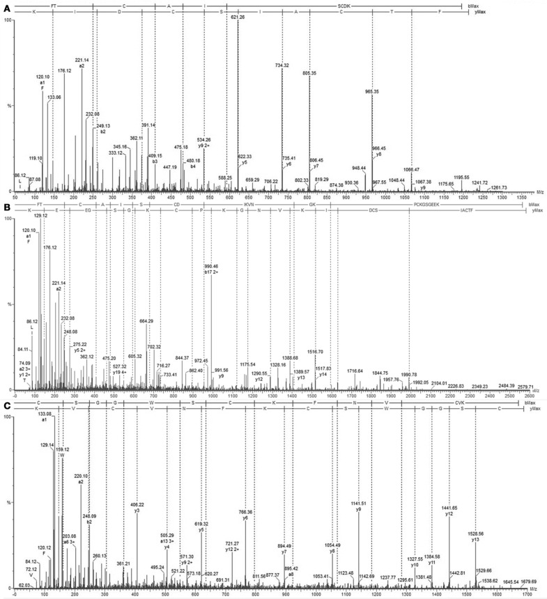
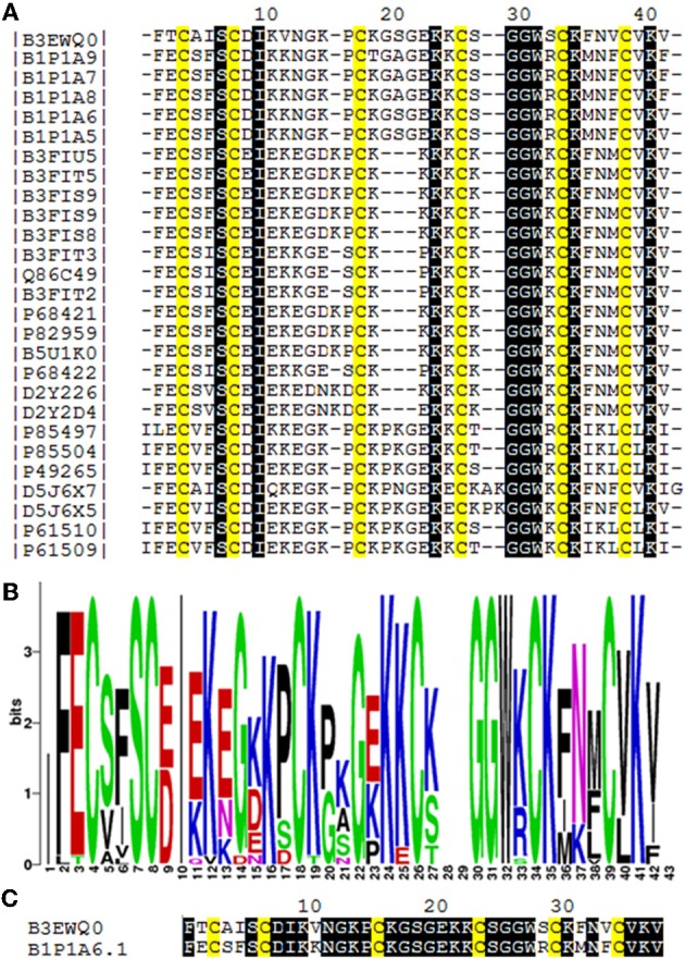
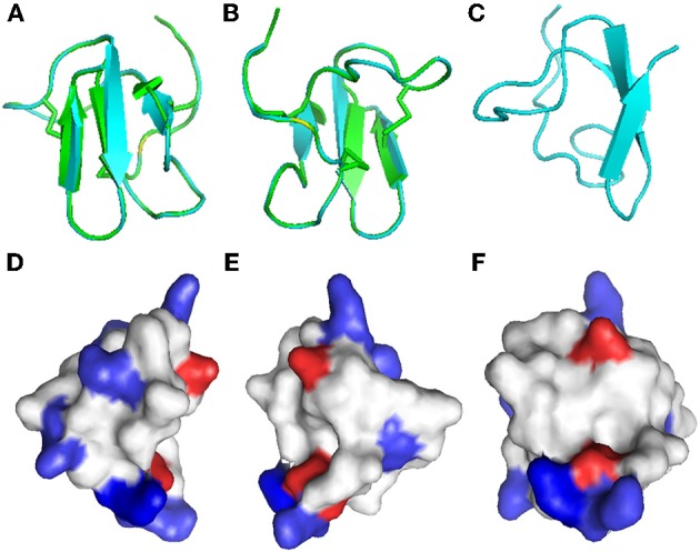
The aim of this study was to screen the venom of the theraposid spider Avicularia juruensis for the identification of antimicrobial peptides (AMPs) which could be further used as prototypes for drug development. Eleven AMPs, named juruentoxins, with molecular weight ranging from 3.5 to 4.5 kDa, were identified by mass spectrometry after the soluble venom was separated by high performance liquid chromatography. Juruentoxins have a putative inhibitory cystine knot (ICK) motif, generally found in neurotoxins, which are also resistant to proteolysis. One juruentoxin that has 38 amino acid residues and three disulfide bonds were characterized, to which we proposed the name Juruin. Based on liquid growth inhibition assays, it has potent antifungal activity in the micromolar range. Importantly, Juruin lacks haemolytic activity on human erythrocytes at the antimicrobial concentrations. Based on the amino acid sequence, it is highly identical to the insecticidal peptides from the theraposid spiders Selenocosmia huwena, Chilobrachys jingzhao, and Haplopelma schmidti from China, indicating they belong to a group of conserved toxins which are likely to inhibit voltage-gated ion channels. Juruin is a cationic AMP, and Lys22 and Lys23 show maximum positive charge localization that might be important for receptor recognition. Although it shows marked sequence similarity to neurotoxic peptides, Juruin is a novel exciting molecule with potent antifungal activity, which could be used as a novel template for development of drugs against clinical resistant fungi strains.
Keywords: Avicularia juruensis; Juruin; Theraphosidae venom; antimicrobial peptides; inhibitory cystine knot motif; juruentoxins.
Figures
References
-
- Bertani R., Fukushima C. S. (2009). Description of two new species of Avicularia Lamarck 1818 and redescription of Avicularia diversipes (C. L. Koch 1842) (Araneae, Theraphosidae, Aviculariinae)-three possibly threatened Brazilian species. Zootaxa 2223, 25–47
-
- Bradford M. M. (1976). A rapid and sensitive for the quantitation of microgram quantitites of protein utilizing the principle of protein-dye binding. Anal. Biochem. 72, 248–254 - PubMed
LinkOut - more resources
Full Text Sources