. 2012 Sep 11:3:285.
doi: 10.3389/fimmu.2012.00285. eCollection 2012.

Positive and negative regulation of T cell responses by fibroblastic reticular cells within paracortical regions of lymph nodes

Affiliations

Positive and negative regulation of T cell responses by fibroblastic reticular cells within paracortical regions of lymph nodes

Stefanie Siegert et al. Front Immunol. .

Abstract

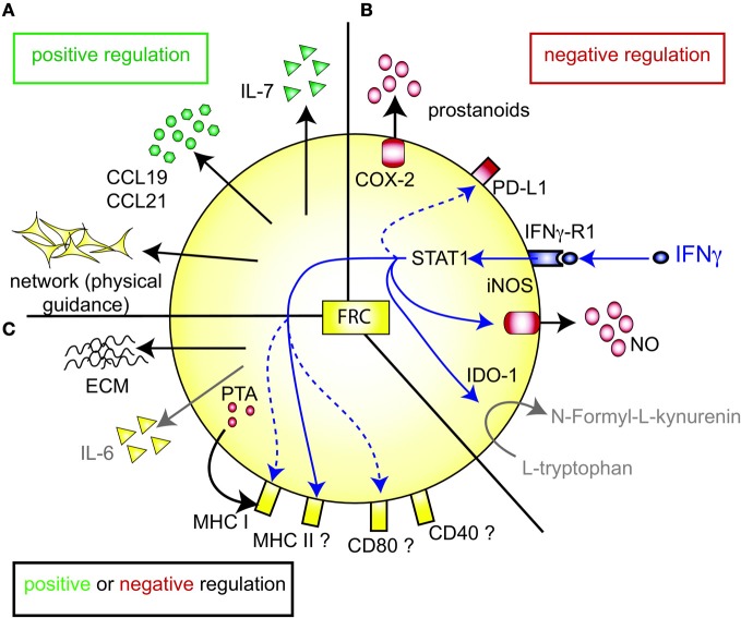
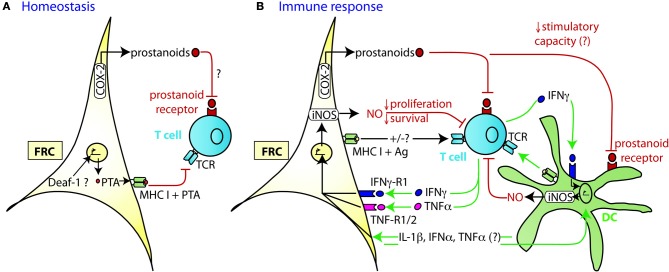
Fibroblastic reticular cells (FRC) form the structural backbone of the T cell rich zones in secondary lymphoid organs (SLO), but also actively influence the adaptive immune response. They provide a guidance path for immigrating T lymphocytes and dendritic cells (DC) and are the main local source of the cytokines CCL19, CCL21, and IL-7, all of which are thought to positively regulate T cell homeostasis and T cell interactions with DC. Recently, FRC in lymph nodes (LN) were also described to negatively regulate T cell responses in two distinct ways. During homeostasis they express and present a range of peripheral tissue antigens, thereby participating in peripheral tolerance induction of self-reactive CD8(+) T cells. During acute inflammation T cells responding to foreign antigens presented on DC very quickly release pro-inflammatory cytokines such as interferon γ. These cytokines are sensed by FRC which transiently produce nitric oxide (NO) gas dampening the proliferation of neighboring T cells in a non-cognate fashion. In summary, we propose a model in which FRC engage in a bidirectional crosstalk with both DC and T cells to increase the efficiency of the T cell response. However, during an acute response, FRC limit excessive expansion and inflammatory activity of antigen-specific T cells. This negative feedback loop may help to maintain tissue integrity and function during rapid organ growth.

Keywords: T lymphocyte activation; cyclooxygenase 2; fibroblastic reticular cell (FRC); immune tolerance; inducible nitric oxide synthase; lymph node stromal cells; mesenchymal stem cells; suppression.


Figures

Figure 1
Figure 1
FRC-expressed molecules that positively or negatively regulate the T cell response. (A) FRC are thought to positively regulate T cell immunity in several ways. Throughout the T zone of SLO FRC form a three-dimensional network that serves as a scaffold for DC adhesion and T cell migration, thereby increasing DC-T interaction. In addition, FRC constitutively produce CCL19 and CCL21 that retain T cells in the T zone while increasing their motility. FRC also constitutively produce IL-7 that promotes T cell survival. CCL19, CCL21, and IL-7 may all serve as co-stimulatory signals in case of weak T cell receptor triggering. (B) FRC express several molecules that may negatively regulate the immune response: COX-2 and PD-L1 are constitutively expressed, while iNOS and IDO-1 are only expressed after induction by IFNγ. iNOS expression in FRC is also induced by other pro-inflammatory cytokines, such as TNFα, IL-1β and IFNα (not shown). PD-L1 expression on FRC is increased by LCMV infection, IFNγ or TLR3 stimulation (Mueller et al., ; Fletcher et al., ; Ng et al., 2012). While blocking iNOS and COX improves T cell proliferation in the presence of FRC, no impact of FRC-derived PD-L1, IDO-1, CD80/86, or IL-2 on T cell proliferation has been shown. It is unclear whether FRC can express L-arginase, but arginase inhibitors showed no effect in vitro (Khan et al., ; Lukacs-Kornek et al., ; Siegert et al., 2011). (C) FRC produce several factors which may regulate adaptive immunity in a positive or negative way. They secrete extracellular matrix (ECM) proteins some of which can bind IL-7 and CCL21 and thereby regulate cytokine availability and localization (Katakai et al., ; Förster et al., ; Huang and Luther, 2012). ECM molecules may also directly modulate T cell and DC behavior. Microarray data suggest that FRC express constitutively IL-6, which is both anti- and pro-inflammatory depending on the context. FRC express MHC I and low levels of MHC II, CD80, and CD40. IFNγ or TLR3 stimulation enhances the expression of MHC I, MHC II and CD80. In addition, LCMV infection or inflammation evoked by LPS injection in vivo can induce MHC II expression on FRC. LN FRC express PTA, which are presented in the context of MHC I, leading to T cell proliferation followed possibly by clonal deletion of self-reactive T cells. Therefore, in certain settings FRC may act as APC.
Figure 2
Figure 2
Mechanism of TRC-mediated negative regulation of T cell expansion. (A) During homeostasis LN FRC express self-antigens (PTA), possibly in a Deaf-1 dependent fashion. PTA presented in the context of MHC I may lead to peripheral tolerance induction via clonal deletion of self-reactive T cells. Cox-2 is constitutively expressed in FRC, presumably leading to the production of PGE2 or other prostanoids that may attenuate T cell priming or proliferation. (B) During immune response DC capture antigen (Ag) in the periphery, home to the T zone of SLO and present peptides of foreign antigens to recirculating T cells. Within the first 20 h upon TCR triggering, T cells produce a first wave of IFNγ and TNFα cytokines, before entering cell cycle. In case of a very strong immune response, the high level of pro-inflammatory cytokines induces transient iNOS expression in the neighboring FRC and DC. The resulting NO release then acts as a negative feedback loop slowing down T cell proliferation over the following days presumably to ensure that the strong T cell response does not endanger tissue integrity. High concentrations of NO may also induce T cell apoptosis. The attenuating effect by FRC can occur in trans, without cognate interaction with T cells. Constitutive expression of COX-2 dependent factors, such as PGE2, may contribute to this attenuation of T cell expansion, by acting directly or indirectly (via DC) on the proliferating T cells. Not only is there crosstalk between FRC and T cells, but also between FRC and DC which leads to a much stronger release of NO by these two cell types if IFNγ is present. While the factors exchanged between FRC and DC have not yet been identified, the may include IL-1β, IFNα, and TNFα, all of which can trigger iNOS expression.

References

    1. Akiyama K., Chen C., Wang D., Xu X., Qu C., Yamaza T., Cai T., Chen W., Sun L., Shi S. (2012). Mesenchymal-stem-cell-induced immunoregulation involves FAS-ligand-/FAS-mediated T cell apoptosis. Cell Stem Cell 10, 544–555 10.1016/j.stem.2012.03.007 - DOI - PMC - PubMed
    1. Albina J. E., Abate J. A., Henry W. L. (1991). Nitric oxide production is required for murine resident peritoneal macrophages to suppress mitogen-stimulated T cell proliferation. Role of IFN-gamma in the induction of the nitric oxide-synthesizing pathway. J. Immunol. 147, 144–148 - PubMed
    1. Asperti-Boursin F., Real E., Bismuth G., Trautmann A., Donnadieu E. (2007). CCR7 ligands control basal T cell motility within lymph node slices in a phosphoinositide 3-kinase-independent manner. J. Exp. Med. 204, 1167–1179 10.1084/jem.20062079 - DOI - PMC - PubMed
    1. Bajenoff M., Egen J. G., Koo L. Y., Laugier J. P., Brau F., Glaichenhaus N., Germain R. N. (2006). Stromal cell networks regulate lymphocyte entry, migration, and territoriality in lymph nodes. Immunity 25, 989–1001 10.1016/j.immuni.2006.10.011 - DOI - PMC - PubMed
    1. Beuneu H., Lemaître F., Deguine J., Moreau H. D., Bouvier I., Garcia Z., Albert M. L., Bousso P. (2010). Visualizing the functional diversification of CD8+ T cell responses in lymph nodes. Immunity 33, 412–423 10.1016/j.immuni.2010.08.016 - DOI - PubMed