. 2012 Sep 11:3:177.
doi: 10.3389/fgene.2012.00177. eCollection 2012.

Rapamycin has a biphasic effect on insulin sensitivity in C2C12 myotubes due to sequential disruption of mTORC1 and mTORC2

Affiliations

Rapamycin has a biphasic effect on insulin sensitivity in C2C12 myotubes due to sequential disruption of mTORC1 and mTORC2

Lan Ye et al. Front Genet. .

Abstract

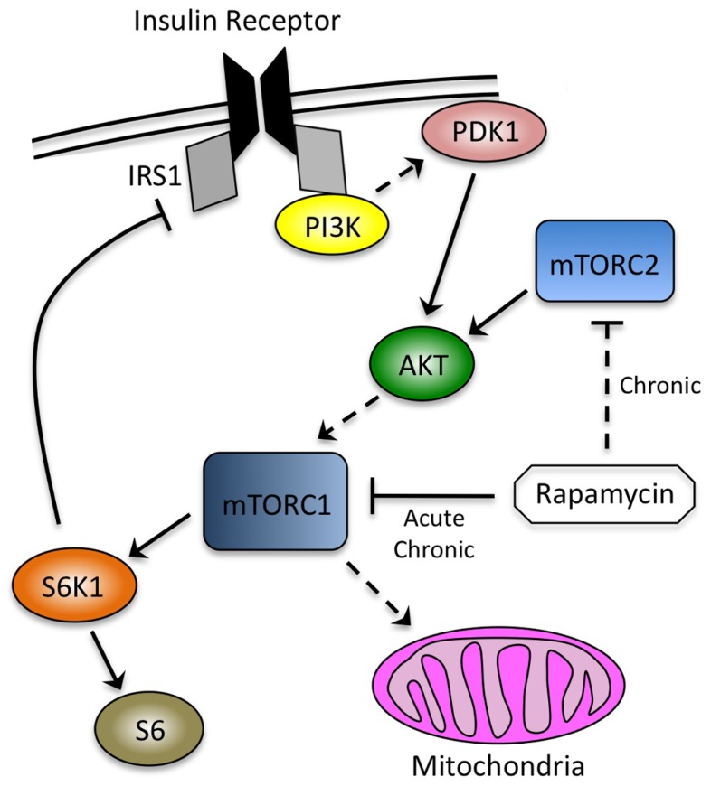
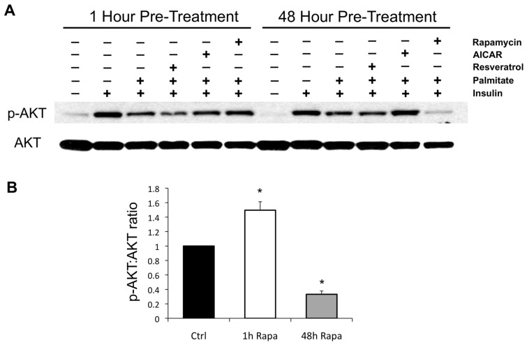
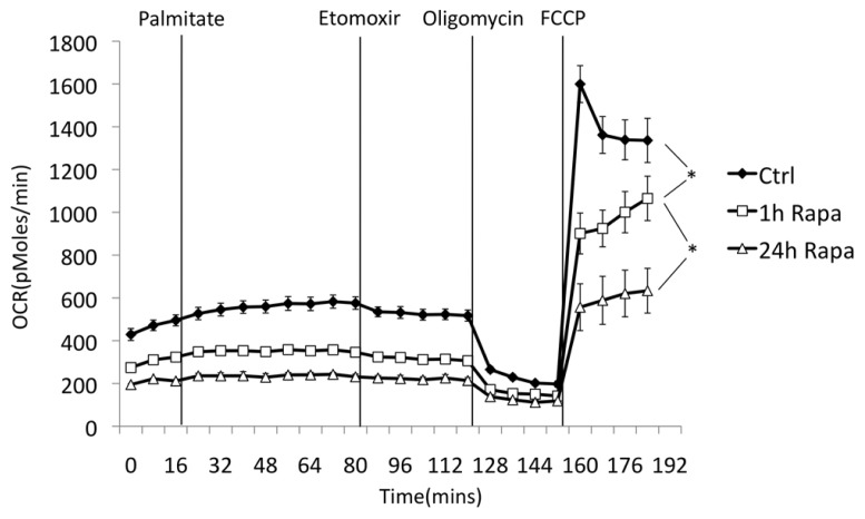
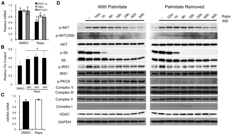
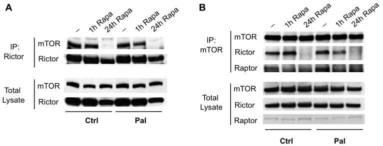
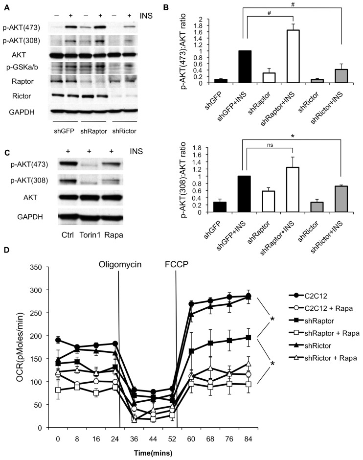
Rapamycin, an inhibitor of mTOR complex 1 (mTORC1), improves insulin sensitivity in acute studies in vitro and in vivo by disrupting a negative feedback loop mediated by S6 kinase. We find that rapamycin has a clear biphasic effect on insulin sensitivity in C2C12 myotubes, with enhanced responsiveness during the first hour that declines to almost complete insulin resistance by 24-48 h. We and others have recently observed that chronic rapamycin treatment induces insulin resistance in rodents, at least in part due to disruption of mTORC2, an mTOR-containing complex that is not acutely sensitive to the drug. Chronic rapamycin treatment may also impair insulin action via the inhibition of mTORC1-dependent mitochondrial biogenesis and activity, which could result in a buildup of lipid intermediates that are known to trigger insulin resistance. We confirmed that rapamycin inhibits expression of PGC-1α, a key mitochondrial transcription factor, and acutely reduces respiration rate in myotubes. However, rapamycin did not stimulate phosphorylation of PKCθ, a central mediator of lipid-induced insulin resistance. Instead, we found dramatic disruption of mTORC2, which coincided with the onset of insulin resistance. Selective inhibition of mTORC1 or mTORC2 by shRNA-mediated knockdown of specific components (Raptor and Rictor, respectively) confirmed that mitochondrial effects of rapamycin are mTORC1-dependent, whereas insulin resistance was recapitulated only by knockdown of mTORC2. Thus, mTORC2 disruption, rather than inhibition of mitochondria, causes insulin resistance in rapamycin-treated myotubes, and this system may serve as a useful model to understand the effects of rapamycin on mTOR signaling in vivo.

Keywords: chronic; diabetes; insulin sensitivity; mTOR; mTORC1; mTORC2; mitochondrial biogenesis; rapamycin; raptor; respiration; rictor.


Figures

FIGURE 1
FIGURE 1
Interdependence of signaling through the insulin receptor and mTOR. mTORC1, the canonical target of rapamycin, is downstream of the insulin signaling cascade and mediates negative feedback through S6K1. mTORC2 is an AKT kinase that is not sensitive to acute rapamycin treatment (~ h), but can be inhibited by chronic exposure (~24 h). Phosphorylation of AKT by PDK1 and mTORC2 occurs at discrete sites (threonine 308 and serine 473, respectively). mTORC1 promotes mitochondrial biogenesis, although the relevance of this observation to its effects on insulin sensitivity has not been established. Solid lines indicate direct effects, whereas dashed lines indicate intermediate steps, or in the case of rapamycin inhibiting mTORC2, the requirement for chronic exposure.
FIGURE 2
FIGURE 2
Effects of rapamycin, AICAR, and resveratrol on palmitate-induced insulin resistance. (A) Palmitate renders C2C12 myotubes refractory to insulin stimulation, as reflected by downstream phosphorylation of AKT. One hour of pre-treatment with resveratrol mildly increases insulin resistance, consistent with previous reports of acute inhibition of insulin signaling (Zhang, 2006), whereas the AMPK activator AICAR is without effect, and rapamycin slightly improves insulin signaling, likely due to disruption of negative feedback through S6K1 (Um et al., 2004). After 48 h of pre-treatment, AICAR rescues insulin signaling, while rapamycin renders myotubes almost completely resistant. The effects of AICAR and rapamycin on insulin signaling were each verified in at least three independent experiments and representative blots are shown. (B) Quantification of the effects of short- (1 h) and long-term (48 h) exposure to rapamycin on the insulin-stimulated phosphorylation of AKT in palmitate-treated C2C12 myotubes. *p < 0.05. Error bars show SEM.
FIGURE 3
FIGURE 3
Rapamycin suppresses oxygen consumption in myotubes. Oxygen consumption was measured using a Seahorse XF24 Flux Analyzer. C2C12 myoblasts were differentiated on Seahorse plates and pre-treated with rapamycin or vehicle (DMSO) for the indicated times. Glucose was maintained at 25 mM throughout the experiment. Acute addition of palmitate (0.5 mM final concentration, in complex with BSA) had only a minor effect on total oxygen consumption, which was reversed with etomoxir (50 μM), an inhibitor of carnitine palmitoyl transferase 1. Oligomycin was then added to inhibit ATP synthase and determine the rate of proton leak across the mitochondrial membrane. Finally, FCCP was added to uncouple mitochondria and measure maximal respiration rate. Decreased respiration in rapamycin-treated cells was verified in three independent experiments, consistent with previous reports (Schieke et al., 2006; Ramanathan and Schreiber, 2009). The length of rapamycin pre-treatment (1 h vs. 24 h) had a significant effect on respiration rate under basal, but not uncoupled conditions. *p < 0.05. Error bars show SEM.
FIGURE 4
FIGURE 4
Rapamycin inhibits mitochondrial transcription factors, but has no major effect on mitochondrial protein levels or PKCθ phosphorylation. (A) Forty-eight hours of rapamycin treatment decreases expression of PGC-1α and TFAM, two of the major transcription factors involved in mitochondrial biogenesis, as well as the level of MT-CO1, an mtDNA-encoded transcript. (B) Rapamycin mildly increases intracellular triglyceride levels in myotubes. The result show is representative of three separate experiments. (C) Forty-eight hours of rapamycin treatment has no effect on mitochondrial DNA copy number, as reflected by the ratio between MT-CO1 (mitochondrial DNA-encoded) and Ndufv1 (nuclear-encoded) DNA. (D) Preincubation of myotubes with rapamycin for 24 h or more induces insulin resistance irrespective of the presence of palmitate. Rapamycin-induced insulin resistance is not associated with major increases in serine phosphorylation of IRS1 or phosphorylation of PKCθ, two hallmarks of insulin resistance caused by insufficient oxidation of fatty acids. Moreover, there is no change in the expression of several proteins involved in electron transport complexes or in the level of VDAC, a structural protein in the mitochondria. *p < 0.05. Error bars show SEM.
FIGURE 5
FIGURE 5
Rapamycin disrupts mTORC2 in myotubes. mTORC2 was immunoprecipitated from myotubes that were pre-treated with vehicle, rapamycin for 1 h, or rapamycin for 24 h, using antibodies to Rictor (A) or mTOR (B). The integrity of the complex was assessed by blotting for the opposite subunit. In each case, mTORC2 formation was disrupted at 24 h despite only minor changes in total protein levels. The mTORC1 subunit Raptor was also absent from mTOR immunoprecipitates at either 1 or 24 h.
FIGURE 6
FIGURE 6
Knockdown of mTORC1 reduces oxygen consumption while knockdown of mTORC2 causes insulin resistance in myotubes. (A) C2C12 myoblasts were infected with shRNAs targeting Raptor (mTORC1) or Rictor (mTORC2) and differentiated. Cells were harvested with or without insulin stimulation and probed for phosphorylation of AKT at threonine 308 and serine 473. Results shown are representative of three independent experiments. (B) Quantification of changes in the ratio of phosphorylated to total AKT following knockdown of mTOR complex subunits. (C) Torin1, an inhibitor of both mTORC1 and mTORC2 is sufficient to block phosphorylation of AKT at both sites, although direct inhibition of upstream phosphoinositide 3-kinase may contribute to this effect (Liu et al., 2012). C2C12 myoblasts were differentiated and treated with Torin1 (250 nM for 4 h) or rapamycin (500 nM for 24 h) (D) Knockdown of Raptor partially suppresses oxygen consumption in the absence of rapamycin. C2C12 myoblasts were pre-treated with rapamycin or vehicle (DMSO) for 24 h and then analyzed on the Seahorse XF24 Flux Analyzer in unbuffered DMEM (25 mM glucose) in the presence of rapamycin or DMSO, as indicated. Oligomycin was added to inhibit ATP synthase in order to determine the rate of proton leak across the mitochondrial membrane, and FCCP was subsequently added to uncouple mitochondria and measure maximal respiration rate. #p < 0.05 one-tailed; *p < 0.05 two-tailed; ns, not significant. Error bars show SEM.

References

    1. Baur J. A. (2009). “Metabolic effects of caloric restriction,” in Encyclopedia of Life Sciences (Chichester: John Wiley & Sons, Ltd).
    1. Bentzinger C. F., Romanino K., Cloetta D., Lin S., Mascarenhas J. B., Oliveri F., Xia J., Casanova E., Costa C. F., Brink M., Zorzato F., Hall M. N, Rüegg M. A. (2008). Skeletal muscle-specific ablation of raptor, but not of rictor, causes metabolic changes and results in muscle dystrophy. Cell Metab. 8 411–424 - PubMed
    1. Blattler S. M., Cunningham J. T., Verdeguer F., Chim H., Haas W., Liu H., Romanino K., Ruegg M. A., Gygi S. P., Shi Y., Puigserver P. (2012). Yin Yang 1 deficiency in skeletal muscle protects against rapamycin-induced diabetic-like symptoms through activation of insulin/IGF signaling. Cell Metab. 15 505–517 - PMC - PubMed
    1. Chavez J. A., Holland W. L., Bar J., Sandhoff K., Summers S. A. (2005). Acid ceramidase overexpression prevents the inhibitory effects of saturated fatty acids on insulin signaling. J. Biol. Chem. 280 20148–20153 - PubMed
    1. Chen C., Liu Y., Zheng P. (2009). mTOR regulation and therapeutic rejuvenation of aging hematopoietic stem cells. Sci. Signal. 2 ra75 - PMC - PubMed