

Pyoverdine, the Major Siderophore in Pseudomonas aeruginosa, Evades NGAL Recognition
- PMID: 22973307
- PMCID: PMC3438788
- DOI: 10.1155/2012/843509
Pyoverdine, the Major Siderophore in Pseudomonas aeruginosa, Evades NGAL Recognition
Abstract
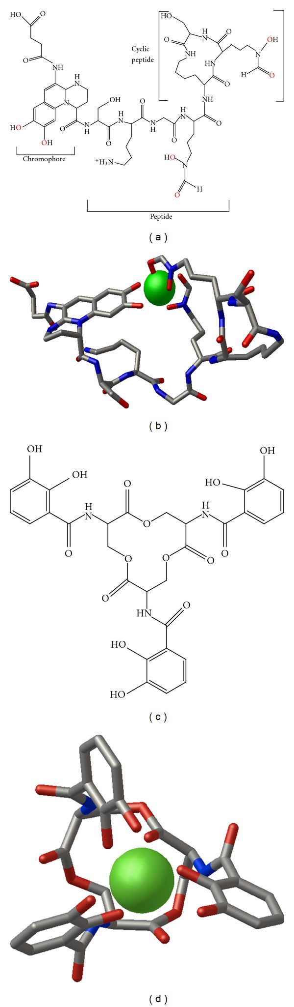
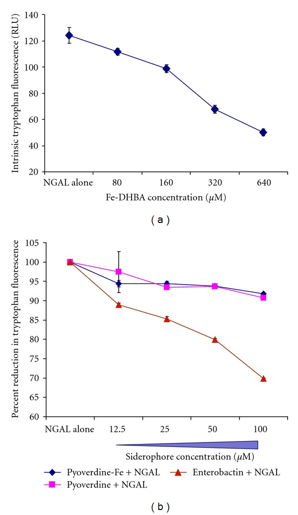
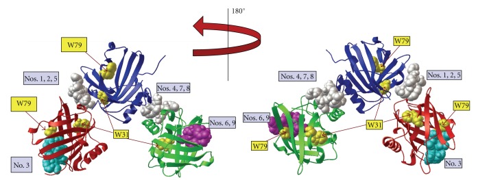
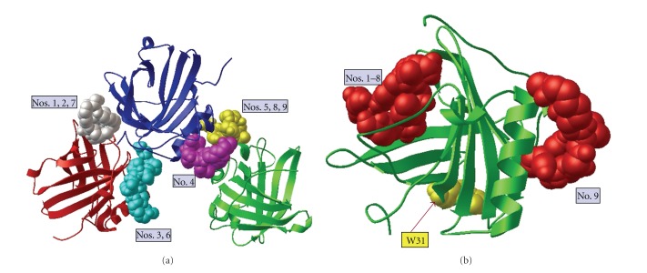
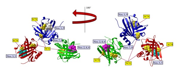
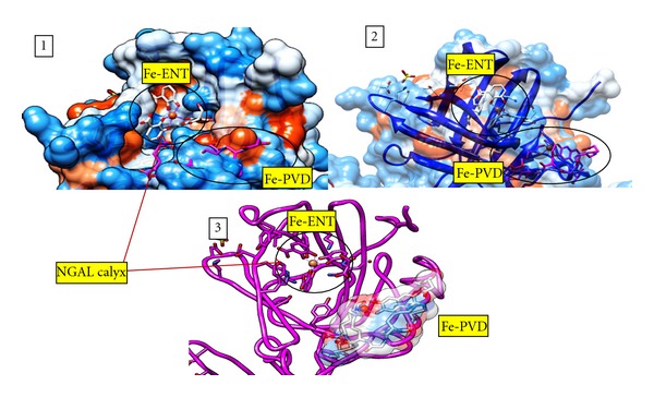
Pseudomonas aeruginosa is the most common pathogen that persists in the cystic fibrosis lungs. Bacteria such as P. aeruginosa secrete siderophores (iron-chelating molecules) and the host limits bacterial growth by producing neutrophil-gelatinase-associated lipocalin (NGAL) that specifically scavenges bacterial siderophores, therefore preventing bacteria from establishing infection. P. aeruginosa produces a major siderophore known as pyoverdine, found to be important for bacterial virulence and biofilm development. We report that pyoverdine did not bind to NGAL, as measured by tryptophan fluorescence quenching, while enterobactin bound to NGAL effectively causing a strong response. The experimental data indicate that pyoverdine evades NGAL recognition. We then employed a molecular modeling approach to simulate the binding of pyoverdine to human NGAL using NGAL's published crystal structures. The docking of pyoverdine to NGAL predicted nine different docking positions; however, neither apo- nor ferric forms of pyoverdine docked into the ligand-binding site in the calyx of NGAL where siderophores are known to bind. The molecular modeling results offer structural support that pyoverdine does not bind to NGAL, confirming the results obtained in the tryptophan quenching assay. The data suggest that pyoverdine is a stealth siderophore that evades NGAL recognition allowing P. aeruginosa to establish chronic infections in CF lungs.
Figures
Similar articles
-
Parsing the functional specificity of Siderocalin/Lipocalin 2/NGAL for siderophores and related small-molecule ligands.J Struct Biol X. 2019 May 30;2:100008. doi: 10.1016/j.yjsbx.2019.100008. eCollection 2019 Apr-Jun. J Struct Biol X. 2019. PMID: 32647813 Free PMC article.
-
In vitro lung epithelial cell model reveals novel roles for Pseudomonas aeruginosa siderophores.Microbiol Spectr. 2024 Mar 5;12(3):e0369323. doi: 10.1128/spectrum.03693-23. Epub 2024 Feb 5. Microbiol Spectr. 2024. PMID: 38311809 Free PMC article.
-
Chemistry and biology of pyoverdines, Pseudomonas primary siderophores.Curr Med Chem. 2015;22(2):165-86. doi: 10.2174/0929867321666141011194624. Curr Med Chem. 2015. PMID: 25312210
-
Pseudomonas aeruginosa virulence attenuation by inhibiting siderophore functions.Appl Microbiol Biotechnol. 2023 Feb;107(4):1019-1038. doi: 10.1007/s00253-022-12347-6. Epub 2023 Jan 12. Appl Microbiol Biotechnol. 2023. PMID: 36633626 Review.
-
A Review of Pseudomonas aeruginosa Metallophores: Pyoverdine, Pyochelin and Pseudopaline.Biology (Basel). 2022 Nov 25;11(12):1711. doi: 10.3390/biology11121711. Biology (Basel). 2022. PMID: 36552220 Free PMC article. Review.
Cited by
-
Inflammation and ER stress downregulate BDH2 expression and dysregulate intracellular iron in macrophages.J Immunol Res. 2014;2014:140728. doi: 10.1155/2014/140728. Epub 2014 Dec 1. J Immunol Res. 2014. PMID: 25762501 Free PMC article.
-
An intra-abdominal abscess or "rind" as a consequence of peritoneal dialysis-associated pseudomonas peritonitis.Clin Nephrol Case Stud. 2013 Feb 26;1:1-6. doi: 10.5414/CNCS107951. eCollection 2013. Clin Nephrol Case Stud. 2013. PMID: 29043117 Free PMC article.
-
Recent advances in graphene-based nanobiosensors for salivary biomarker detection.Biosens Bioelectron. 2021 Jan 1;171:112723. doi: 10.1016/j.bios.2020.112723. Epub 2020 Oct 13. Biosens Bioelectron. 2021. PMID: 33096432 Free PMC article. Review.
-
Potential Therapeutic Targets for Combination Antibody Therapy against Pseudomonas aeruginosa Infections.Antibiotics (Basel). 2021 Dec 14;10(12):1530. doi: 10.3390/antibiotics10121530. Antibiotics (Basel). 2021. PMID: 34943742 Free PMC article. Review.
-
Microbial siderophores and their potential applications: a review.Environ Sci Pollut Res Int. 2016 Mar;23(5):3984-99. doi: 10.1007/s11356-015-4294-0. Epub 2015 Mar 12. Environ Sci Pollut Res Int. 2016. PMID: 25758420 Review.
References
-
- Visca P, Imperi F, Lamont IL. Pyoverdine siderophores: from biogenesis to biosignificance. Trends in Microbiology. 2007;15(1):22–30. - PubMed
-
- Lamont IL, Konings AF, Reid DW. Iron acquisition by Pseudomonas aeruginosa in the lungs of patients with cystic fibrosis. BioMetals. 2009;22(1):53–60. - PubMed
-
- Saiga H, Nishimura J, Kuwata H, et al. Lipocalin 2-dependent inhibition of mycobacterial growth in alveolar epithelium. Journal of Immunology. 2008;181(12):8521–8527. - PubMed
LinkOut - more resources
Full Text Sources
Other Literature Sources
Molecular Biology Databases
Miscellaneous
