

Brain miffed by macrophage migration inhibitory factor
- PMID: 22973314
- PMCID: PMC3438795
- DOI: 10.1155/2012/139573
Brain miffed by macrophage migration inhibitory factor
Abstract
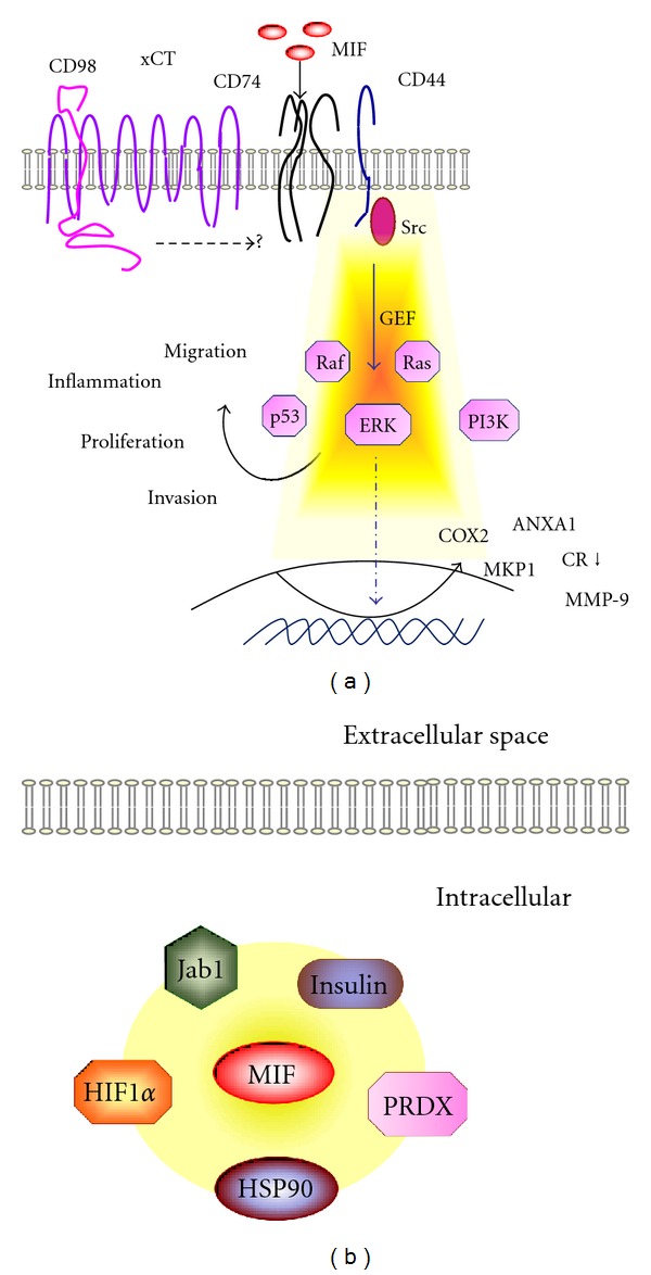
Macrophage migration inhibitory factor (MIF) is a cytokine which also exhibits enzymatic properties like oxidoreductase and tautomerase. MIF plays a pivotal role in innate and acquired immunity as well as in the neuroendocrine axis. Since it is involved in the pathogenesis of acute and chronic inflammation, neoangiogenesis, and cancer, MIF and its signaling components are considered suitable targets for therapeutic intervention in several fields of medicine. In neurodegenerative and neurooncological diseases, MIF is a highly relevant, but still a hardly investigated mediator. MIF operates via intracellular protein-protein interaction as well as in CD74/CXCR2/CXCR4 receptor-mediated pathways to regulate essential cellular systems such as redox balance, HIF-1, and p53-mediated senescence and apoptosis as well as multiple signaling pathways. Acting as an endogenous glucocorticoid antagonist, MIF thus represents a relevant resistance gene in brain tumor therapies. Alongside this dual action, a functional homolog-annotated D-dopachrome tautomerase/MIF-2 has been uncovered utilizing the same cell surface receptor signaling cascade as MIF. Here we review MIF actions with respect to redox regulation in apoptosis and in tumor growth as well as its extracellular function with a focus on its potential role in brain diseases. We consider the possibility of MIF targeting in neurodegenerative processes and brain tumors by novel MIF-neutralizing approaches.
Figures
Similar articles
-
D-dopachrome tautomerase in adipose tissue inflammation and wound repair.J Cell Mol Med. 2017 Jan;21(1):35-45. doi: 10.1111/jcmm.12936. Epub 2016 Sep 7. J Cell Mol Med. 2017. PMID: 27605340 Free PMC article.
-
Studying Plant MIF/D-DT-Like Genes and Proteins (MDLs).Methods Mol Biol. 2020;2080:249-261. doi: 10.1007/978-1-4939-9936-1_22. Methods Mol Biol. 2020. PMID: 31745887
-
MIF, CD74 and other partners in kidney disease: tales of a promiscuous couple.Cytokine Growth Factor Rev. 2013 Feb;24(1):23-40. doi: 10.1016/j.cytogfr.2012.08.001. Epub 2012 Sep 7. Cytokine Growth Factor Rev. 2013. PMID: 22959722 Review.
-
A selective small-molecule inhibitor of macrophage migration inhibitory factor-2 (MIF-2), a MIF cytokine superfamily member, inhibits MIF-2 biological activity.J Biol Chem. 2019 Dec 6;294(49):18522-18531. doi: 10.1074/jbc.RA119.009860. Epub 2019 Oct 2. J Biol Chem. 2019. PMID: 31578280 Free PMC article.
-
D-dopachrome tautomerase in cardiovascular and inflammatory diseases-A new kid on the block or just another MIF?FASEB J. 2022 Nov;36(11):e22601. doi: 10.1096/fj.202201213R. FASEB J. 2022. PMID: 36269019 Review.
Cited by
-
Identification of Iguratimod as an Inhibitor of Macrophage Migration Inhibitory Factor (MIF) with Steroid-sparing Potential.J Biol Chem. 2016 Dec 16;291(51):26502-26514. doi: 10.1074/jbc.M116.743328. Epub 2016 Oct 28. J Biol Chem. 2016. PMID: 27793992 Free PMC article.
-
Transcriptomic analyses and leukocyte telomere length measurement in subjects exposed to severe recent stressful life events.Transl Psychiatry. 2017 Feb 21;7(2):e1042. doi: 10.1038/tp.2017.5. Transl Psychiatry. 2017. PMID: 28221367 Free PMC article.
-
MIF: mood improving/inhibiting factor?J Neuroinflammation. 2014 Jan 21;11:11. doi: 10.1186/1742-2094-11-11. J Neuroinflammation. 2014. PMID: 24447830 Free PMC article. Review.
-
Macrophage Migration Inhibitory Factor and microRNA-451a in Response to Mindfulness-based Therapy or Treatment as Usual in Patients with Depression, Anxiety, or Stress and Adjustment Disorders.Int J Neuropsychopharmacol. 2018 Jun 1;21(6):513-521. doi: 10.1093/ijnp/pyy001. Int J Neuropsychopharmacol. 2018. PMID: 29373661 Free PMC article. Clinical Trial.
-
Deletion of macrophage migration inhibitory factor worsens stroke outcome in female mice.Neurobiol Dis. 2013 Jun;54:421-31. doi: 10.1016/j.nbd.2013.01.016. Epub 2013 Jan 30. Neurobiol Dis. 2013. PMID: 23376686 Free PMC article.
References
-
- Isaacs A, Lindenmann J. Virus interference. I. The interferon. Proceedings of the Royal Society B. 1957;147(927):258–267. - PubMed
-
- Bloom BR, Bennett B. Mechanism of a reaction in vitro associated with delayed-type hypersensitivity. Science. 1966;153(3731):80–82. - PubMed
-
- Bernhagen J, Calandra T, Mitchell RA, et al. MIF is a pituitary-derived cytokine that potentiates lethal endotoxaemia. Nature. 1993;365(6448):756–759. - PubMed
LinkOut - more resources
Full Text Sources
Other Literature Sources
Research Materials
Miscellaneous
