. 2012;7(9):e44400.
doi: 10.1371/journal.pone.0044400. Epub 2012 Sep 4.

Galectin-8 promotes cytoskeletal rearrangement in trabecular meshwork cells through activation of Rho signaling

Affiliations

Galectin-8 promotes cytoskeletal rearrangement in trabecular meshwork cells through activation of Rho signaling

Shiri Diskin et al. PLoS One. 2012.

Abstract

Purpose: The trabecular meshwork (TM) cell-matrix interactions and factors that influence Rho signaling in TM cells are thought to play a pivotal role in the regulation of aqueous outflow. The current study was designed to evaluate the role of a carbohydrate-binding protein, galectin-8 (Gal8), in TM cell adhesion and Rho signaling.

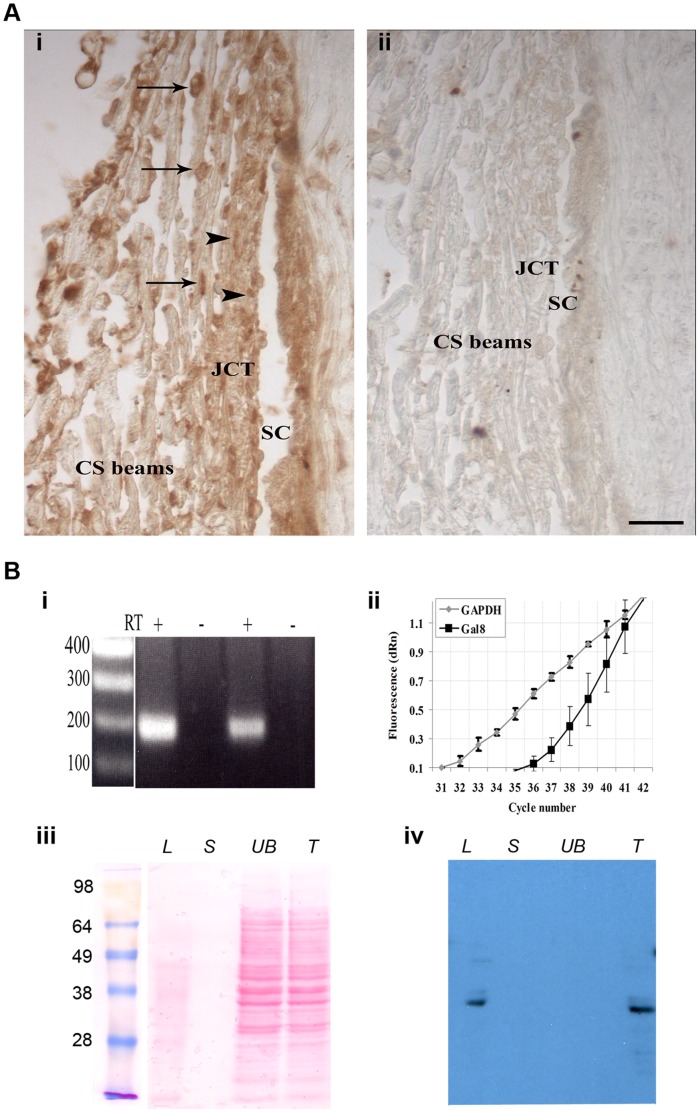
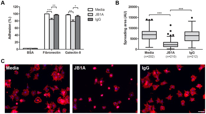
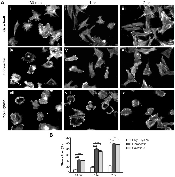
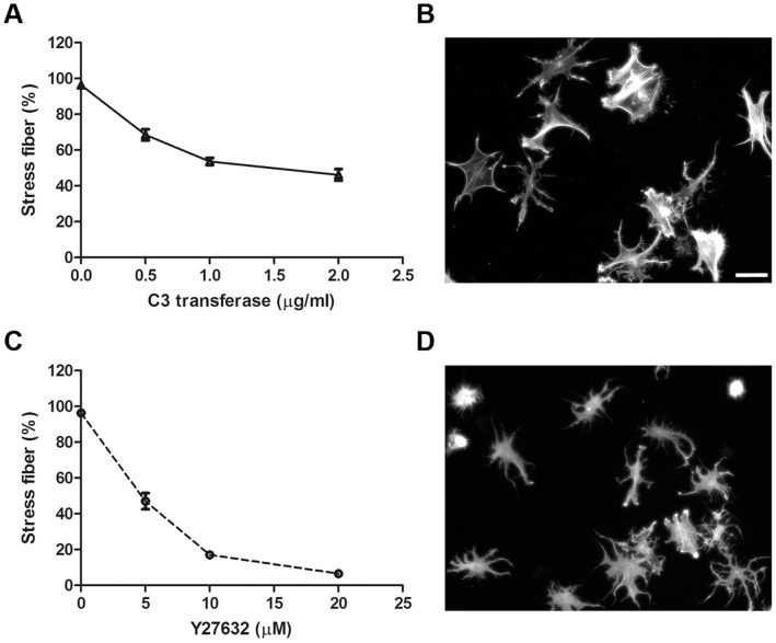
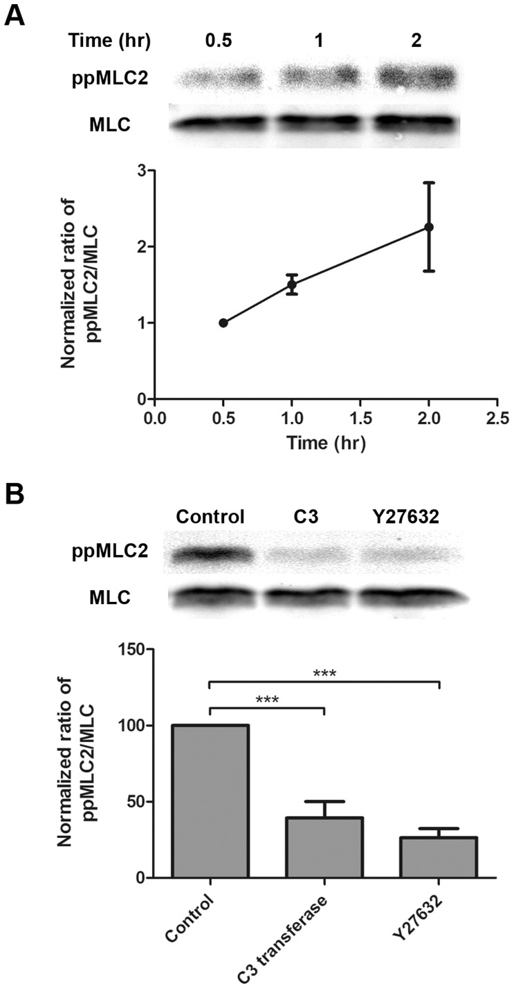
Methods: Normal human TM cells were assayed for Gal8 expression by immunohistochemistry and Western blot analysis. To assess the role of Gal8 in TM cell adhesion and Rho signaling, the cell adhesion and spreading assays were performed on Gal8-coated culture plates in the presence and the absence of anti-β₁ integrin antibody and Rho and Rho-kinase inhibitors. In addition, the effect of Gal8-mediated cell-matrix interactions on TM cell cytoskeleton arrangement and myosin light chain 2 (MLC2) phosphorylation was examined.

Principal findings: We demonstrate here that Gal8 is expressed in the TM and a function-blocking anti-β₁ integrin antibody inhibits the adhesion and spreading of TM cells to Gal8-coated wells. Cell spreading on Gal8 substratum was associated with the accumulation of phosphorylated myosin light chain and the formation of stress fibers that was inhibited by the Rho inhibitor, C3 transferase, as well as by the Rho-kinase inhibitor, Y27632.

Conclusions/significance: The above findings present a novel function for Gal8 in activating Rho signaling in TM cells. This function may allow Gal8 to participate in the regulation of aqueous outflow.


Conflict of interest statement

Competing Interests: The authors have declared that no competing interests exist.

Figures

Figure 1
Figure 1. Galectin-8 is expressed in the Trabecular Meshwork (TM).
A: paraffin sections of anterior chamber angle from a normal human eye were immunostained with anti-Gal8 antibody. (i) anti-Gal8 IgG reacted intensely with cells on the trabecular beams (arrows) and with cells in the juxtacanalicular portion of TM (arrowheads). Staining was also observed in the ECM of both portions of TM (JCT and CS) and in the wall of Schlemm’s canal. (ii) No staining was observed when the sections were not exposed to the primary antibody. SC: Schlemm’s canal, JCT: juxtacanalicular TM; CS beams: corneoscleral beams. Bar: 25 µm. B: (i) RT-PCR. Total RNA (1.0 µg) from confluent cultures of normal human TM cells was subjected to RT-PCR. The expected 191 bp fragment was amplified using Gal8- specific-primers. In each case, no components were amplified when reaction mixtures lacked reverse transcriptase (RT). (ii) qRT-PCR. Total RNA was subjected to Taq-Man RT-PCR using Gal8 specific primers. Original amplification plots of Gal8 and GAPDH mRNAs genes are shown (Ct 37.37 and 33.38 for Gal8 and GAPDH, respectively). N = 3 for each experiment; all experiments were performed twice using TM cells from two different donors with reproducible results. (iii and iv) Western Blot Analysis. Protein extracts from confluent cultures of normal human TM cells were incubated with lactogel beads and eluted first with sucrose, and then with lactose. Eluted proteins were electrophoresed, the protein blot of the gel was stained with Ponceau S (iii) and was then processed for immunostaining with goat anti-Gal8 (iv). Both the total cell extract (T) and the lactose eluate (L) contained a major 36-kDa anti-Gal8 reactive component. This component was not detected in the unbound fraction (UB) and in the sucrose eluate (S).
Figure 2
Figure 2. TM cell adhesion and spreading on Gal8 is mediated by β1 integrins. A:
Normal human TM cells were incubated on microtiter wells coated with BSA, fibronectin, or Gal8 in DPBS, in the presence and absence of a function-blocking anti-β1 integrin antibody (JB1A), or control mouse IgG. Following incubation at 37°C for 30 min, cells were fixed and stained with crystal violet. Attached cells in fibronectin-coated wells are set as 100% (positive control); attached cells in other wells are presented as percent of positive control. Data are expressed as mean±SEM and analyzed with one-way ANOVA. *P<0.05 vs IgG; **P<0.01 vs media or IgG; ***P<0.001 vs media. B and C: Cell spreading assay. TM cells were fixed with 4% paraformaldehyde after adhesion for 30 min. F-actin was stained with rhodamine-labeled phalloidin and cell nuclei were labeled with DAPI. Random fields of each experimental condition were photographed, and spread areas of individual cells were quantified with ImageJ. Representative micrographs of TM cells incubated in the presence and the absence of anti-β1 integrin antibody are shown in C. Data are presented as Box–whisker plot (after Tukey) and analyzed with one-way ANOVA. ***P<0.001 vs media or IgG. This experiment was performed three times with reproducible results. Bar: 100 µm.
Figure 3
Figure 3. Galectin-8 promotes cytoskeletal rearrangement in TM cells.
A: Normal human TM cells were plated on eight-chamber glass slides coated with 20 µg/ml of recombinant human Gal8 (i–iii), 20 µg/ml of fibronectin (iv–vi), or 100 µg/ml of poly-L-lysine (vii–ix) in serum-free DMEM at 37°C for 30 min (i, iv, vii), 1 hr (ii, v, viii), and 2 hr (iii, vi, ix). Following the incubation period, cells were fixed with 4% paraformaldehyde and stained with rhodamine-labeled phalloidin. Bar: 50 µm. B: Quantification of stress fiber formation. Random fields were photographed, and cells with robust stress fibers were counted. N = 225 to 362. Data are expressed as mean±SEM and analyzed with one-way ANOVA. ***P<0.001 vs poly-L-lysine at different time points. This experiment was performed three times with reproducible results.
Figure 4
Figure 4. Rho and ROCK inhibitors inhibit Galectin-8 induced stress fiber formation.
A and C: Serum-starved human TM cells were incubated on chamber glass slides coated with recombinant human Gal8 in the presence of the Rho inhibitor, C3 transferase or ROCK inhibitor, Y27632, at different concentrations. After 2 hr, cells were stained with rhodamine-labeled phalloidin, and cells with robust stress fibers were enumerated. Data are expressed as mean±SEM. B and D: Cells were treated C3 transferase at 2 µg/ml (B) or Y27632 at 20 µM (D), stained with rhodamine-labeled phalloidin, and random fields were photographed. Note that cells treated with Y27632 or C3 transferase are not spread and exhibit dendrite-like structures. Bar: 50 µm.
Figure 5
Figure 5. Galectin-8 promotes phosphorylation of myosin light chain 2 in a Rho- and ROCK- dependent manner.
A: Gal8 induces phosphorylation of MLC2 in a time-dependent manner. Normal human TM cells were incubated on 100-mm dishes coated with Gal8 for 0.5, 1, and 2 hr. Following incubation, cells were lysed, and protein extracts were subjected to electrophoresis in 12% SDS-PAGE gels. Blots were probed with anti-phosphorylated myosin light chain 2 (ppMLC2) (Thr18/Ser19) antibody. The blots were subsequently stripped and reprobed with anti-MLC antibody. A representative Western blot is shown in the top panel. Images were acquired by Odyssey Infrared Imaging System, and band intensity was quantified by ImageJ (bottom panel). N = 3. B: Phosphorylation of MLC2 is inhibited by Rho and ROCK inhibitors. Normal human TM cells were serum-starved overnight and treated with C3 transferase (2 µg/ml) and Y27632 (20 µM) for 4 hr. Treated cells were detached and plated on Gal8-coated dishes for 2 hr in the presence or absence of inhibitors and were then examined for the expression levels of ppMLC2 as described in the legend to panel A. Top: A representative Western blot; bottom: quantification of ppMLC2. Data are expressed as mean±SEM and analyzed with one-way ANOVA. ***P<0.001 vs control. N = 3.

References

    1. Weinreb RN, Khaw PT (2004) Primary open-angle glaucoma. Lancet 363: 1711–1720. - PubMed
    1. Vittitow JL, Garg R, Rowlette LL, Epstein DL, O'Brien ET, et al. (2002) Gene transfer of dominant-negative RhoA increases outflow facility in perfused human anterior segment cultures. Mol Vis 8: 32–44. - PubMed
    1. Johnson M (2006) 'What controls aqueous humour outflow resistance?'. Exp Eye Res 82: 545–557. - PMC - PubMed
    1. Rao PV, Deng P, Maddala R, Epstein DL, Li CY, et al. (2005) Expression of dominant negative Rho-binding domain of Rho-kinase in organ cultured human eye anterior segments increases aqueous humor outflow. Mol Vis 11: 288–297. - PubMed
    1. Polansky J, Alvarado J (1994) Cellular mechanisms influencing the aqueous humor outflow pathway. In: DM A, FA J, editors. Principles and practice of ophthalmology: basic science. Philadelphia: W. B. Saunders Co. 226–251.

Publication types

MeSH terms