. 2013 May;20(5):573-83.
doi: 10.2174/0929866511320050011.

Functional and structural analysis of the conserved EFhd2 protein

Affiliations

Functional and structural analysis of the conserved EFhd2 protein

Yancy Ferrer-Acosta et al. Protein Pept Lett. 2013 May.

Abstract

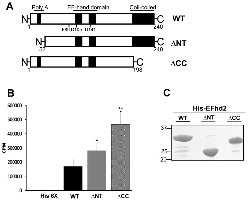
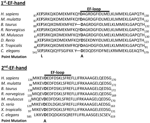
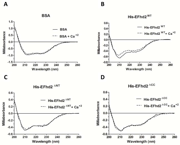
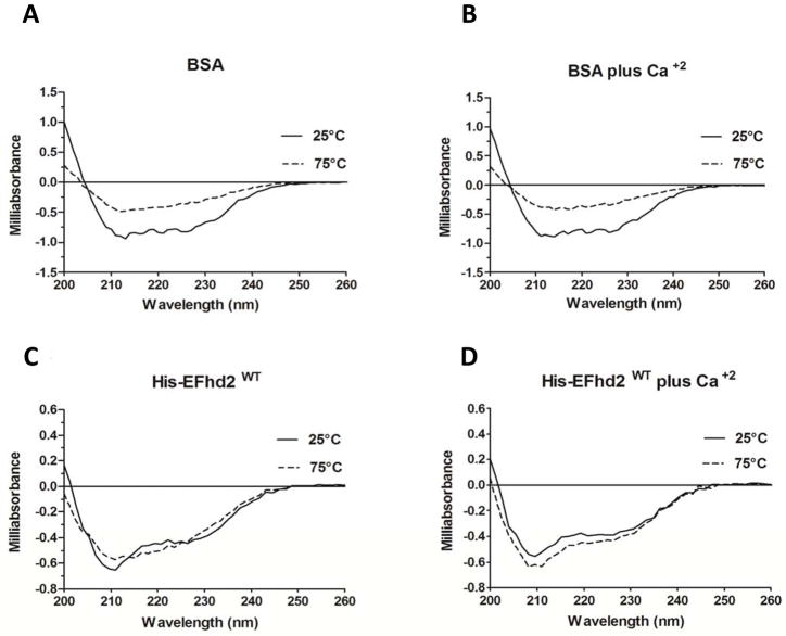
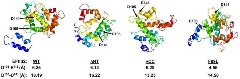
EFhd2 is a novel protein conserved from C. elegans to H. sapiens. This novel protein was originally identified in cells of the immune and central nervous systems. However, it is most abundant in the central nervous system, where it has been found associated with pathological forms of the microtubule-associated protein tau. The physiological or pathological roles of EFhd2 are poorly understood. In this study, a functional and structural analysis was carried to characterize the molecular requirements for EFhd2's calcium binding activity. The results showed that mutations of a conserved aspartate on either EF-hand motif disrupted the calcium binding activity, indicating that these motifs work in pair as a functional calcium binding domain. Furthermore, characterization of an identified single-nucleotide polymorphisms (SNP) that introduced a missense mutation indicates the importance of a conserved phenylalanine on EFhd2 calcium binding activity. Structural analysis revealed that EFhd2 is predominantly composed of alpha helix and random coil structures and that this novel protein is thermostable. EFhd2's thermo stability depends on its N-terminus. In the absence of the N-terminus, calcium binding restored EFhd2's thermal stability. Overall, these studies contribute to our understanding on EFhd2 functional and structural properties, and introduce it into the family of canonical EF-hand domain containing proteins.


Conflict of interest statement

Conflict of Interest: The authors declare that there are no conflicts of interest.

Figures

Fig. 1
Fig. 1. His-EFhd2WT and truncation mutants His-EFhd2ΔNT and His-EFhd2ΔCC bind calcium in vitro
A) Illustrated representation of EFhd2 domains under study, the N-terminus, the polyalanine domain, within the central region of the protein, the two EF-hand motifs and the coiled-coil domain at the C-terminus of the protein. The illustration is not at scale. B) Recombinant His-EFhd2WT, His-EFhd2ΔNT and His-EFhd2ΔCC were purified and incubated with radioactive 45CaCl2 to determine their calcium binding activity. Nickel-column beads were incubated with un-induced bacterial extract as negative control (His 6X). The radioactivity remaining in the beads was measured on a scintillation counter in counts per minute (CPM). C) A gel representative of the proteins used in this assay is illustrated. Protein amount was used to normalize the level of calcium binding detected. Significance was determined by a student’s T-test (two-tailed, paired). Statistical significance of * p < 0.01 and ** p < 0.001 is indicated.
Fig. 2
Fig. 2. EFhd2 EF-hand motifs are conserved throughout evolution
Sequence alignment of EFhd2 calcium binding motifs (1st and 2nd EF-hand motif) was performed using sequences from various species. The aspartates (D) in bold represent the first amino acid within each EF-hand motif loop (EF-loop) that is essential for the domain’s calcium-binding activity. These aspartate residues were changed to alanine (A) by introducing a point mutation in the cDNA sequence. The phenylalanine residue (F) in the first EF-hand represents a conserved amino acid affected by the identified SNP rs12131549. The SNP introduces a missense mutation that changes the phenylalanine to a leucine (L).
Fig. 3
Fig. 3. Both EF-hand domains are required for EFhd2 calcium-binding affinity
A) Recombinant proteins HIS-EFhd2WT, HIS-EFhd2F89L, HIS-EFhd2D105A and HIS-EFhd2D141A were subjected to in vitro calcium binding assay using radioactive 45CaCl2. Nickel-column beads were incubated with un-induced bacterial extract as negative control (His 6X). The amount of radioactive calcium was measured with a scintillation counter in counts per minute (CPM). B) The amount of recombinant protein was used to normalize the level of calcium binding. Significance was determined by a student’s T-test (two-tailed, paired). Statistical significance of * p< 0.02 and **p<0.01 is indicated.
Fig. 4
Fig. 4. His-EFhd2WT, His-EFhd2ΔNT and His-EFhd2ΔCC secondary structure revealed by circular dichroism
AD) Circular dichroism was used to determine changes upon calcium binding in the secondary structure of recombinant His-EFhd2WT and truncation mutants at 25°C. The secondary structure of BSA (A), His-EFhd2WT (B), His-EFhd2ΔNT (C) and EFhd2ΔCC (D) was analyzed in the far-UV region (200–260 nm) without calcium (black solid line) and with 1mM of CaCl2 (dashed line). Bovine serum albumin was used as control for instrument precision and as a negative control for structural changes after addition of calcium.
Fig. 5
Fig. 5. Thermal stability of EFhd2WT
AD) Thermal stability studies on His-EFhd2WT were conducted using circular dichroism. Measurements of the protein in the far-UV region (200–260 nm presented) were taken every 10°C from 25°C to 75°C. Two representative temperature spectra at 25°C (solid line) and 75°C (dashed line) are shown. The thermal stability of BSA (used as positive denaturation control) was measured in absence (A) or presence (B) of 1mM CaCl2. His-EFhd2WT was analyzed in the absence (C) or presence (D) of 1mM CaCl2 using the same temperature range. The representative spectra at 25°C (solid line) and 75°C (dashed line) are shown.
Fig. 6
Fig. 6. The N-terminus and calcium binding are required for EFhd2’s thermal stability
Recombinant His-EFhd2ΔNT and His-EFhd2ΔCC secondary structures were analyzed by circular dichroism to determine the contribution of the N- and C-terminus on EFhd2 thermal stability. Measurements were taken every 10°C from 25°C to 75°C in the far-UV region (200–260 nm). Two representative spectra, 25°C (solid line) and 75°C (dashed line) are shown. The effect of temperature on the structure of His-EFhd2ΔNT was determined in the absence (a) or presence (b) of 1mM CaCl2. The same analyses were performed for His-EFhd2ΔCC without calcium (c) or with (d) 1mM CaCl2. Deconvolution of the CD spectra obtained for His-EFhd2ΔNT and His-EFhd2ΔCC spectra are presented in Tables 3 (without calcium) and 4 (plus calcium)
Fig. 7
Fig. 7. Structural prediction of the EFhd2 protein
The mouse protein sequence of EFhd2 was analyzed using the protein structure prediction algorithms Phyre2. The predicted structures of EFhd2 wild-type (WT) protein, N- (ΔNT) and C-terminus (ΔCC) deletion mutants and EFhd2F89L mutant are illustrated. The position of the conserved aspartates (D105 and D141), is indicated (arrows). Dashed arrows indicate that D105 and D141 are behind the plane. The distance (Å) between amino acids in the same EF-hand loop (D105-D116) and adjacent EF-hand motifs (D105-D141) is illustrated.

Similar articles

Cited by

References

    1. Dütting S, Brachs S, Mielenz D. Fraternal twins: Swiprosin-1/EFhd2 Swiprosin-2/EFhd1 two homologous EF-hand containing calcium binding adaptor proteins with distinct functions. Cell Commun Signal. 2011;9:2–14. - PMC - PubMed
    1. Vega IE, Traverso EE, Ferrer-Acosta Y, Matos E, Colón M, González J, Dickson D, Hutton M, Lewis J, Yen SH. A novel calcium binding protein is associated with tau proteins in tauopathy. J Neurochem. 2008;106:96–106. - PMC - PubMed
    1. Nelson MR, Thulin E, Fagan PA, Forsén S, Chazin WJ. The EF-hand domain: A globally cooperative structural unit. Protein Sci. 2002;11:198–205. - PMC - PubMed
    1. Woolfson DN. The Design of Coiled-Coiled Structures and assemblies. Adv Prot Chem Rev. 2005;70:79–111. - PubMed
    1. Avramidou A, Kroczek C, Lang C, Schuh W, Jäck HM, Mielenz D. The novel adaptor protein Swiprosin 1 enhances BCR signals and contributes to BCR-induced apoptosis. Cell Death Differ. 2007;14:1936–1947. - PubMed

Publication types

MeSH terms