

Chronic leg ulcers as a rare cause for the first diagnosis of epidermolysis bullosa dystrophica
- PMID: 22974048
- PMCID: PMC7950492
- DOI: 10.1111/j.1742-481X.2012.01087.x
Chronic leg ulcers as a rare cause for the first diagnosis of epidermolysis bullosa dystrophica
Abstract
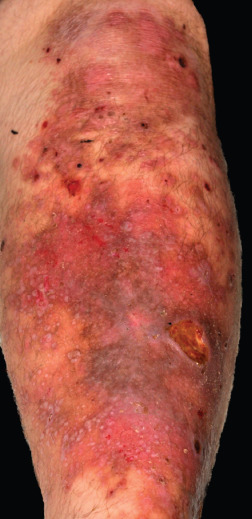
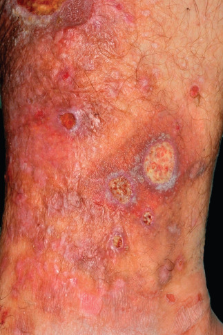
Chronic leg ulcers occur most frequently in the elderly population as a result of an underlying vascular disease especially chronic venous insufficiency. But it also occurs less commonly in younger people due to other aetiologies, for example, infections, vasculitis, neoplasia or genetic diseases. The following case report presents chronic leg ulcers as a rare cause for the first diagnosis of dystrophic epidermolysis bullosa. We report about a 21-year-old man with painful chronic leg ulcers resistant to different wound treatments for 4 months. After exclusion of the more common vascular aetiologies and reviewing the patient's family history, we considered an epidermolysis bullosa dystrophica which could be confirmed by genetic analyses. We treated the patient with debridement, modified negative pressure therapy with non-adhesive foil and skin grafting. The chronic leg ulcers healed completely. This case report demonstrates that the family history and genetic diseases should be considered as rare causes for therapy-refractory chronic leg ulcers, especially in young patients.
Keywords: Chronic wound; Epidermolysis bullosa; Genetic disorder; Leg ulcer.
© 2012 The Authors. International Wound Journal © 2012 Medicalhelplines.com Inc and John Wiley & Sons Ltd.
Figures
References
-
- Zeegelaar JE, Faber WR. Imported tropical infectious ulcers in travelers. Am J Clin Dermatol 2008;9:219–32. - PubMed
-
- Körber A, Klode J, Al‐Benna S, Wax C, Schadendorf D, Steinsträsser L, Dissemond J. Genese des chronischen Ulcus cruris bei 31.619 Patienten im Rahmen einer Expertenbefragung in Deutschland. J Dtsch Dermatol Ges 2011;9:116–22. - PubMed
-
- Mittler N, Grabbe S, Dissemond J. Verwendung von Folienverbänden für die Vakuumversiegelung chronischer Wunden mit irritierter umgebender Haut. Z Flugwiss 2006; 4:178–81.
-
- Furue M, Ando I, Inoue Y, Tamaki K, Oohara K, Kukita A. Pretibial epidermolysis bullosa: successful therapy with a skin graft. Arch Dermatol 1986;122:310–13. - PubMed
-
- Carterg DM, Lin AN, Varghese MC, Caldwell D, Pratt LA, Eisinger M. Treatment of junctional epidermolysis bullosa with epidermal autografts. J Am Acad Dermatol 1987;17:246–50. - PubMed
Publication types
MeSH terms
LinkOut - more resources
Full Text Sources
Medical
