

Keratins in colorectal epithelial function and disease
- PMID: 22974212
- PMCID: PMC3444987
- DOI: 10.1111/j.1365-2613.2012.00830.x
Keratins in colorectal epithelial function and disease
Abstract
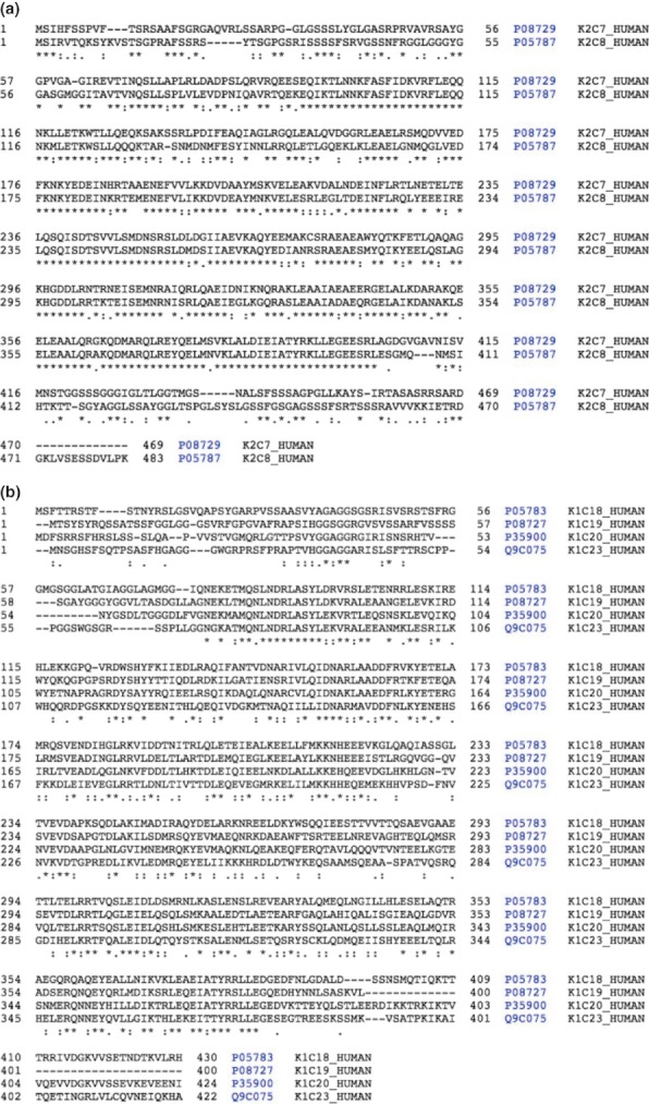
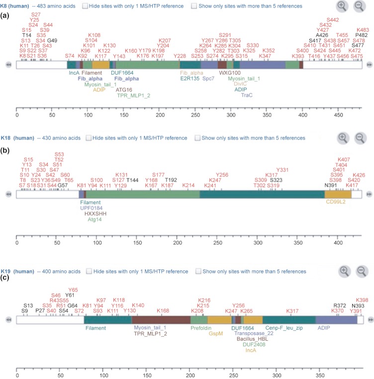
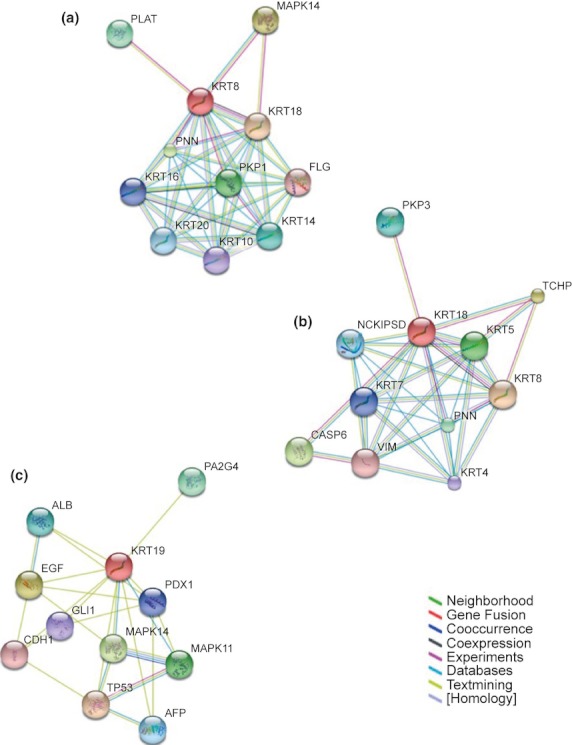
Keratins are the largest subgroup of intermediate filament proteins, which are an important constituent of the cellular cytoskeleton. The principally expressed keratins (K) of the intestinal epithelium are K8, K18 and K19. The specific keratin profile of a particular epithelium provides it with strength and integrity. In the colon, keratins have been shown to regulate electrolyte transport, likely by targeting ion transporters to their correct location in the colonocytes. Keratins are highly dynamic and are subject to post-translational modifications including phosphorylation, acetylation and glycosylation. These affect the filament dynamics and hence solubility of keratins and may contribute to protection against degradation. Keratin null mice (K8(-/-) ) develop colitis, and abnormal keratin mutations have been shown to be associated with inflammatory bowel disease (IBD). Abnormal expression of K7 and K20 has been noted in colitis-associated dysplasia and cancers. In sporadic colorectal cancers (CRCs) may be useful in predicting tumour prognosis; a low K20 expression is noted in CRCs with high microsatellite instability; and keratins have been noted as dysregulated in peri-adenomatous fields. Caspase-cleaved fragment of K18 (M30) in the serum of patients with CRC has been used as a marker of cancer load and to assess response to therapy. These data suggest an emerging importance of keratins in maintaining normal function of the gastrointestinal epithelium as well as being a marker of various colorectal diseases. This review will primarily focus on the biology of these proteins, physiological functions and alterations in IBD and CRCs.
© 2012 The Authors. International Journal of Experimental Pathology © 2012 International Journal of Experimental Pathology.
Figures

Similar articles
-
Keratin modifications and solubility properties in epithelial cells and in vitro.Subcell Biochem. 1998;31:105-40. Subcell Biochem. 1998. PMID: 9932491 Review.
-
Keratin-8 null mice have different gallbladder and liver susceptibility to lithogenic diet-induced injury.J Cell Sci. 2003 Nov 15;116(Pt 22):4629-38. doi: 10.1242/jcs.00782. J Cell Sci. 2003. PMID: 14576356
-
Keratin hypersumoylation alters filament dynamics and is a marker for human liver disease and keratin mutation.J Biol Chem. 2011 Jan 21;286(3):2273-84. doi: 10.1074/jbc.M110.171314. Epub 2010 Nov 9. J Biol Chem. 2011. PMID: 21062750 Free PMC article.
-
Keratin 8-deletion induced colitis predisposes to murine colorectal cancer enforced by the inflammasome and IL-22 pathway.Carcinogenesis. 2016 Aug;37(8):777-786. doi: 10.1093/carcin/bgw063. Epub 2016 May 27. Carcinogenesis. 2016. PMID: 27234655
-
Keratins: Biomarkers and modulators of apoptotic and necrotic cell death in the liver.Hepatology. 2016 Sep;64(3):966-76. doi: 10.1002/hep.28493. Epub 2016 Apr 4. Hepatology. 2016. PMID: 26853542 Free PMC article. Review.
Cited by
-
Analysis of mRNA expression differences in bladder cancer metastasis based on TCGA datasets.J Int Med Res. 2021 Mar;49(3):300060521996929. doi: 10.1177/0300060521996929. J Int Med Res. 2021. PMID: 33787386 Free PMC article.
-
Intermediate Filaments and Polarization in the Intestinal Epithelium.Cells. 2016 Jul 15;5(3):32. doi: 10.3390/cells5030032. Cells. 2016. PMID: 27429003 Free PMC article. Review.
-
Transcriptomic Maps of Colorectal Liver Metastasis: Machine Learning of Gene Activation Patterns and Epigenetic Trajectories in Support of Precision Medicine.Cancers (Basel). 2023 Jul 28;15(15):3835. doi: 10.3390/cancers15153835. Cancers (Basel). 2023. PMID: 37568651 Free PMC article.
-
Epithelial to mesenchymal transition influences fibroblast phenotype in colorectal cancer by altering miR-200 levels in extracellular vesicles.J Extracell Vesicles. 2022 May;11(5):e12226. doi: 10.1002/jev2.12226. J Extracell Vesicles. 2022. PMID: 35595718 Free PMC article.
-
Integrated multi-omics analyses on patient-derived CRC organoids highlight altered molecular pathways in colorectal cancer progression involving PTEN.J Exp Clin Cancer Res. 2021 Jun 21;40(1):198. doi: 10.1186/s13046-021-01986-8. J Exp Clin Cancer Res. 2021. PMID: 34154611 Free PMC article.
References
-
- Achtstaetter T, Hatzfeld M, Quinlan RA, Parmelee DC, Franke WW. Separation of cytokeratin polypeptides by gel electrophoretic and chromatographic techniques and their identification by immunoblotting. Methods Enzymol. 1986;134:355–371. - PubMed
-
- Ameen NA, Figueroa Y, Salas PJ. Anomalous apical plasma membrane phenotype in CK8-deficient mice indicates a novel role for intermediate filaments in the polarization of simple epithelia. J. Cell Sci. 2001;114:563–575. - PubMed
-
- Arnold CN, Goel A, Blum HE, Boland CR. Molecular pathogenesis of colorectal cancer: implications for molecular diagnosis. Cancer. 2005;104:2035–2047. - PubMed
-
- Askling J, Dickman PW, Karlen P, et al. Family history as a risk factor for colorectal cancer in inflammatory bowel disease. Gastroenterology. 2001;120:1356–1362. - PubMed
-
- Ausch C, Buxhofer-Ausch V, Olszewski U, et al. Circulating cytokeratin 18 fragment m65-a potential marker of malignancy in colorectal cancer patients. J. Gastrointest. Surg. 2009;13:2020–2026. - PubMed
Publication types
MeSH terms
Substances
LinkOut - more resources
Full Text Sources
Other Literature Sources
Research Materials
