. 2012 Sep 13;9(1):33.
doi: 10.1186/1476-9255-9-33.

Human umbilical cord mesenchymal stem cells reduce systemic inflammation and attenuate LPS-induced acute lung injury in rats

Affiliations

Human umbilical cord mesenchymal stem cells reduce systemic inflammation and attenuate LPS-induced acute lung injury in rats

Jianjun Li et al. J Inflamm (Lond). .

Abstract

Background: Mesenchymal stem cells (MSCs) possess potent immunomodulatory properties and simultaneously lack the ability to illicit immune responses. Hence, MSCs have emerged as a promising candidate for cellular therapeutics for inflammatory diseases. Within the context of this study, we investigated whether human umbilical cord-derived mesenchymal stem cells (UC-MSCs) could ameliorate lipopolysaccharide- (LPS-) induced acute lung injury (ALI) in a rat model.

Methods: ALI was induced via injection of LPS. Rats were divided into three groups: (1) saline group(control), (2) LPS group, and (3) MSC + LPS group. The rats were sacrificed at 6, 24, and 48 hours after injection. Serum, bronchoalveolar lavage fluid (BALF), and lungs were collected for cytokine concentration measurements, assessment of lung injury, and histology.

Results: UC-MSCs increased survival rate and suppressed LPS-induced increase of serum concentrations of pro-inflammatory mediators TNF-α, IL-1β, and IL-6 without decreasing the level of anti-inflammatory cytokine IL-10. The MSC + LPS group exhibited significant improvements in lung inflammation, injury, edema, lung wet/dry ratio, protein concentration, and neutrophil counts in the BALF, as well as improved myeloperoxidase (MPO) activity in the lung tissue. Furthermore, UC-MSCs decreased malondialdehyde (MDA) production and increased Heme Oxygenase-1 (HO-1) protein production and activity in the lung tissue.

Conclusion: UC-MSCs noticeably increased the survival rate of rats suffering from LPS-induced lung injury and significantly reduced systemic and pulmonary inflammation. Promoting anti-inflammatory homeostasis and reducing oxidative stress might be the therapeutic basis of UC-MSCs.


Figures

Figure 1
Figure 1
UC-derived MSC-like cells in passaged cultures. (A) H&E staining of UC-derived MSC-like cells. Osteogenic differentiation as indicated by (B) the formation of mineralized matrix shown by alizarin red staining and (C) alkaline phosphatase expression. Adipocytic differentiation was noted by the presence of broadened morphology and formation of lipid vacuoles (D) (positive oil-red O staining). Scale bars = 80 μm. (E) Immunophenotype of UC-derived MSC-like cells.
Figure 2
Figure 2
(A-C) LPS caused a significant acute systemic inflammatory response as early as 6 hours. Administration of UC-MSCs reduced pro-inflammatory cytokines at each time point. (D) LPS also caused an increase of anti-inflammatory cytokine IL-10, but this change was not altered by intravenous administration of UC-MSCs (*, p < 0.05 compared with healthy controls; #, p < 0.05 comparing LPS plus UC-MSCs with LPS alone).
Figure 3
Figure 3
(A) Histological analysis indicated LPS injection caused capillary expansion and congestion, as well as neutrophil infiltration into the lung tissue. In addition, lung septae was noticeably thickened. Administration of UC-MSCs improved the lung injury at all time points. (B) Lung injury score decreased significantly in the MSC + LPS group at all time points (*, p < 0.05 compared with healthy controls; #, p < 0.05 comparing LPS plus UC-MSCs with LPS alone.). Fibroblast injections have no effect on improvement of both pathological morphology and lung injury score.
Figure 4
Figure 4
(A) Injection of UC-MSCs reduced pulmonary edema induced by LPS. Pulmonary edema was measured as the wet-dry ratio. (B): BALF protein concentration significantly increased after LPS injection and reached its peak at 24 h, while UC-MSCs caused a non-satistically significant decrease in the protein concentration. (*, p < 0.05 compared with healthy controls; #, p < 0.05 comparing LPS plus UC-MSCs with LPS alone.).
Figure 5
Figure 5
(A) BALF neutrophil counts and (B) lung MPO activity. Neutrophil counts and lung tissue MPO activity were significantly higher in the LPS group compared to the control group. Treatment with UC-MSCs significantly reduced the LPS-induced increase in BALF neutrophil counts and lung MPO activity at 24 and 48 hours (*, p < 0.05 compared with healthy controls; #, p < 0.05 comparing LPS plus UC-MSCs with LPS alone).
Figure 6
Figure 6
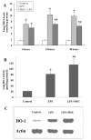
(A) MDA levels were significantly increased in LPS-treated rats compared to the control group. This increase was significantly reduced at 24 and 48 hours by treatment with UC-MSCs. (B) Activity of HO-1 in the lungs of LPS-treated rats at 24 hours. (C) Expression of HO-1 as measured by Western blotting analysis at 24 hours. LPS induced HO-1 protein expression and stimulated HO-1 activity in the lung tissues. Treatment with UC-MSCs further increased the expression and activity of HO-1 (*, p < 0.05 compared with healthy controls; #, p < 0.05 comparing LPS plus UC-MSCs with LPS alone).
Figure 7
Figure 7
The survival rate of the LPS + MSC group over 48 hours is significantly higher than that of the LPS group (87% vs 60%; * p < 0.05). UC-MSCs treatment improved survival rates at all time-points.

Similar articles

Cited by

References

    1. Wang HM, Bodenstein M, Markstaller K. Overview of the pathology of three widely used animal models of acute lung injury. Eur Surg Res. 2008;40:305–316. doi: 10.1159/000121471. - DOI - PubMed
    1. Weil BR, Markel TA, Herrmann JL, Abarbanell AM, Kelly ML, Meldrum DR. Stem cells in sepsis. Ann Surg. 2009;250:19–27. doi: 10.1097/SLA.0b013e3181a77b9c. - DOI - PubMed
    1. Nauta AJ, Fibbe WE. Immunomodulatory properties of mesenchymal stromal cells. Blood. 2007;110:3499–3506. doi: 10.1182/blood-2007-02-069716. - DOI - PubMed
    1. Weiss DJ, Sueblinvong V. Cell therapy approaches for lung diseases:current status. Curr Opin Phamacol. 2009;9:268–273. doi: 10.1016/j.coph.2009.03.002. - DOI - PMC - PubMed
    1. Gupta N, Su X, Popov B, Lee JW, Serikov V, Matthay MA. Intrapulmonary delivery of bone marrow-derived mesenchymal stemcells improves survival and attenuates endotoxin-induced acute lung injury in mice. J Immunol. 2007;179:1855–1863. - PubMed