Review
. 2012:46:537-60.
doi: 10.1146/annurev-genet-110711-155454. Epub 2012 Sep 4.

Dosage compensation of the sex chromosomes

Affiliations
Review

Dosage compensation of the sex chromosomes

Christine M Disteche. Annu Rev Genet. 2012.

Abstract

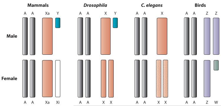
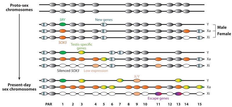
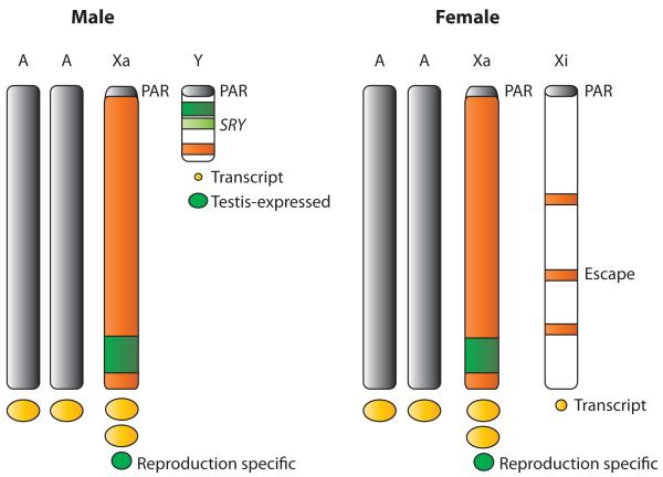
Differentiated sex chromosomes evolved because of suppressed recombination once sex became genetically controlled. In XX/XY and ZZ/ZW systems, the heterogametic sex became partially aneuploid after degeneration of the Y or W. Often, aneuploidy causes abnormal levels of gene expression throughout the entire genome. Dosage compensation mechanisms evolved to restore balanced expression of the genome. These mechanisms include upregulation of the heterogametic chromosome as well as repression in the homogametic sex. Remarkably, strategies for dosage compensation differ between species. In organisms where more is known about molecular mechanisms of dosage compensation, specific protein complexes containing noncoding RNAs are targeted to the X chromosome. In addition, the dosage-regulated chromosome often occupies a specific nuclear compartment. Some genes escape dosage compensation, potentially resulting in sex-specific differences in gene expression. This review focuses on dosage compensation in mammals, with comparisons to fruit flies, nematodes, and birds.


Figures

Figure 1
Figure 1
Main types of sex chromosome dosage compensation. Male and female sexes are indicated. From left to right: Mammals, in which expressed genes on the active X (Xa) are upregulated in both sexes and genes on the inactive X (Xi) are silenced in females; Drosophila, in which the X is upregulated in males only; Caenorhabditis elegans, in which the X is upregulated in both sexes and downregulated in hermaphrodites (mottled); birds, in which there is apparent partial upregulation of the Z chromosome in both sexes and partial or stochastic downregulation in males. Dots represent both increased expression and repression. Abbreviation: AA, autosomes.
Figure 2
Figure 2
Schematic of potential evolutionary pathways of mammalian sex-linked genes. From top to bottom: evolution of homologous genes originally located on the proto-sex chromosomes ( gray) to their full differentiation into Y-linked genes (Y) and genes on the active X (Xa) and inactive X (Xi). New genes have been acquired by the X and Y (mottled gray) and thus are not remnants of the proto-sex chromosomes; some may be unique to the Y (gene 7) or the X (gene 15), others may be acquired by both sex chromosomes (genes 5-6), depending on how much recombination between the sex chromosomes still occurred at that time. Pseudoautosomal region (PAR) genes are homologous on the sex chromosomes; however, note that PAR genes can also derive from additions to the sex chromosomes later in evolution. Gene 1 represents the sex determinant SRY derived from the X-linked gene SOX3, which becomes upregulated on the Xa (orange) and silenced on the Xi (white). Genes 2, 4-6, 8, 10-12, and 14-15 are examples of genes progressively lost from the Y. Genes 3 and 13 evolved from X/Y pairs by acquiring a testis function on the Y (mottled yellow/green). Their X paralog becomes silenced (gene 3) or escapes X inactivation (gene 13). Most genes on the Xa become upregulated (orange), but some do not (gene 12), and others acquire reproduction-related functions (mottled yellow/green) (genes 5, 6, 14, 15). A majority of genes (genes 1-6, 8, 10, 12, 14, 15) on the Xi become silenced (empty). A few genes escape X inactivation on the Xi (genes 9, 11, 13); some of these retain a functional Y paralog (gene 9, X/Y gene pair), resulting in equal sex expression, although expression is usually lower on the Xi and Y than on the Xa ( pale orange); others have lost the Y paralog (gene 11) or have a differentiated Y paralog expressed in testis (gene 13). Escape genes may acquire a female advantageous role (genes 11 and 13) (mottled purple/pink). Overall, both the Y and X chromosomes have acquired a number of new testis-specific (or reproduction-related) genes (mottled yellow/green).
Figure 3
Figure 3
Schematic of the mammalian sex chromosomes in males and females. Expressed genes on the active X (Xa) produce a higher level of transcripts compared with genes on the autosomes (AA). The pseudoautosomal region (PAR) is not upregulated. The Y and X chromosomes contain many testis-expressed (or reproduction-specific) genes ( green). The dots under the chromosomes represent the amount of gene product. Genes that escape X inactivation on the inactive X (Xi) and their few paralogs on the Y produce a small number of transcripts (small yellow dots).

Similar articles

Cited by

References

    1. Adler DA, Rugarli EI, Lingenfelter PA, Tsuchiya K, Poslinski D, et al. Evidence of evolutionary up-regulation of the single active X chromosome in mammals based on Clc4 expression levels in Mus spretus and Mus musculus. Proc. Natl. Acad. Sci. USA. 1997;94:9244–48. - PMC - PubMed
    1. Agrelo R, Souabni A, Novatchkova M, Haslinger C, Leeb M, et al. SATB1 defines the developmental context for gene silencing by Xist in lymphoma and embryonic cells. Dev. Cell. 2009;16:507–16. - PMC - PubMed
    1. Al Nadaf S, Deakin JE, Gilbert C, Robinson TJ, Graves JA, Waters PD. A cross-species comparison of escape from X inactivation in Eutheria: implications for evolution of X chromosome inactivation. Chromosoma. 2011;121:71–78. - PMC - PubMed
    1. Al Nadaf S, Waters PD, Koina E, Deakin JE, Jordan KS, Graves JA. Activity map of the tammar X chromosome shows that marsupial X inactivation is incomplete and escape is stochastic. Genome Biol. 2010;11:R122. - PMC - PubMed
    1. Andina RJ. A study of X chromosome regulation during oogenesis in the mouse. Exp. Cell Res. 1978;111:211–18. - PubMed

Publication types

Substances

LinkOut - more resources