. 2012 Nov 12;13(11):3494-502.
doi: 10.1021/bm300942c. Epub 2012 Sep 28.

Self-assembly of filamentous amelogenin requires calcium and phosphate: from dimers via nanoribbons to fibrils

Affiliations

Self-assembly of filamentous amelogenin requires calcium and phosphate: from dimers via nanoribbons to fibrils

Olga Martinez-Avila et al. Biomacromolecules. .

Abstract

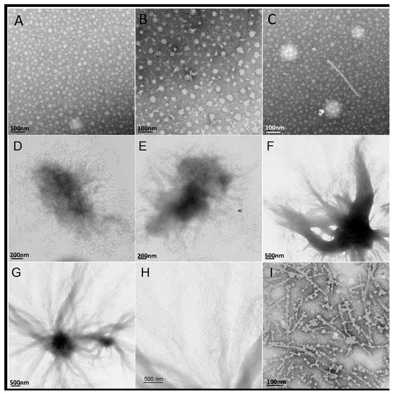
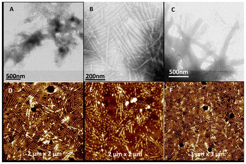
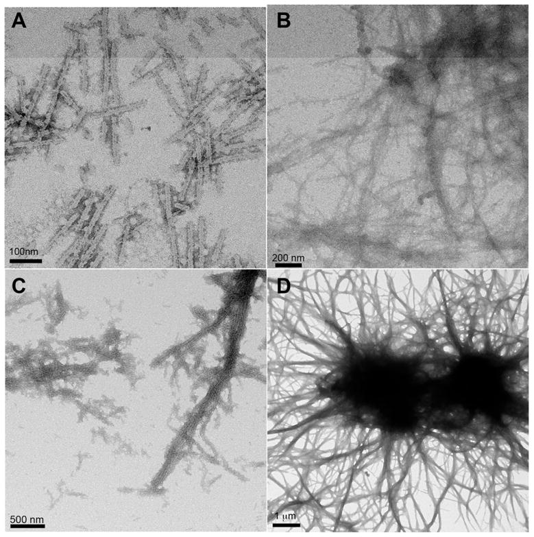
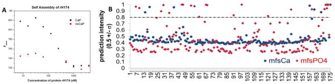
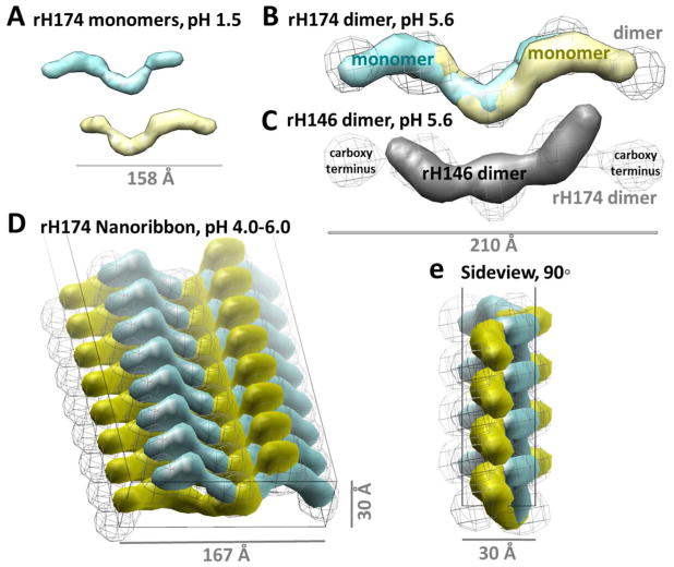
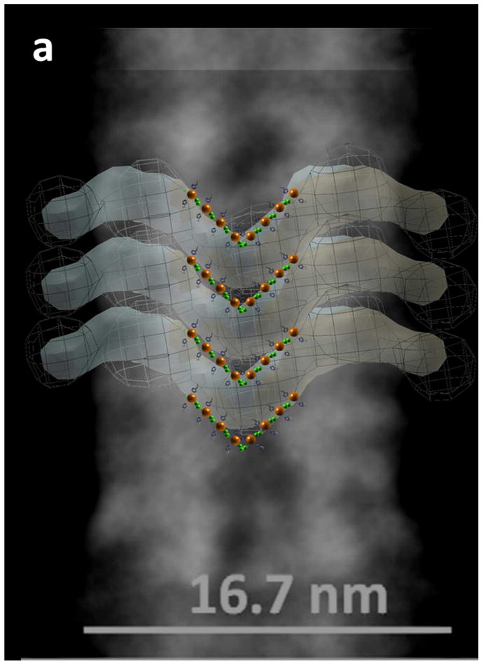
Enamel matrix self-assembly has long been suggested as the driving force behind aligned nanofibrous hydroxyapatite formation. We tested if amelogenin, the main enamel matrix protein, can self-assemble into ribbon-like structures in physiologic solutions. Ribbons 17 nm wide were observed to grow several micrometers in length, requiring calcium, phosphate, and pH 4.0-6.0. The pH range suggests that the formation of ion bridges through protonated histidine residues is essential to self-assembly, supported by a statistical analysis of 212 phosphate-binding proteins predicting 12 phosphate-binding histidines. Thermophoretic analysis verified the importance of calcium and phosphate in self-assembly. X-ray scattering characterized amelogenin dimers with dimensions fitting the cross-section of the amelogenin ribbon, leading to the hypothesis that antiparallel dimers are the building blocks of the ribbons. Over 5-7 days, ribbons self-organized into bundles composed of aligned ribbons mimicking the structure of enamel crystallites in enamel rods. These observations confirm reports of filamentous organic components in developing enamel and provide a new model for matrix-templated enamel mineralization.


Figures

Figure 1
Figure 1
TEM analysis of self-assembled structures of rH174 (0.4 mg/mL) observed at pH 4.5. (A) in the absence of calcium and phosphate ions at day 1 and (B) day 7 (C–H) compared to structures which develop over the same time period in the presence of calcium and phosphate: (C) day 1, shows both nanospheres and nanoribbons; (D) day 4, short ribbons of about 200–800 nm length are the only structural feature observed; (E) day 5, domains of aligned ribbons appear; (F and G) day 7, bundle-like structures containing numerous aligned ribbons appear; (H) high-resolution of (G) showing that shorter ribbon segments are present at the end of the ribbons and in process to be added to existing longer and aligned ribbons; I) ribbons exposed to 0.5 M EDTA for 2h at pH 6, showing disintegration of ribbons, resulting in the appearance of nanospheres.
Figure 2
Figure 2
TEM and AFM images of supramolecular structures of rH174 at day 7 self-assembled at different pH. (A) pH 4.0, showing short randomly oriented ribbons; (B) pH 5.5 and C) pH 6.0 ribbons have aligned and formed bundles. AFM images of (D) amelogenin nanoribbons formed at pH 4.5 and immobilized onto glass slide; E) same ribbons after rinsing with HCl-solution at pH 2, causing the ribbons to disintegrate; F) ribbons remained unchanged when exposed to buffered solutions at pH 7.0 for three days.
Figure 3
Figure 3
TEM images of supramolecular structures of rH174 present at day 6, as a function of protein concentration at pH 4.5: (A) at 0.4 mg/mL; (B) at 0.8 mg/mL; (C) at 1.2 mg/mL and (D) at 1.6 mg/mL. Size and alignment including the formation of bundles are enhanced with increasing protein concentration.
Figure 4
Figure 4
Structural binding analysis of amelogenin (H175) to calcium and phosphate ions. (A) Microscale Thermophoresis (MST) experiments show a strong change in fluorescence intensity in the presence of calcium and phosphate ions when unlabeled rH174 is titrated into labeled rH174 sols at pH 4.5 (black curve); indicating strong protein-protein interaction. The change in fluorescence was not quantifiable in the absence of calcium and phosphate (red data points). (B) Meta-functional signatures (MFS) showing the probability for each residue in amelogenin H175 to bind soluble phosphate ions (mfsPO4) or soluble calcium ions (mfsCa). Scores above threshold (horizontal line) indicate high binding probablity. Threshold was set at least three standard deviations higher than the mean for all scores of non-calcium or non-phosphate binding.
Figure 5
Figure 5
Multiscale modeling of amelogenin ribbon assembly. Ab-initio shapes of (A) the rH174 monomer (cyan and yellow; Rg 46.4Å, Dmax 158Å) and (B) rH174 dimer (gray wire; Rg 64.1Å, Dmax 209Å) were derived from SAXS profiles measured in the presence of calcium and phosphate at pH 1.5 and pH 5.6, respectively. (B) Superimposition of SAXS-derived rH174 monomers within a rH174 dimer supports antiparallel symmetry (cross correlation coefficient = 0.82). (C) Antiparallel monomer orientation is also supported by comparison of an rH146 dimer (solid gray) superposed to an H174 dimer (CCC = 0.76), which also reveals the carboxy termini lying on the distant ends of the H174 dimer. (D), (E) Building out these dimers as units of a multimer into a flat columnar template derived from TEM and AFM measurements of the ribbons presents a mechanistic molecular model of amelogenin self-assembly.
Figure 6
Figure 6
Comparison of the dimer-based model to a consensus TEM overlay for amelogenin nanoribbons. The apparent repeating units observed by TEM overlay are supported by the size and shape of the SAXS dimer model. The electron dense negative stain shown in the TEM, obtained in a previous study, suggests buildup of calcium and phosphate along the center of the ribbon. Thus phosphates (orange) and histidines thought to carry out the pH dependent phosphate interactions are shown at the intersection of this region, with calcium ions (green) interspersed to represent potential counter ions for phosphate bridging. The coarse localization for histidines provides probabilistic tethers for future modeling efforts. The stacked orientation of amelogenin, calcium, and phosphate facilitates an explanation for the linear scaling of multimerization observed in this study. The SAXS based models of dimeric building blocks exceed the width of the ribbon as observed in the TEM by about 2nm on both sides, suggesting that the non-overlapping portion at the C-terminus may be folded inwards.

Similar articles

Cited by

References

    1. Smith KH, Tejeda-Montes E, Poch M, Mata A. Chem Soc Rev. 2011;40:4563–4577. - PubMed
    1. Ingber DE. Sci Am. 1998;278:48–57. - PubMed
    1. Warshawsky H. Anat Rec. 1989;224:242–262. - PubMed
    1. White SN, Luo W, Paine ML, Fong H, Sarikaya M, Snead ML. J Dent Res. 2001;80:321–326. - PubMed
    1. Smith CE. Crit Rev Oral Biol Med. 1998;9:128–161. - PubMed

Publication types