. 2012 Sep 13;2(1):19.
doi: 10.1186/2044-5040-2-19.

Eps homology domain endosomal transport proteins differentially localize to the neuromuscular junction

Affiliations

Eps homology domain endosomal transport proteins differentially localize to the neuromuscular junction

Suzanne E Mate et al. Skelet Muscle. .

Abstract

Background: Recycling of endosomes is important for trafficking and maintenance of proteins at the neuromuscular junction (NMJ). We have previously shown high expression of the endocytic recycling regulator Eps15 homology domain-containing (EHD)1 proteinin the Torpedo californica electric organ, a model tissue for investigating a cholinergic synapse. In this study, we investigated the localization of EHD1 and its paralogs EHD2, EHD3, and EHD4 in mouse skeletal muscle, and assessed the morphological changes in EHD1-/- NMJs.

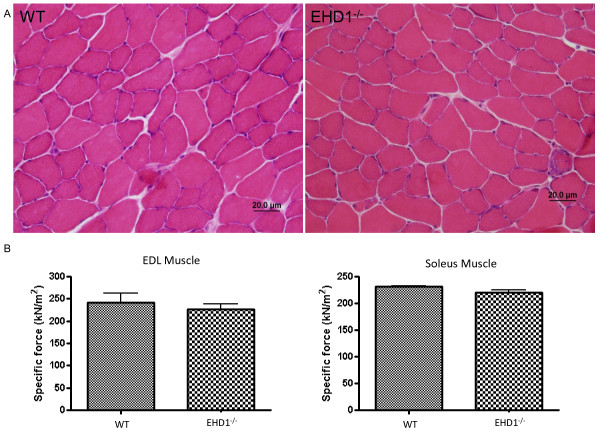
Methods: Localization of the candidate NMJ protein EHD1 was assessed by confocal microscopy analysis of whole-mount mouse skeletal muscle fibers after direct gene transfer and immunolabeling. The potential function of EHD1 was assessed by specific force measurement and α-bungarotoxin-based endplate morphology mapping in EHD1-/- mouse skeletal muscle.

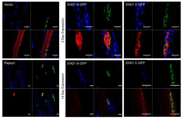
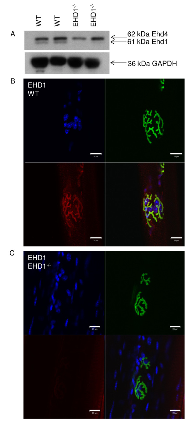
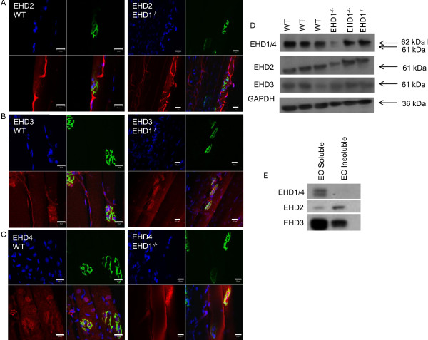
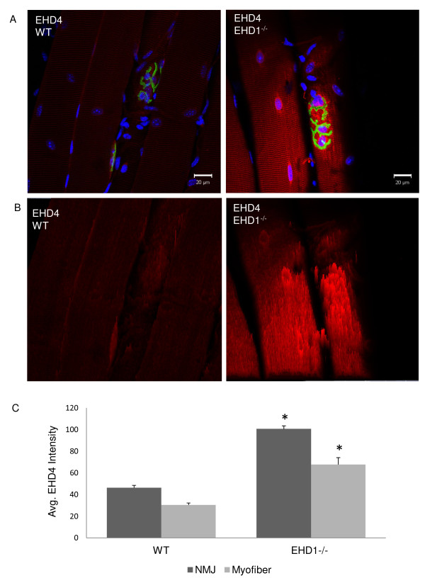
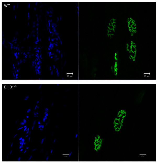
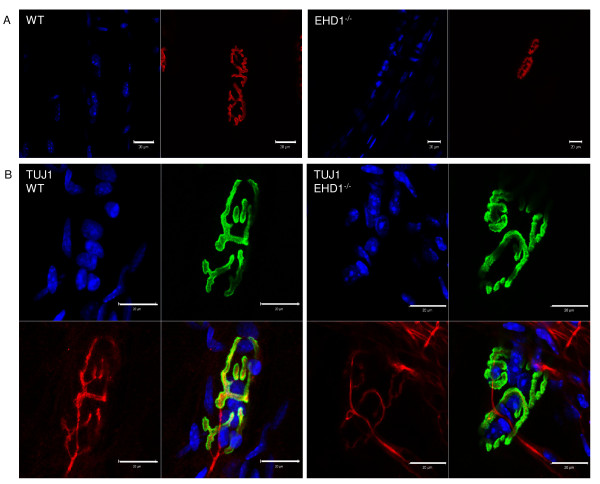
Results: Endogenous EHD1 localized to primary synaptic clefts of murine NMJ, and this localization was confirmed by expression of recombinant green fluorescent protein labeled-EHD1 in murine skeletal muscle in vivo. EHD1-/- mouse skeletal muscle had normal histology and NMJ morphology, and normal specific force generation during muscle contraction. The EHD 1-4 proteins showed differential localization in skeletal muscle: EHD2 to muscle vasculature, EHD3 to perisynaptic regions, and EHD4 to perinuclear regions and to primary synaptic clefts, but at lower levels than EHD1. Additionally, specific antibodies raised against mammalian EHD1-4 recognized proteins of the expected mass in the T. californica electric organ. Finally, we found that EHD4 expression was more abundant in EHD1-/- mouse skeletal muscle than in wild-type skeletal muscle.

Conclusion: EHD1 and EHD4 localize to the primary synaptic clefts of the NMJ. Lack of obvious defects in NMJ structure and muscle function in EHD1-/- muscle may be due to functional compensation by other EHD paralogs.


Figures

Figure 1
Figure 1
Recombinant green fluorescent protein (GFP)-tagged Eps15 homology domain-containing (EHD) protein localizes to the neuromuscular junction (NMJ). Control and EHD1 proteins were expressed for 3 or 14 days in 6- to 10-week-old C57Bl/6 J mouse tibialis anterior (TA) skeletal muscle as C-terminal and N-terminal GFP recombinant proteins. Four mice were injected for each recombinant protein expressed. The TA muscle was fixed in situ and teased into small bundles for immunofluorescence and visualization via confocal microscopy, referenced to α-bungarotoxin (BTX) labeling of AChRs at the NMJ. Expression of GFP (vector) was seen as a diffuse signal throughout the myofiber and did not concentrate at the NMJ (n = 7). Rapsyn-GFP expression localized exclusively to the NMJ of transfected fibers (n = 2). Both transfected and non-transfected fibers are shown in the bottom left panel. N- and C-terminal GFP recombinant EHD1 proteins localized to the NMJ and to the primary synaptic cleft with BTX after 3 or 14 days of transfection (n = 24). Each image consists of separate blue (DAPI), green (BTX Alexa Fluor 488 conjugate), and red (anti-GFP Alexa Fluor 555 conjugate) channels in addition to a merged image. Scale bars: 20 μm. The larger reference bar was added in powerpoint to make the 20 μm bar visible in the images. It reflects the 20 μm scale bar that was added within the Zen software upon image acquisition.
Figure 2
Figure 2
Eps15 homology domain-containing (EHD)1 is expressed in mouse skeletal muscle and localizes to the primary synaptic clefts of neuromuscular junctions. (A) Western blot of two WT and two EHD1−/− skeletal muscle lysates probed with peptide-specific antibodies against EHD1 and EHD4 showedexpression in skeletal muscle and verified the loss of EHD1 in null mice. Glyceraldehyde 3-phosphate dehydrogenase (GAPDH) served as a loading control. (B)The tibialis anterior (TA) skeletal muscles of three wild-type (WT) and three EHD1−/− mice were teased into small bundles for immunofluorescence and visualization via confocal microscopy. α-bungarotoxin (BTX) labeling of AChRs denote NMJs in all images. EHD1 localized to the NMJ and delineated the primary synaptic cleft similar to BTX labeling. (C) Because the EHD1 antibody crossreacts with EHD4, imparting low background signal, EHD1−/− fibers were labeled with anti-EHD1 to show the specificity of the localization. Blue, DAPI; green,BTXAlexa Fluor 488 conjugate; red, anti-green fluorescent protein Alexa Fluor 555 conjugate. Scale bars: 20 μm. A larger reference bar was added above the scale bars.
Figure 3
Figure 3
Eps15 homology domain-containing (EHD) 1–4 proteins show differential localization in skeletal muscle. The tibialis anterior (TA) skeletal muscles of three wild-type (WT) and three EHD1−/− mice were teased into small bundles for immunofluorescence and visualization via confocal microscopy. α-bungarotoxin (BTX) labeling of AChRs denote neuromuscular junctions (NMJs) in all images. EHD2, 3, and 4 are expressed in WT and EHD1−/− mice. Panel (A) EHD2 localized to skeletal muscle vasculature endothelium. (B) EHD3 localized to synaptic regions adjacent to theBTX labeling and in the vasculature, but to a lesser extent than EHD2. (C) EHD4 localized to the synaptic cleft with BTX in addition to adjacent extrasynaptic regions and perinuclear regions. (D) EHD 1–4 expression in skeletal muscle lysates was analyzed by western blotting. (E) EHD1-4 antibodies also recognized proteins of similar mass in Torpedo californica electric organ lysates. Blue, DAPI; green, BTX Alexa Fluor 488 conjugate; red, anti-rabbit Alexa Fluor 555 conjugate.Scale bars: 20 μm. A larger reference bar was added above the scale bars.
Figure 4
Figure 4
Eps15 homology domain-containing (EHD)4 expression is increased in EHD1−/−muscle. The tibialis anterior (TA) skeletal muscle of three wild-type (WT) and three EHD1−/− mice were teased into small bundles for immunofluorescence labeling with anti-EHD4 antibody and z-stacks were acquired to assess signal throughout the entire myofiber diameter/thickness via confocal microscopy. α-bungarotoxin (BTX) labeling of AChRs denote NMJs. EHD4 signal intensities were compared in myofibers (n = 5) and at neuromuscular junctions (NMJs) (n = 10) from WT and EHD1−/− mice (n = 3 per group) using identical acquisition parameters. (A) The z-section with the highest signal for EHD4 (red channel). (B) Intensity projections of the EHD4 signal from representative NMJs in (A). Blue, DAPI; green, BTX Alexa Fluor 488 conjugate; red, anti-rabbit Alexa Fluor 555 conjugate.Scale bars: 20 μm. A larger reference bar was added above the scale bars. (C) The average intensity of EHD4 expressed in WT and EHD1−/− mice. The bars represent the average vector measurements of the intensity with standard error from myofibers and NMJs in each group. Statistical significance was measured by Student’s t-test (P < 0.05).
Figure 5
Figure 5
Neuromuscular junctions (NMJs) in Eps15 homology domain-containing (EHD)1−/−mice show no morphological defects in synaptic endplate structure. (A) The tibialis anterior (TA) skeletal muscle fibers from three EHD1−/− and three wild-type (WT) mice were labeled with α-bungarotoxin (BTX) Alexa Fluor 555 or 488 conjugate for morphological mapping of endplates using confocal microscopy. NMJs were seen in both WT (n = 260) and in EHD1−/− (n = 275) fibers. Blue, DAPI; green, BTX Alexa Fluor 488 conjugate; red, anti-rabbit Alexa Fluor 555 conjugate. Scale bars: 20 μm. A larger reference bar was added above the scale bars.
Figure 6
Figure 6
Neuromuscular junctions (NMJs) of Eps15 homology domain-containing (EHD)1−/−mice are innervated by a single motor axon. Panel (A) Isolated tibialis anterior (TA) skeletal muscle fibers from three EHD1−/− and three wild-type (WT) mice were labeled with (BTX) Alexa Fluor 555 conjugate for morphological mapping of endplates using confocal microscopy. Rare EHD1−/−endplates (n = 2) consisted of two discreet structures, with one being smaller and less developed than the other. (B) Isolated TA skeletal muscle fibers from three EHD1−/− and three WT mice were immunolabeled with neuronal class III β-tubulin monoclonal antibody (TUJ1) antibody, a neuronal marker, and visualized using confocal microscopy. WT (n = 13) and EHD1−/− (n = 12) endplates with partitioning were seen. EHD1−/− endplates were singly innervated, even when partitioned. Blue, DAPI; green, α-BTXAlexa Fluor 488 conjugate; red, anti-mouse Alexa Fluor 568 conjugate. Scale bars: 20 μm. A larger reference bar was added above the scale bars.
Figure 7
Figure 7
Eps15 homology domain-containing (EHD)1−/−skeletal muscle shows normal pathology and generates average specific force. Panel (A) Stainnig of tibialis anterior (TA) skeletal muscle cryosections (8 μm) from three WT and three EHD1−/− mice did not show no degeneration or regeneration of fibers in EHD1−/− mice, as shown by the absence of centralized nuclei. There was an absence of infiltrating white blood cells, also suggesting normal pathology (hematoxylin and eosin). (B) The force generated during a muscle contraction was measured in vivo in three WT and three EHD1−/− EDL and soleus muscles each. EHD1−/− EDL and soleus muscles showed no deficit in the amount of force generated during a muscle contraction (n = 3 per group).

Similar articles

Cited by

References

    1. Mate SE, Brown KJ, Hoffman EP. Integrated genomics and proteomic of theTorpedo californicaelectric organ: concordance with the mammalian neuromuscular junction. Skeletal Muscle. 2011;1:20. doi: 10.1186/2044-5040-1-20. - DOI - PMC - PubMed
    1. Grant BD, Donaldson JG. Pathways and mechanisms of endocytic recycling. Nat Rev Mol Cell Biol. 2009;10:597–608. doi: 10.1038/nrm2755. - DOI - PMC - PubMed
    1. Mintz L, Galperin E, Pasmanik-Chor M, Tulzinsky S, Bromberg Y, Kozak CA, Joyner A, Fein A, Horowitz M. EHD1—An EH-domain-containing protein with a specific expression pattern. Genomics. 1999;59:66–76. doi: 10.1006/geno.1999.5800. - DOI - PubMed
    1. George M, Ying G, Rainey MA, Solomon A, Parikh PT, Gao Q, Band V, Band H. Shared as well as distinct roles of EHD proteins revealed by biochemical and functional comparisons in mammalian cells and C. elegans. BMC Cell Biol. 2007;8:3. doi: 10.1186/1471-2121-8-3. - DOI - PMC - PubMed
    1. Pohl U, Smith JS, Tachibana I, Ueki K, Lee HK, Ramaswamy S, Wu Q, Mohrenweiser HW, Jenkins RB, Louis DN. EHD2, EHD3, and EHD4 encode novel members of a highly conserved family of EH domain-containing proteins. Genomics. 2000;63:255–262. doi: 10.1006/geno.1999.6087. - DOI - PubMed