. 2012 Dec;12(6):454-62.
doi: 10.2174/156652312803519760.

In vivo, cardiac-specific knockdown of a target protein, malic enzyme-1, in rat via adenoviral delivery of DNA for non-native miRNA

Affiliations

In vivo, cardiac-specific knockdown of a target protein, malic enzyme-1, in rat via adenoviral delivery of DNA for non-native miRNA

J Michael O'Donnell et al. Curr Gene Ther. 2012 Dec.

Abstract

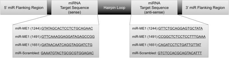
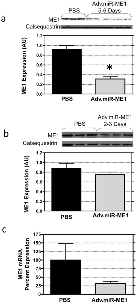
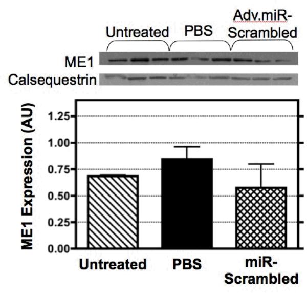
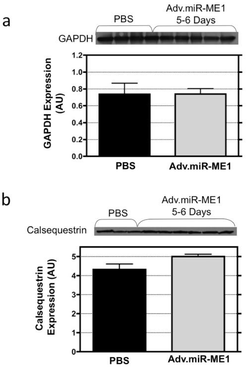
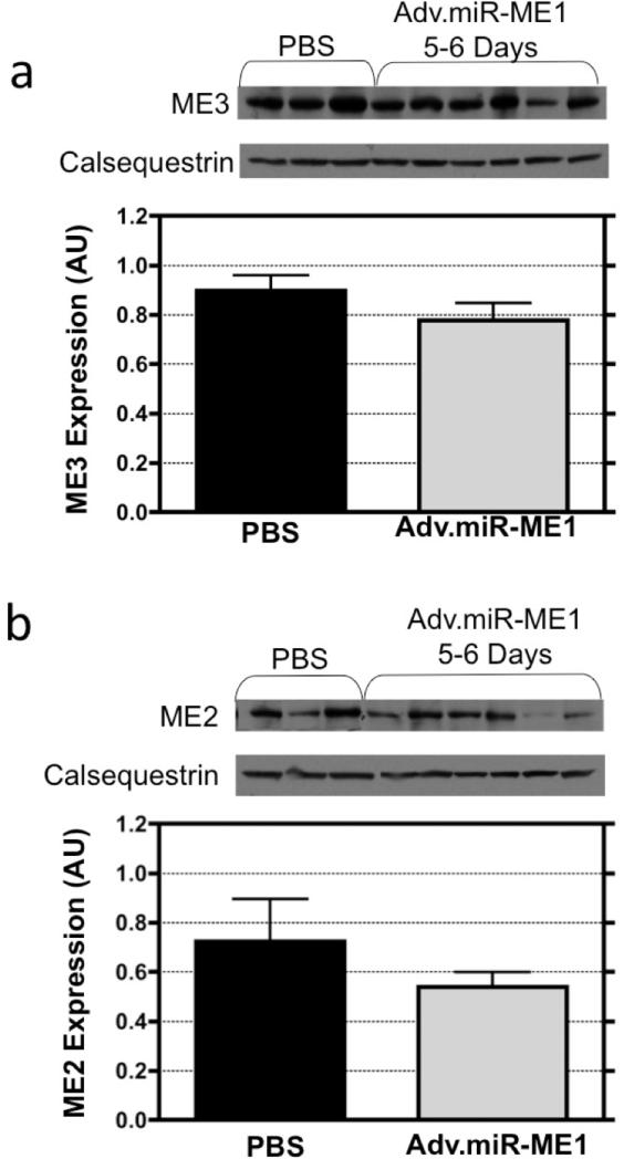
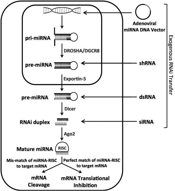
This study examines the feasibility of using the adenoviral delivery of DNA for a non-native microRNA to suppress expression of a target protein (cytosolic NADP(+)-dependent malic-enzyme 1, ME1) in whole heart in vivo, via an isolated-heart coronary perfusion approach. Complementary DNA constructs for ME1 microRNA were inserted into adenoviral vectors. Viral gene transfer to neonatal rat cardiomyocytes yielded 65% suppression of ME1 protein. This viral package was delivered to rat hearts in vivo (Adv.miR_ME1, 10(13) vp/ml PBS) via coronary perfusion, using a cardiac-specific isolation technique. ME1 mRNA was reduced by 73% at 2-6 days post-surgery in heart receiving the Adv.miR_ME1. Importantly, ME1 protein was reduced by 66% (p < 0.0002) at 5-6 days relative to sham-operated control hearts. Non-target protein expression for GAPDH, calsequestrin, and mitochondrial malic enzyme, ME3, were all unchanged. The non-target isoform, ME2, was unchanged at 2-5 days and reduced at day 6. This new approach demonstrates for the first time significant and acute silencing of target RNA translation and protein content in whole heart, in vivo, via non-native microRNA expression.


Figures

Figure 1
Figure 1
The general structure of the miRNA sequences placed into the adenoviral vector include a 5′ and 3′ flanking region, hairpin loop, and the target sequence. Three separate target sequences were designed and examined in this study to target ME1 knockdown. Preliminary studies in isolated neonatal cardiomyocyte indicated sequence #1244 resulted in the greatest knockdown of ME1, and was selected for in vivo studies. A fourth sequence was designed as a scrambled control.
Figure 2
Figure 2
Protein and mRNA expression of ME1 in heart (a) 5-6 days and (b) 2-3 days following delivery of Adv.miR-ME1 to rat heart via isolated-heart retrograde perfusion of the coronaries, in vivo. ME1 content was not significantly reduced at 2-3 days post-treatment (n=3) compared to PBS controls (n=3). However, at 5-6 days post-treatment, ME1 expression was reduced by 65% (n=6, p<0.0002). (c) mRNA content was reduced at both 2-3 days and 5-6 days post Adv.miR-ME1 treatment.
Figure 3
Figure 3
ME1 expression was measured by Western blot analysis for three different control conditions: untreated rat hearts, sham operated control rat hearts retrograde perfused with a bolus of virus-free PBS, and sham operated control rat hearts retrograde perfused with a bolus of PBS containing Adv.miR-scrambled. At 5-6 days post-treatment, the expression of ME1 was not significantly different between control groups.
Figure 4
Figure 4
The expression of the non-target proteins, calsequestrin and GAPDH, were assessed by western blot analysis from heart receiving Adv.miR-ME1 or PBS treatment. At 5-6 day post treatment, calsequestrin and GAPDH protein expression were not different between groups. (The calsequestrin blot shown in panel b is the loading control shown in Figure 2a).
Figure 5
Figure 5
The expression of the non-target mitochondrial isoforms of malic enzyme (a) ME3, a mitochondrial NADP+ protein, and (b) ME2, also a mitochondrial NAD+ protein, were assessed in heart tissue by western blot analysis 5-6 days following Adv.miR-ME1 or PBS treatment. The expression of ME3 was not different between the two treatment groups. ME2 expression was also not statistically different between groups, though the expression was reduced at day 6.
Figure 6
Figure 6
While a variety of approaches can be used to introduce RNAi into the cell, all are processed similarly and incorporate into the RNAi protein machinery. microRNAs start the furthest upstream in the pathway; DNA is taken up into the nucleus and transcribed into the primary miRNA (pri-miRNA) strand which contains the target strand, hairpin loop, and a 5′ and 3′ flanking region. The flanking regions are cleaved off by DROSHA/DGCR8, into the precursor miRNA (pre-miRNA). This structure resembles shRNAs introduced into the nucleus. The pre-miRNA is then exported out of the nucleus by Exportin-5 into the cytoplasm where it is cleaved again into its characteristic 19-23nt structure. Argonaute-2 facilitates unwinding of the duplex, and the functional guide strand is incorporated into the RISC complex. Perfect complementary of the guide strand to the mRNA target leads to mRNA cleavage by RISC. Partial complementary leads to suppression of mRNA translation.

Similar articles

Cited by

References

    1. Gray SJ, Samulski RJ. Optimizing gene delivery vectors for the treatment of heart disease. Expert Opin Biol Ther. 2008;8(7):911–22. - PubMed
    1. Muller OJ, Katus HA, Bekeredjian R. Targeting the heart with gene therapy-optimized gene delivery methods. Cardiovasc Res. 2007;73(3):453–62. - PubMed
    1. Poller W, Fechner H. Development of novel cardiovascular therapeutics from small regulatory RNA molecules—an outline of key requirements. Curr Pharm Des. 2010;16(20):2252–68. - PubMed
    1. Du J, Gao S, Lou J, Zhang G, Cong G, Shao J, Lin T, Cai X, Chang H. Effective inhibition of foot-and-mouth disease virus (FMDV) replication in vitro by vector-delivered microRNAs targeting the 3D gene. Virol J. 2011;8:292–303. - PMC - PubMed
    1. Lv Z, Xu L. Salvianolic acid B inhibits ERK and p38 MAPK signaling in TGF-b1-stumulated human hepatic stellate cell line (LX-2) via distinct pathways. Evid Based Complement Alternat Med. 2012;2012:960128. - PMC - PubMed

Publication types

MeSH terms

Substances