Review
. 2013:75:313-37.
doi: 10.1146/annurev-physiol-030212-183711. Epub 2012 Sep 4.

Functional insights from glutamate receptor ion channel structures

Affiliations
Review

Functional insights from glutamate receptor ion channel structures

Janesh Kumar et al. Annu Rev Physiol. 2013.

Abstract

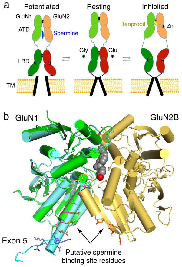
X-ray crystal structures for the soluble amino-terminal and ligand-binding domains of glutamate receptor ion channels, combined with a 3.6-Å-resolution structure of the full-length AMPA receptor GluA2 homotetramer, provide unique insights into the mechanisms of the assembly and function of glutamate receptor ion channels. Increasingly sophisticated biochemical, computational, and electrophysiological experiments are beginning to reveal the mechanism of action of partial agonists and suggest new models for the mechanism of action of allosteric modulators. Newly identified NMDA receptor ligands acting at novel sites offer hope for the development of subtype-selective modulators. The many unresolved issues include the role of the amino-terminal domain in AMPA receptor signaling and the mechanisms by which auxiliary proteins regulate receptor activity. The structural basis for ion permeation and ion channel block also remain areas of uncertainty, and despite substantial progress, molecular dynamics simulations have yet to reveal how glutamate binding opens the ion channel pore.


Figures

Figure 1
Figure 1
Domain organization and mechanism of iGluR activation and desensitization. (a) An iGluR subunit, with extracellular amino terminal (ATD) R1 R2 and ligand binding (LBD) S1 S2 domains interrupted by insertion of the ion channel pore; the highly conserved “SYTAN” motif present on the M3 TM helix is shaded in red. (b) Crystal structure of the full length GluA2 tetramer (3KG2) with each subunit colored differently. The extracellular and ion channel domains are arranged as layers, with subunit crossover taking place at the LBD layer for the B and D subunits. (c) Least squares superpositions of the AC and BD subunit pairs; distinct A/C and B/D subunit conformers result from subunit crossover. (d) Crystal structures of the GluA2 LBD dimer with a cartoon representation of the ion channel in resting (1FTO), glutamate activated (1FTJ) and desensitized (2I3V) states. A 21° rotation of the lower lobe on ligand binding increases separation of the S2 domains by ~5 Å compared to the apo structure, leading to ion channel opening, as shown by the position of glycine residue Cα atoms in the GT linker (red spheres). In the desensitized state, the dimer interface is broken due to a 14° rotation of domain S1, resulting in ~ 13Å separation of the upper lobes, depicted by blue spheres for the Ser741 Cα atoms on the top of helix J, which decouples LBD domain closure from channel opening.
Figure 2
Figure 2
ATD structure and function. (a) Superimposed size exclusion chromatography profiles for the isolated GluR6 and KA2 ATD domains injected separately (dashed lines) or as a mixture of the two proteins at equal concentrations (solid lines); the increase in dimer peak and corresponding decrease in monomer peak indicates formation of GluR6/KA2 ATD heterodimers; red data points show mass values of the eluted peaks estimated by multi angle light scattering. (b) Crystal structure of GluR6/KA2 ATD heterotetramer assembly; two GluR6/KA2 heterodimer pairs are arranged such that the dimer of dimers interface is mediated by the GluR6 subunit (green), while KA2 subunits (red) occupy the lateral edges of the tetramer. (c) Stereoview of the cleft between the R1 and R2 domains of the GluA2 ATD (3HSY) showing an omit map contoured at 7σ revealing a characteristic tetrahedral density in which a phosphate ion present in crystallization condition could be easily modeled along with a well ordered water molecule.
Figure 3
Figure 3
Models for allosteric modulation by NMDA receptor ATD domains. (a) Schematic representation of a model for bidirectional allosteric regulation of NMDA receptors; spermine binding to the lower lobes of the ATD heterodimer interface stabilizes the open channel state, potentiating receptor function, whereas ifenprodil binding to the upper lobe dimer interface leads to channel inhibition. (b) Crystal structure of the GluN1b (exon5 variant) and GluN2B heterodimer complexed with ifenprodil (3QEL); a composite model generated after least-squares superpositions of GluN1 subunit homodimer crystal structures (3QEK) shows the position of Arg and Lys residues inserted due to exon 5 splicing (blue sticks), preceded by Glu and Asp residues which form the putative polyamine binding site.
Figure 4
Figure 4
Partial agonists induce nearly full LBD closure. (a) Plot of mean domain closure for the GluA2 LBD for either WT or disulfide trapping mutants in complex with glutamate, kainate, 5-iodowillardine and CNQX, measured with respect to the apo LBD crystal structure (1FTO); the “error” bars indicate minimum and maximum domain closure values in individual subunits. (b) Least squared superimposition of GluA2 LBD disulfide trapping mutant in complex with CNQX, crystallized in either oxidizing (yellow 3T9U) or reducing conditions (dark brown 3T9V), showing an average cleft closure of ~17° in the oxidizing condition. (c) Stereoview of the oxidized state ligand binding cavities for the glutamate (green 3T93), kainate (cyan 3T9H), 5-iodowillardine (red 3T96) and CNQX (yellow 3T9U) complexes; note that despite similar domain closure the cavity sizes differ to accommodate diverse ligand molecules, and that in the kainate complex the cavity is continuous with bulk solvent; sticks show the location of the A452C and S652C mutant disulfide bond that traps the closed cleft conformation.

References

LITERATURE CITED

    1. Petralia RS, Wang YX, Wenthold RJ. Histological and ultrastructural localization of the kainate receptor subunits, KA2 and GluR6/7, in the rat nervous system using selective antipeptide antibodies. J Comp Neurol. 1994;349:85–110. - PubMed
    1. Matsubara A, Laake JH, Davanger S, Usami S, Ottersen OP. Organization of AMPA receptor subunits at a glutamate synapse: a quantitative immunogold analysis of hair cell synapses in the rat organ of Corti. J Neurosci. 1996;16:4457–67. - PMC - PubMed
    1. Petralia RS, Sans N, Wang YX, Wenthold RJ. Ontogeny of postsynaptic density proteins at glutamatergic synapses. Mol Cell Neurosci. 2005;29:436–52. - PMC - PubMed
    1. Chen X, Winters C, Azzam R, Li X, Galbraith JA, et al. Organization of the core structure of the postsynaptic density. Proc Natl Acad Sci U S A. 2008;105:4453–8. - PMC - PubMed
    1. Traynelis SF, Wollmuth LP, McBain CJ, Menniti FS, Vance KM, et al. Glutamate receptor ion channels: structure, regulation, and function. Pharmacol Rev. 2010;62:405–96. A comprehensive compilation of recent iGluR literature and a good reference resource. - PMC - PubMed

Related articles

    1. Jackson AC, Nicoll RA. The expanding social network of ionotropic glutamate receptors: TARPs and other transmembrane auxiliary subunits. Neuron. 2011;70:178–99. - PMC - PubMed
    1. Straub C, Hunt DL, Yamasaki M, Kim KS, Watanabe M, et al. Distinct functions of kainate receptors in the brain are determined by the auxiliary subunit Neto1. Nat Neurosci. 2011;14:866–73. - PMC - PubMed
    1. Zhang W, St-Gelais F, Grabner CP, Trinidad JC, Sumioka A, et al. A transmembrane accessory subunit that modulates kainate-type glutamate receptors. Neuron. 2009;61:385–96. - PMC - PubMed
    1. Yuzaki M. Cbln1 and its family proteins in synapse formation and maintenance. Curr Opin Neurobiol. 2011;21:215–20. - PubMed
    1. McMahon SA, Diaz E. Mechanisms of excitatory synapse maturation by trans-synaptic organizing complexes. Curr Opin Neurobiol. 2011;21:221–7. - PMC - PubMed

Publication types

MeSH terms

LinkOut - more resources