. 2012 Dec 7:314:57-68.
doi: 10.1016/j.jtbi.2012.08.034. Epub 2012 Sep 6.

A mechanistic compartmental model for total antibody uptake in tumors

Affiliations

A mechanistic compartmental model for total antibody uptake in tumors

Greg M Thurber et al. J Theor Biol. .

Abstract

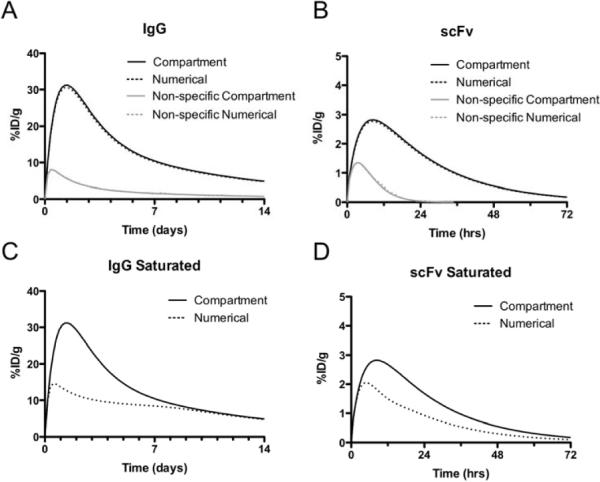
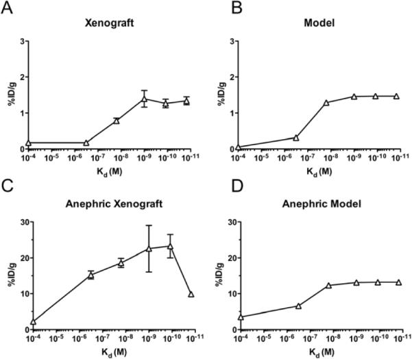
Antibodies are under development to treat a variety of cancers, such as lymphomas, colon, and breast cancer. A major limitation to greater efficacy for this class of drugs is poor distribution in vivo. Localization of antibodies occurs slowly, often in insufficient therapeutic amounts, and distributes heterogeneously throughout the tumor. While the microdistribution around individual vessels is important for many therapies, the total amount of antibody localized in the tumor is paramount for many applications such as imaging, determining the therapeutic index with antibody drug conjugates, and dosing in radioimmunotherapy. With imaging and pretargeted therapeutic strategies, the time course of uptake is critical in determining when to take an image or deliver a secondary reagent. We present here a simple mechanistic model of antibody uptake and retention that captures the major rates that determine the time course of antibody concentration within a tumor including dose, affinity, plasma clearance, target expression, internalization, permeability, and vascularization. Since many of the parameters are known or can be estimated in vitro, this model can approximate the time course of antibody concentration in tumors to aid in experimental design, data interpretation, and strategies to improve localization.


Figures

Figure 1
Figure 1
Diagram of the Compartmental Model and Krogh Cylinder Simulations. A) Arrows indicate transport between compartments. The dotted line surrounds the two-compartment model for normal tissue. B) Diagram of the Krogh cylinder model with the equations and boundary conditions within the tissue.
Figure 2
Figure 2
Low Biot Number Justifies Compartmental Model Effects of Vessel and Tissue Heterogeneity. The ratio of unbound interstitial antibody versus the plasma concentration is shown for a range of Biot numbers (A). A comparison between the current compartmental model and a spatially distributed Krogh cylinder numerical simulation is shown with varying values of the Biot number (B).
Figure 3
Figure 3
Time profiles for specific and non-specific scFv and IgG in mouse Time profiles for specific and non-specific IgGs (A, C) and scFvs (B, D) under subsaturating (A, B) and saturating (C, D) conditions in mice. Profiles are shown for the spatially distributed Krogh cylinder numerical simulation (dotted lines) and the current compartmental model (solid lines).
Figure 4
Figure 4
Effects of Affinity and Clearance on HER2 Binding scFvs. Experimental data from Adams and colleagues (Adams et al., 2001) with anti-ER2 scFvs in normal (A) and anephric (C) mice. Model predictions for both cases (B and D) are shown on the right. The control antibody was modeled as a 100 μM Kd binder.
Figure 5
Figure 5
Comparison to IgG Uptake – Melanoma Xenografts Time Course of Four Different Anti-Melanoma IgGs in Small Xenografts. Experimental data from Shockely and colleagues (Shockley et al., 1992) is shown for four different antibodies in SK-MEL-2 xenografts (A) and M21 xenografts (C). Using parameter values from the literature, the model predictions for these two cell lines are given for the SK-MEL-2 (B) and M21 (D) xenografts.

References

    1. Ackerman M, Chalouni C, Schmidt M, Raman V, Ritter G, Old L, Mellman I, Wittrup K. A33 antigen displays persistent surface expression. Cancer Immunology, Immunotherapy. 2008a - PMC - PubMed
    1. Ackerman ME, Chalouni C, Schmidt MM, Raman VV, Ritter G, Old LJ, Mellman I, Wittrup KD. A33 antigen displays persistent surface expression. Cancer Immunology Immunotherapy. 2008b;57(7):1017–1027. - PMC - PubMed
    1. Ackerman ME, Pawlowski D, Wittrup KD. Effect of antigen turnover rate and expression level on antibody penetration into tumor spheroids. Molecular Cancer Therapeutics. 2008c;7(7):2233–2240. - PMC - PubMed
    1. Adams G, Schier R, McCall A, Simmons H, Horak E, Alpaugh K, Marks J, Weiner L. High Affinity Restricts the Localization and Tumor Penetration of Single-Chain Fv Antibody Molecules. Cancer Research. 2001;61:4750–4755. - PubMed
    1. Ahlstrom H, Christofferson R, Lorelius L. Vascularization of the continuous human colonic cancer cell line LS 174 T deposited subcutaneously in nude rats. APMIS. 1988;96:701–710. - PubMed

Publication types

LinkOut - more resources