. 2012 Sep 11;23(3):600-10.
doi: 10.1016/j.devcel.2012.07.015.

Flow-regulated endothelial S1P receptor-1 signaling sustains vascular development

Affiliations

Flow-regulated endothelial S1P receptor-1 signaling sustains vascular development

Bongnam Jung et al. Dev Cell. .

Abstract

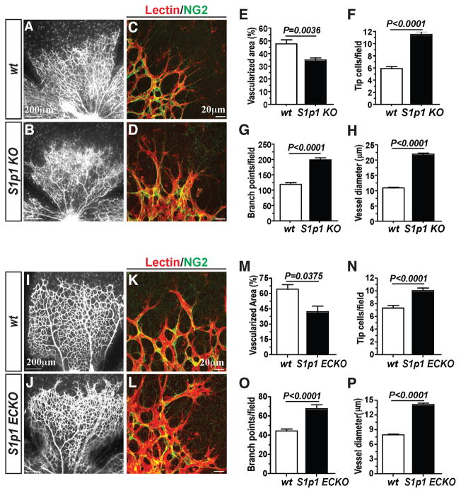
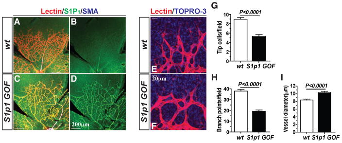
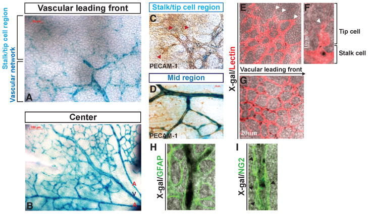
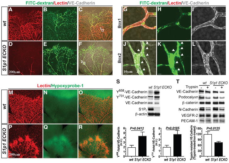
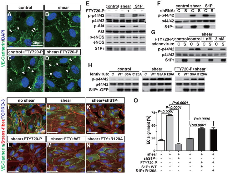
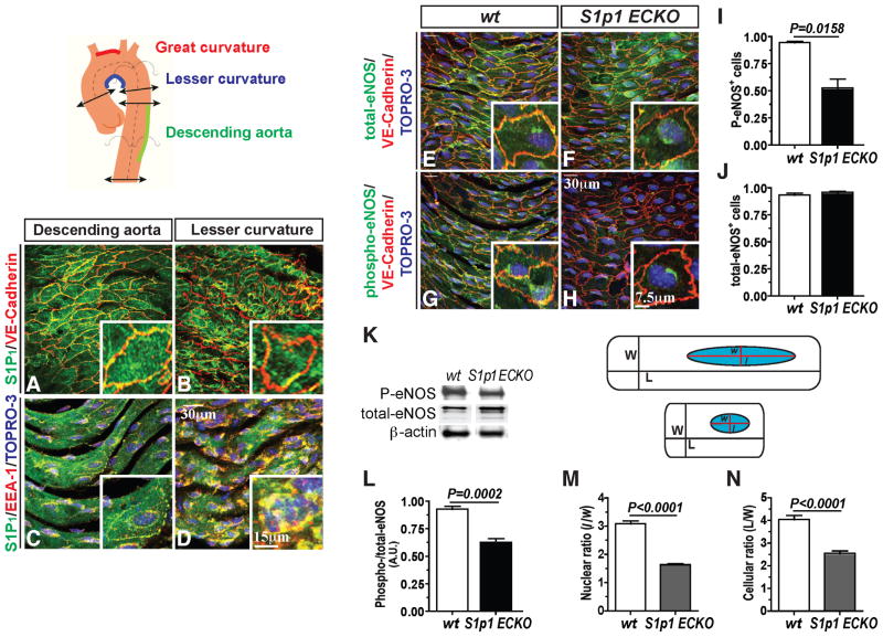
During angiogenesis, nascent vascular sprouts fuse to form vascular networks, enabling efficient circulation. Mechanisms that stabilize the vascular plexus are not well understood. Sphingosine 1-phosphate (S1P) is a blood-borne lipid mediator implicated in the regulation of vascular and immune systems. Here we describe a mechanism by which the G protein-coupled S1P receptor-1 (S1P1) stabilizes the primary vascular network. A gradient of S1P1 expression from the mature regions of the vascular network to the growing vascular front was observed. In the absence of endothelial S1P1, adherens junctions are destabilized, barrier function is breached, and flow is perturbed, resulting in abnormal vascular hypersprouting. Interestingly, S1P1 responds to S1P as well as laminar shear stress to transduce flow-mediated signaling in endothelial cells both in vitro and in vivo. These data demonstrate that blood flow and circulating S1P activate endothelial S1P1 to stabilize blood vessels in development and homeostasis.


Figures

Figure 1
Figure 1. Endothelial S1P1 regulates retinal vascular development
(A–D) Retinas from postnatal day 4 (P4) S1p1 KO animals [WT (n=6) and S1p1 KO (n=7)] were stained in lectin to visualize retinal vasculature. Vascularization (E), tip cell numbers (F), branch points (G), and vessel diameter (H) were quantitated as described in Experimental Procedures. (I–L) EC-specific loss of S1P1 (S1p1 ECKO) resulted in retinal hypersprouting. Reduced vascularization (M), increased tip cell numbers (N), reduced branch points (O), and increased vessel diameter (P) were also observed in S1p1 ECKO animals [WT (n=3) and S1p1 ECKO (n=6)]. See also Figures S1 and S2. All values are mean ± SEM.
Figure 2
Figure 2. S1P1 overexpression (S1p1 GOF) suppresses sprouting angiogenesis
(A–F) Retinal vascular plexus formation in S1p1 GOF (P5) mice. S1P1 immunostaining is shown in B and D. Note that the vessel density and the number of tip cells were markedly reduced upon S1P1 overexpression. (E and F) High power images of the growing vessels at the tip. Quantification results from S1p1 GOF mouse retinas displayed reduced sprouting of tip cells (G), reduced branch points (H) and increased dilation of the vessels (I) [WT (n=4) and S1p1 GOF (n=5)]. See also Figure S1. All values are mean ± SEM.
Figure 3
Figure 3. Gradient of S1P1 expression in the developing retinal vasculature
(A and B) The expression of the S1pr1 locus in the retinal vasculature was examined by X-gal staining using P4 S1pr1LacZ/+ mice (shown in blue), in combination with PECAM-1 (C and D), lectin (E–G), GFAP (H) or NG2 (I) co-staining. Note that high LacZ activity was detected in mature vascular ECs compared to the vascular leading front, and that LacZ expression is restricted to endothelium. Red and white arrowheads indicate tip cells (C, E, and F), and black arrowheads denote NG2+ cells (I). See also Figure S3.
Figure 4
Figure 4. S1P1 is critical for stabilization of the flow-competent vessels
(A–J) Retinas from WT and S1p1 ECKO mice were imaged after intracardiac injection of FITC-Dextran (Mr~ 2,000 kDa) and lectin. High power images of the WT (Box1, G–I) and S1p1 ECKO (Box2, J–L) retinas. Arrowheads indicate sites of vascular leakage. (M–R) Retinal hypoxia was visualized by Hypoxyprobe-1 staining as described in Experimental Procedures. (S) Total lung lysates from WT and S1P1 ECKO animals were analyzed for Y658-, and Y731-VE-cadherin levels by immunoblots [WT (n=5) and S1p1 ECKO (n=5)]. (T) Retina explants were treated with 0.05 % trypsin for 60 min and total protein extracts were analyzed for EC markers. Note that VE-cadherin is more sensitive to trypsin in S1p1 ECKO retinas [WT (n=3) and S1p1 ECKO (n=6)]. See also Figure S4. All values are mean ± SEM.
Figure 5
Figure 5. S1P1 is required for shear stress-induced signaling in EC in vitro
(A–D) HUVEC were pretreated with FTY720-P, and laminar shear (8 dynes/cm2) was applied for 1 hr. Adherens junction formation was visualized by VE-cadherin immunofluorescence staining. Arrowheads indicate intercellular gaps (E–G) ERK, Akt and eNOS activation were examined 10 min after laminar shear application in the presence or the absence of FTY720-P (E), shRNA for S1P1 (F), FTY720-P with adenoviral overexpression of S1P1 (G). (H) Short-term laminar shear was applied to WT, S5A, or R120A mutants and immunoblots for ERK activation were carried out. (I–N) shRNA for S1P1-treated cells, FTY720-P-treated WT or R120A mutant cells underwent long-term laminar shear (12 hr), and EC alignment was examined. Percentage of the aligned cell numbers over the total cell numbers were quantified as described in Experimental Procedures (O). See also Figure S5. All values are mean ± SD.
Figure 6
Figure 6. In vivo requirement for S1P1 in shear stress signaling
(A–D) Expression of S1P1 and VE-cadherin in areas of laminar (descending) and turbulent shear (lesser curvature) in the aorta. Sections of the aorta were dissected and stained for S1P1 and VE-cadherin (A and B) or EEA-1 (C and D) in en face preparations. (E–J) Sections of the aorta were stained for total-eNOS (E and F) and phospho-eNOS (G and H). Quantification of phospho-(I) and total-eNOS (J) levels in WT ad S1p1 ECKO descending aortas is shown [WT (n=4) and S1p1 ECKO (n=4)]. (K and L) Retinal tissues from WT and S1p1 ECKO (P5) were analyzed for phospho-eNOS and total-eNOS levels by immunoblots [WT (n=5) and S1p1 ECKO (n=4)]. (M and N) Quantification of the morphological changes observed in the descending aortic region from WT and S1p1 ECKO mice is shown. [WT (n=4) and S1p1 ECKO (n=4)]. All values are mean ± SEM.

Comment in

References

    1. Allende ML, Yamashita T, Proia RL. G-protein-coupled receptor S1P1 acts within endothelial cells to regulate vascular maturation. Blood. 2003;102:3665–3667. - PubMed
    1. Bankovich AJ, Shiow LR, Cyster JG. CD69 suppresses sphingosine 1-phosophate receptor-1 (S1P1) function through interaction with membrane helix 4. J Biol Chem. 2010;285:22328–22337. - PMC - PubMed
    1. Benedito R, Rocha SF, Woeste M, Zamykal M, Radtke F, Casanovas O, Duarte A, Pytowski B, Adams RH. Notch-dependent VEGFR3 upregulation allows angiogenesis without VEGF-VEGFR2 signalling. Nature. 2012;484:110–114. - PubMed
    1. Butler JM, Kobayashi H, Rafii S. Instructive role of the vascular niche in promoting tumour growth and tissue repair by angiocrine factors. Nat Rev Cancer. 2010;10:138–146. - PMC - PubMed
    1. Carlson CM, Endrizzi BT, Wu J, Ding X, Weinreich MA, Walsh ER, Wani MA, Lingrel JB, Hogquist KA, Jameson SC. Kruppel-like factor 2 regulates thymocyte and T-cell migration. Nature. 2006;442:299–302. - PubMed

Publication types

Substances