. 2012 Sep 11;23(3):624-36.
doi: 10.1016/j.devcel.2012.07.012.

Wnt signaling regulates postembryonic hypothalamic progenitor differentiation

Affiliations

Wnt signaling regulates postembryonic hypothalamic progenitor differentiation

Xu Wang et al. Dev Cell. .

Abstract

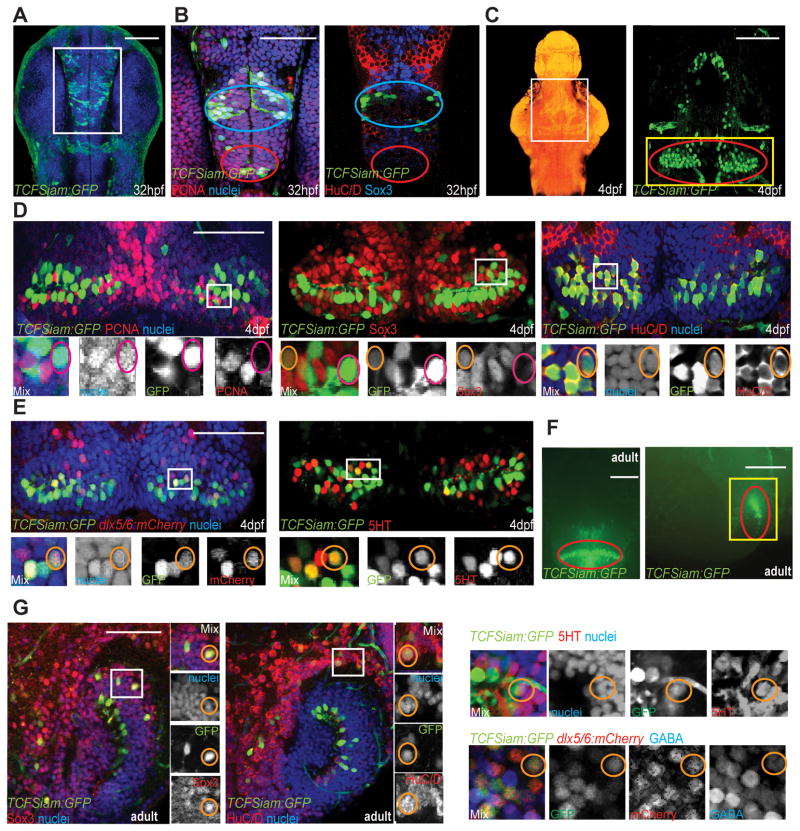
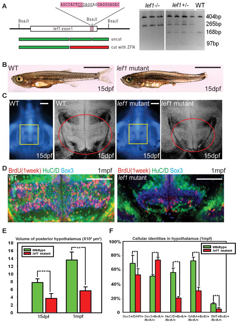
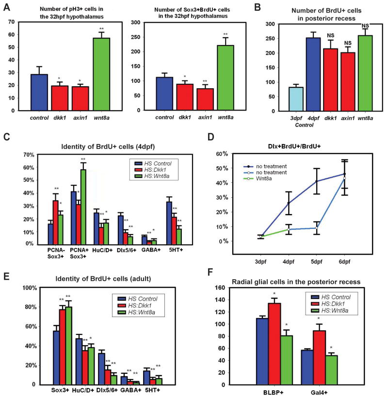
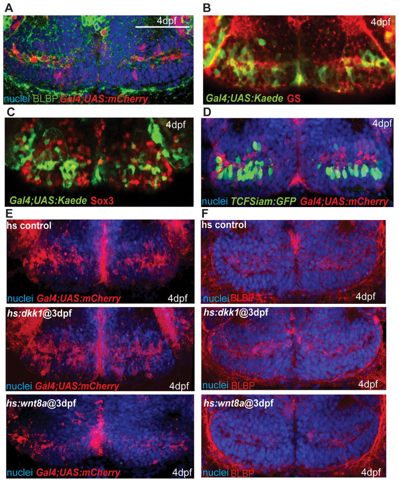
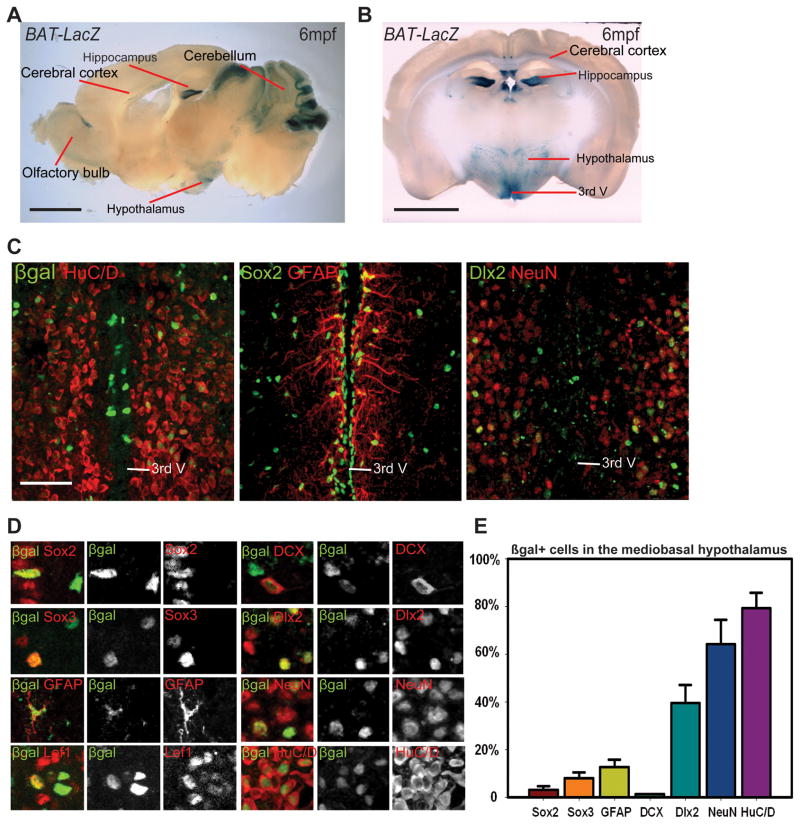
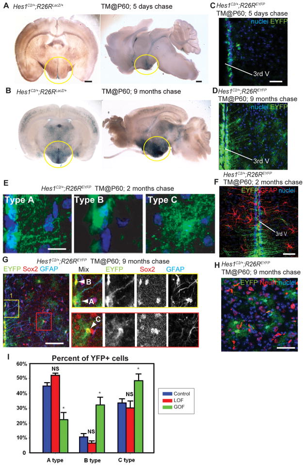
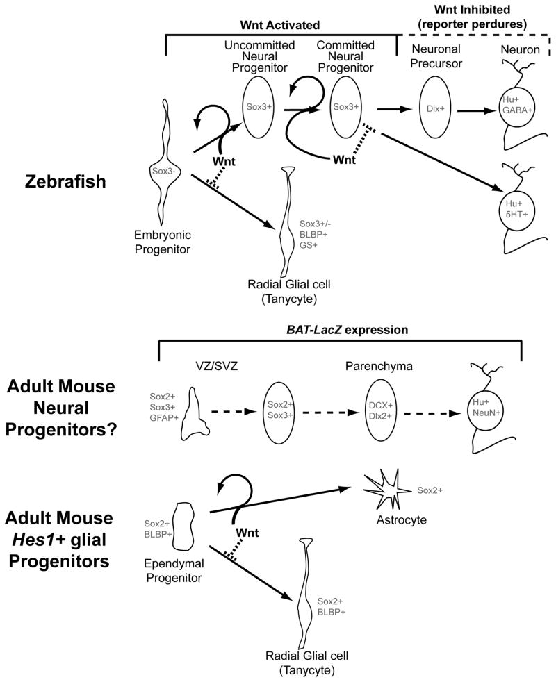
Previous studies have raised the possibility that Wnt signaling may regulate both neural progenitor maintenance and neuronal differentiation within a single population. Here we investigate the role of Wnt/β-catenin activity in the zebrafish hypothalamus and find that the pathway is first required for the proliferation of unspecified hypothalamic progenitors in the embryo. At later stages, including adulthood, sequential activation and inhibition of Wnt activity is required for the differentiation of neural progenitors and negatively regulates radial glia differentiation. The presence of Wnt activity is conserved in hypothalamic progenitors of the adult mouse, where it plays a conserved role in inhibiting the differentiation of radial glia. This study establishes the vertebrate hypothalamus as a model for Wnt-regulated postembryonic neural progenitor differentiation and defines specific roles for Wnt signaling in neurogenesis.


Figures

Figure 1
Figure 1
Identification of Wnt-responsive cells in the zebrafish hypothalamus. (A) Ventral view of TCFSiam:GFP. White box marks the area shown in (B). (B) Co-staining with PCNA, Sox3, and Hu. Blue ovals label the presumptive lateral recess, and red ovals label the presumptive posterior recess. (C) Maximum intensity confocal Z-projection of TCFSiam:GFP at 4dpf. In nuclear stained ventral view of brain on left, white box marks hypothalamic area shown on right. Yellow box marks the area shown in (D, E), and red oval marks the posterior recess. (D) Co-staining of TCFSiam:GFP with PCNA, Sox3, and HuC/D. (E) Co-staining of TCFSiam:GFP with dlx5/6:gfp and serotonin. (F) Dissecting microscope ventral and sagittal views of TCFSiam:GFP in the adult hypothalamus. Yellow box marks the area shown in (G), and red oval labels the presumptive posterior recess. (G) Co-staining of TCFSiam:GFP with Sox3, Hu, 5HT, dlx5/6:mCherry, and GABA in the adult posterior hypothalamus. Single optical sections from ventral views are shown in all panels, unless otherwise indicated. Small orange circles label cells with colocalization, and small magenta circles label cells without colocalization. Scale bars: (A–E, G) 80μm, (F) 250μm. See also Figure S1.
Figure 2
Figure 2
lef1 is required for proliferation and neurogenesis in the post-embryonic hypothalamus. (A) lef1 ZFN target region and genotyping. (B) Whole fish and (C) brain size comparisons of lef1 mutant to wild-type at 15dpf. Boxed region in left panel is enlarged on right, posterior recess is circled. (D) Expression of HuC/D, Sox3, and 7-day BrdU labeling in posterior recess of wild-type and lef1 mutant hypothalamus. (E) Quantification of posterior hypothalamus size. (F) Tracing of proliferating cells. lef1 mutants have a smaller Sox3+ progenitor pool, but a higher percentage of BrdU+ cells express Sox3 and fewer produce HuC/D+,GABA+, or 5HT+ neurons. Single optical sections from ventral views are shown in panels (C) and (D). Scale bars: (B) 2mm. (C) 200μm. (D) 100μm. Brain volumes were calculated using Amira software. Cell counts were collected from maximum intensity Z-projections through the posterior recess of 3 individual samples for each genotype and calculated using Volocity software. *: p<0.05, **: p<0.005. Error=±SD. See also Figure S3.
Figure 3
Figure 3
Wnt signaling regulates hypothalamic progenitor proliferation and differentiation. (A) pH3+ and BrdU+/Sox3+ cell numbers in the 32hpf hypothalamus following 2 hour labeling and Wnt pathway inhibition or activation at 24hpf. (B) BrdU+ cell numbers in the 4dpf posterior recess following 2 hour labeling and Wnt pathway inhibition or activation at 3dpf. (C) BrdU+ cell fates in the 4dpf hypothalamus following labeling and Wnt pathway inhibition or activation at 3dpf. (D) Percentage of BrdU+ cells expressing dlx5/6:gfp following Wnt pathway activation at 3dpf. (E) BrdU+ cell fates in the adult hypothalamus following 2 day labeling and Wnt pathway inhibition or activation for 15 days. (F) Number of Gal4+ and BLBP+ cells in the 4dpf posterior recess following Wnt pathway inhibition or activation at 3dpf. All cell counts were collected from ventral maximum intensity confocal Z-projections through 5 individual brains. The entire hypothalamus was counted at 32hpf, the entire posterior recess was counted at 4dpf, and a hemisphere of the posterior recess was counted in adults. *: p<0.05, **: p<0.005. Error=±SD. See also Figure S4.
Figure 4
Figure 4
Wnt signaling inhibits the formation of radial glia in the posterior recess at 4dpf. (A) Co-expression of mCherry driven by the Gal4 zc1066a insertion with the radial glial marker BLBP. (B) Co-expression of Gal4-driven Kaede with the radial glial marker glutamine synthetase. (C) Co-staining for Sox3 and Gal4-driven Kaede. Most Gal4+ cells have low or absent Sox3 expression. (D) Co-staining for TCFSiam:GFP and Gal4-driven mCherry. Few Gal4+ cells show Wnt reporter activity. (E–F) Gal4 zc1066a-driven mCherry (E) and BLBP (F) expression in the posterior recess of 4dpf embryos following Wnt pathway inhibition or activation at 3dpf. Single ventral confocal optical sections are shown in all panels. Scale bar: 80μm.
Figure 5
Figure 5
The adult mouse hypothalamus has a Wnt-responsive cell population. (A) Sagittal and (B) coronal sections of adult Bat-LacZ mouse brains. (C) β-gal+ cells are distributed in both the ventricular zone (where Sox2+ and GFAP+ cells reside) and the parenchymal zone (where Sox2, HuC/D+, NeuN+, and Dlx2+ cells reside) Coronal 40μm cryosections are shown. (D) Colocalization of β-gal with specific markers in the hypothalamus. (E) Percentage of marker co-expression within the β-gal+ population. Single confocal optical sections are shown in panels (C) and (D). Scale bars: (A–B) 2mm. (C) 80μm. Cell counts were collected from the mediobasal hypothalamus, using six 40μm cryosections each from three mice. Error=±SD.
Figure 6
Figure 6
Wnt signaling inhibits the production of tanycytes from adult Hes1+ progenitors. (A, B) Coronal and sagittal sections of adult Hes1C2/+; R26RLacZ/+ mouse brains, 5 days and 9 months after TM administration at P60. (C, D) Coronal cryosections through the hypothalamus of adult Hes1C2/+; R26REYFP/+ mouse brains, 5 days and 9 months after TM administration at P60. (E) The three types of EYFP+ cells observed at 2 months post TM. (F) Expression of EYFP and GFAP in Hes1C2/+; R26REYFP/+ mice 2 months post TM. (G–H) Marker analysis of Hes1C2/+; R26REYFP/+ mice 9 months post TM. Boxes in (G) show enlarged ventricular (yellow) and parenchymal (red) zones in which cell types are indicated by arrowheads. All EYFP+ cells are Sox2+, and some ventricular Type A cells are also GFAP+. (H) Expression of EYFP and NeuN+ in the parenchymal zone, where no colabeled cells are observed. (I) Percentage of Type A, B, and C EYFP+ cells 2 months following β-catenin inactivation or activation. Single confocal optical sections are shown in panels (C)– (H). Scale bars: (A–B) 1mm. (C, D. F, G, H) 80μm. (E) 10μm. Cell counts were collected from the mediobasal hypothalamus, using six 40μm sections from three mice for each genotype. *: p<0.05. Error=±SD.
Figure 7
Figure 7
Model for the role of Wnt signaling in hypothalamic progenitor differentiation. In zebrafish (above), Wnt signaling is active in unspecified embryonic progenitors and Sox3+ post-embryonic neural progenitors, but is lost as these cells undergo neurogenesis. Wnt signaling promotes mitotic activity in progenitors, and is also required for their ability to undergo neuronal differentiation, which underlies the transition from an “uncommitted” to a “committed” progenitor state. Finally, Wnt signaling must be inhibited for differentiation to proceed. In contrast the formation of radial glia is inhibited by Wnt activity. In the adult mouse (below), Wnt signaling is active in ventricular/subventricular zone cells (VZ/SVZ) that express neural progenitor markers, and in parenchymal zone cells that express neuronal and neuronal precursor markers. Ventricular Hes1+ progenitors do not require Wnt activity to make radial glial tanycytes, and ectopic pathway activity inhibits radial glial formation at the expense of other fates.

Similar articles

Cited by

References

    1. Agathocleous M, Iordanova I, Willardsen MI, Xue XY, Vetter ML, Harris WA, Moore KB. A directional Wnt/beta-catenin-Sox2-proneural pathway regulates the transition from proliferation to differentiation in the Xenopus retina. Development. 2009;136:3289–3299. - PMC - PubMed
    1. Bergsland M, Ramskold D, Zaouter C, Klum S, Sandberg R, Muhr J. Sequentially acting Sox transcription factors in neural lineage development. Genes & development. 2011;25:2453–2464. - PMC - PubMed
    1. Brault V, Moore R, Kutsch S, Ishibashi M, Rowitch DH, McMahon AP, Sommer L, Boussadia O, Kemler R. Inactivation of the beta-catenin gene by Wnt1-Cre-mediated deletion results in dramatic brain malformation and failure of craniofacial development. Development. 2001;128:1253–1264. - PubMed
    1. Chapouton P, Jagasia R, Bally-Cuif L. Adult neurogenesis in non-mammalian vertebrates. Bioessays. 2007;29:745–757. - PubMed
    1. Chapouton P, Webb KJ, Stigloher C, Alunni A, Adolf B, Hesl B, Topp S, Kremmer E, Bally-Cuif L. Expression of hairy/enhancer of split genes in neural progenitors and neurogenesis domains of the adult zebrafish brain. The Journal of comparative neurology. 2011;519:1748–1769. - PubMed

Publication types