. 2012 Sep 11;22(3):318-30.
doi: 10.1016/j.ccr.2012.08.001.

EGF receptor signaling is essential for k-ras oncogene-driven pancreatic ductal adenocarcinoma

Affiliations

EGF receptor signaling is essential for k-ras oncogene-driven pancreatic ductal adenocarcinoma

Carolina Navas et al. Cancer Cell. .

Abstract

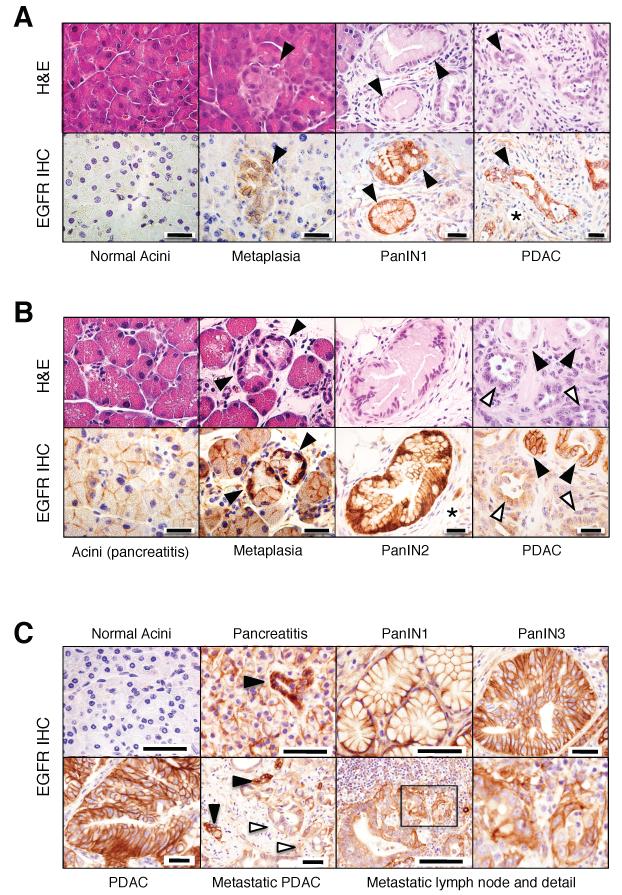
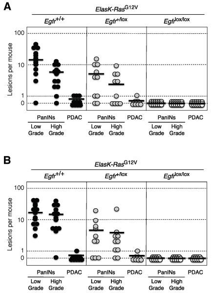
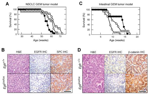
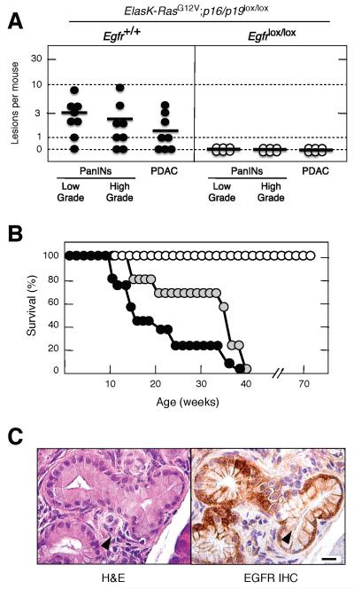
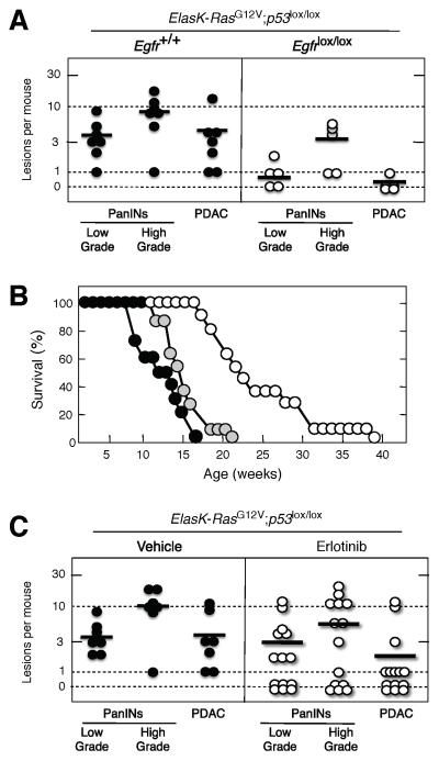
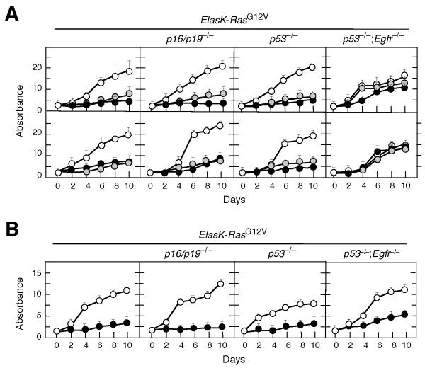
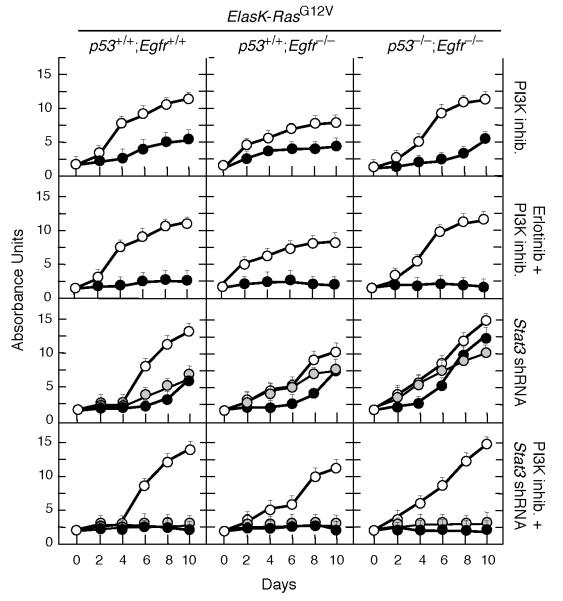
Clinical evidence indicates that mutation/activation of EGF receptors (EGFRs) is mutually exclusive with the presence of K-RAS oncogenes in lung and colon tumors. We have validated these observations using genetically engineered mouse models. However, development of pancreatic ductal adenocarcinomas driven by K-Ras oncogenes are totally dependent on EGFR signaling. Similar results were obtained using human pancreatic tumor cell lines. EGFRs were also essential even in the context of pancreatic injury and absence of p16Ink4a/p19Arf. Only loss of p53 made pancreatic tumors independent of EGFR signaling. Additional inhibition of PI3K and STAT3 effectively prevented proliferation of explants derived from these p53-defective pancreatic tumors. These findings may provide the bases for more rational approaches to treat pancreatic tumors in the clinic.


Figures

Figure 1
Figure 1. Expression of EGFR in pancreas of ElasK-RasG12V mice and of patients with pancreatitis and PDAC
(A) Serial paraffin sections obtained from ElasK-RasG12V mice not exposed to doxycycline depicting normal acini, acinar to ductal metaplasia, PanIN1 and PDAC were stained with hematoxylin and eosin (H&E) or with antibodies against the EGFR (EGFR IHC). Lesions are indicated by solid arrowheads. Asterisk indicates stroma cells positive for EGFR immunostaining. Scale bars represent 20 μm. (B) Serial paraffin sections obtained from ElasK-RasG12V mice exposed to doxycycline from conception to P60 and to caerulein from P90 to P180 depicting acini, acinar to ductal metaplasia, PanIN2 and PDAC were stained with H&E or with antibodies against the EGFR (EGFR IHC). Lesions are indicated by solid arrowheads. Open arrowheads indicate less-differentiated glands within a PDAC. Asterisk indicates stroma cells positive for EGFR expression. Scale bars represent 20 μm. (C) EGFR IHC of human pancreatic biopsies depicting normal pancreata, pancreata from patients with pancreatitis, PanIN lesions (PanIN1 and PanIN3), non metastatic PDAC and a metastatic lymph node with amplified detail. Lesions are indicated by solid arrowheads. Open arrowheads indicate less-differentiated glands within the metastatic PDAC. Scale bars represent 50 μm. See also Figure S1
Figure 2
Figure 2. Induction of PanINs and PDAC tumors by an endogenous K-RasG12V oncogene requires expression of the EGFR
(A) Number of low- and high-grade PanINs and PDACs per mouse in untreated, one year old ElasK-RasG12V mice carrying the indicated Egfr alleles. ElasK-RasG12V;Egfr+/+ (solid circles), ElasK-RasG12V;Egfr+/lox (grey circles) and ElasK-RasG12V;Egfrlox/lox (open circles) mice. In these mice, Cre recombinase-mediated expression of the endogenous K-RasG12V oncogene and ablation of the conditional Egfrlox alleles took place in a percentage (30%) of acinar cells during late embryonic development. (B) Number of low- and high-grade PanINs and PDACs per mouse in 14 month old ElasK-RasG12V mice carrying the indicated Egfr alleles. ElasK-RasG12V;Egfr+/+ (solid circles), ElasK-RasG12V;Egfr+/lox (grey circles) and ElasK-RasG12V;Egfrlox/lox (open circles) mice. These mice were exposed to doxycycline from conception to P60, a time at which Cre recombinase-mediated expression led to the concomitant activation of the resident K-RasG12V oncogene and ablation of the conditional Egfrlox alleles in acinar cells. Mice were subsequently treated with caerulein from P90 to P180. Horizontal bars indicate the average number of lesions per mouse for each genotype. See also Figure S2
Figure 3
Figure 3. Ablation of EGFRs has no effect on K-RasG12V driven lung and intestinal tumors
(A) Survival of RERT;K-RasG12V;Egfr+/+ (solid circles), RERT;K-RasG12V;Egfr+/lox (grey circles) and RERT;K-RasG12V;Egfrlox/lox (open circles) mice treated with a single injection of 4OHT at P21 to induce NSCLCs. (B) H&E staining and EGFR and pro-surfactant protein C (SPC) immunostaining (IHC) of consecutive paraffin sections showing representative adenocarcinoma lesions from nine month old RERTK-RasG12V mice carrying either (top) wild type Egfr or (bottom) conditional Egfr alleles. Scale bar represents 50 μm. (C) Survival of RERT;K-RasG12V;Apclox/lox;Egfr+/+ (solid circles), RERT;K-RasG12V;Apclox/lox;Egfr+/lox (grey circles) and RERT;K-RasG12V;Apclox/lox;Egfrlox/lox (open circles) mice treated with 4OHT (three days per week, for two weeks) at P21 to induce intestinal tumors. (D) H&E staining and EGFR and β-catenin immunostaininig (IHC) of consecutive paraffin sections showing representative intestinal tumor lesions from two month old RERT;K-RasG12V;Apclox/lox mice carrying either (top) wild type Egfr or (bottom) conditional Egfr alleles. Scale bar represents 50 μm.
Figure 4
Figure 4. Loss of p16/p19 tumor suppressors does not abrogate the need for EGFR expression during PanIN and PDAC development
(A) Number of low- and high-grade PanINs and PDAC lesions per mouse in untreated, sixteen week old ElasK-RasG12V;p16/p19lox/lox mice carrying either wild type (solid circles) or conditional (open circles) Egfr alleles. In these mice, expression of a Cre recombinase in pancreatic acinar cells during late embryonic development results in the concomitant expression of the endogenous K-RasG12V oncogene and in the ablation of the conditional p16/p19 and Egfr alleles. PanIN lesions positive for EGFR expression in ElasK-RasG12V;p16/p19lox/lox;Egfrlox/lox mice (see below) were not scored. Horizontal bars indicate the average number of lesions per mouse. (B) Survival of untreated ElasK-RasG12V;p16/p19lox/lox; Egfr+/+ (solid circles), ElasK-RasG12V;p16/p19lox/lox;Egfr+/lox (grey circles) and ElasK-RasG12V;p16/p19lox/lox;Egfrlox/lox (open circles) mice. (C) H&E (left) and EGFR IHC (right) of consecutive paraffin sections showing an occasional PanIN lesion observed in ElasK-RasG12V;p16/p19lox/lox;Egfrlox/lox animals. Scale bar represents 50 μm.
Figure 5
Figure 5. Loss of EGFRs delays but does not prevent PanIN and PDAC development in the absence of p53
(A) Number of low- and high-grade PanINs and PDACs per mouse in untreated, ten week old ElasK-RasG12V;p53lox/lox mice carrying either wild type (solid circles) or conditional (open circles) Egfr alleles. In these mice, expression of a Cre recombinase in pancreatic acinar cells during late embryonic development results in the concomitant expression of the endogenous K-RasG12V oncogene and in the ablation of the conditional p53 and Egfr alleles. Horizontal bars indicate the average number of lesions per mouse. (B) Survival of untreated ElasK-RasG12V;p53lox/lox;Egfr+/+ (solid circles), ElasK-RasG12V;p53lox/lox;Egfr+/lox (grey circles) and ElasK-RasG12V;p53lox/lox;Egfrlox/lox (open circles) mice. (C) Inhibition of EGFR signaling by Erlotinib treatment reduces the number of PanIN and PDAC lesions. Number of low- and high-grade PanINs and PDACs per mouse in six week old ElasK-RasG12V;p53lox/lox mice treated for four weeks with vehicle (solid circles) or Erlotinib (open circles). Horizontal bars indicate the average number of lesions per mouse. The decrease in the number of PDAC tumors in the Erlotinib treated cohort was statistically significant (p<0.05). See also Figure S4
Figure 6
Figure 6. EGFR expression is required for proliferation of PDAC tumor explants in vitro
(A) PDAC cell explants derived from tumors present in mice with the indicated genotypes were infected with lentiviral vectors expressing two independent shRNAs against the Egfr (solid and grey circles) or shRNA control (open circles). Results are the average of two experiments carried out with two independent cell explants done in triplicate. Errors bars mean SD. (B) PDAC cell explants derived from tumors present in mice with the indicated genotypes were either untreated (open circles) or treated with Erlotinib (solid circles). Erlotinib was used at a final concentration of 50 μM, a concentration that corresponds with the average IC90 for these cell explants. Results are the average of two experiments carried out with two independent cell explants done in triplicate. Errors bars mean SD. See also Figure S5
Figure 7
Figure 7. Loss of p53 activates STAT3 and PI3K pathways
PDAC cell explants derived from tumors present in mice with the indicated genotypes were treated with the indicated inhibitor(s) or infected with lentiviral vectors expressing two independent shRNA against Stat3 (solid and grey circles). Control cells were either left untreated or infected with a shRNA control (open circles). Erlotinib was used at a final concentration of 50 μM, a concentration that corresponds with the average IC90 for these cell explants. ETP-46992, a selective PI3Kp110α and p110δ inhibitor, was used at a final concentration of 20 μM, a concentration that corresponds to the average IC90 for these tumor explants. Each graph represents the average of two experiments carried out with two independent cell explants. Each sample was carried out in triplicate. Errors bars mean SD See also Figure S6

Comment in

References

    1. Aguirre AJ, Bardeesy N, Sinha M, Lopez L, Tuveson DA, Horner J, Redston MS, DePinho RA. Activated Kras and Ink4a/Arf deficiency cooperate to produce metastatic pancreatic ductal adenocarcinoma. Genes Dev. 2003;17:3112–3126. - PMC - PubMed
    1. Burris HA, 3rd, Moore MJ, Andersen J, Green MR, Rothenberg ML, Modiano MR, Cripps MC, Portenoy RK, Storniolo AM, Tarassoff P, et al. Improvements in survival and clinical benefit with gemcitabine as first-line therapy for patients with advanced pancreas cancer: a randomized trial. J Clin Oncol. 1997;15:2403–2413. - PubMed
    1. Campbell PJ, Yachida S, Mudie LJ, Stephens PJ, Pleasance ED, Stebbings LA, Morsberger LA, Latimer C, McLaren S, Lin ML, et al. The patterns and dynamics of genomic instability in metastatic pancreatic cancer. Nature. 2010;467:1109–1113. - PMC - PubMed
    1. Collado M, Gil J, Efeyan A, Guerra C, Schuhmacher AJ, Barradas M, Benguría A, Zaballos A, Flores JM, Barbacid M, Beach D, Serrano M. Tumour biology: senescence inpremalignant tumours. Nature. 2005;436:642. - PubMed
    1. Cook N, Frese KK, Bapiro TE, Jacobetz MA, Gopinathan A, Miller JL, Rao SS, Demuth T, Howat WJ, Jodrell DI, Tuveson DA. Gamma secretase inhibition promotes hypoxic necrosis in mouse pancreatic ductal adenocarcinoma. J. Exp. Med. 2012;209:437–444. - PMC - PubMed

Publication types

MeSH terms