Review
. 2012 Dec 3:1487:3-15.
doi: 10.1016/j.brainres.2012.08.042. Epub 2012 Sep 10.

Connexin and pannexin hemichannels in inflammatory responses of glia and neurons

Affiliations
Review

Connexin and pannexin hemichannels in inflammatory responses of glia and neurons

Michael V L Bennett et al. Brain Res. .

Abstract

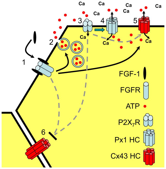
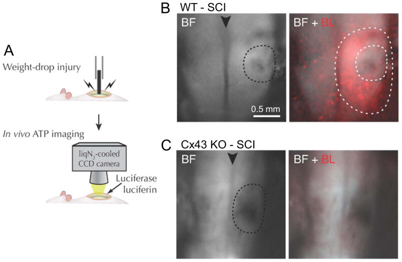
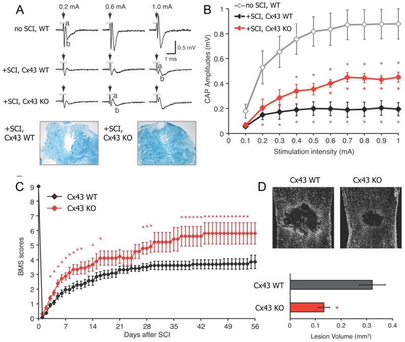
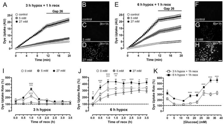
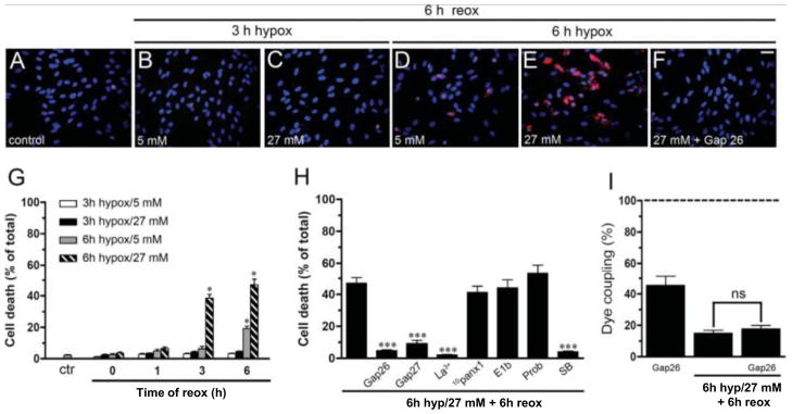
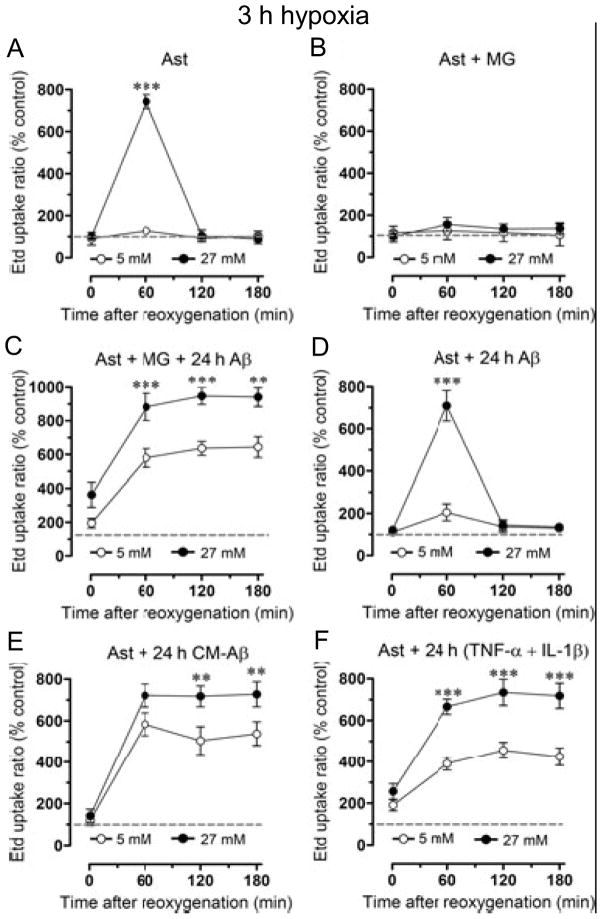
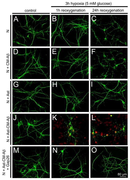
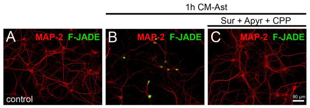
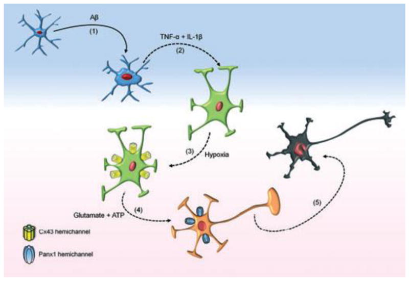
Mammals express ∼20 different connexins, the main gap junction forming proteins in mammals, and 3 pannexins, homologs of innexins, the main gap junction forming proteins in invertebrates. In both classes of gap junction, each channel is formed by two hemichannels, one contributed by each of the coupled cells. There is now general, if not universal, agreement that hemichannels of both classes can open in response to various physiological and pathological stimuli when they are not apposed to another hemichannels and face the external milieu. Connexin (and likely pannexin) hemichannel permeability is consistent with that of the cell-cell channels and open hemichannels can be a release site for relatively large molecules such as ATP and glutamate, which can serve as transmitters between cells. Here we describe three experimental paradigms in which connexin and pannexin hemichannel signaling occurs. (1) In cultures of spinal astrocytes FGF-1 causes the release of ATP, and ATP causes opening of pannexin hemichannels, which then release further ATP. Subsequently, several hours later, connexin hemichannels are also opened by an unknown mechanism. Release of ATP appears to become self sustaining through action of P2X7 receptors to open pannexin hemichannels and then connexin hemichannels, both of which are ATP permeable. (2) Spinal cord injury by dropping a small weight on the exposed cord is followed by release of ATP in the region surrounding the primary lesion. This release is greatly reduced in a mouse in which Cx43 is knocked down in the astrocytes. Application of FGF-1 causes a similar release of ATP in the uninjured spinal cord, and an inhibitor of the FGF-1 receptor, PD173074, inhibits both FGF-1 and injury-induced release. Reduction in ATP release is associated with reduced inflammation and less secondary expansion of the lesion. (3) Cortical astrocytes in culture are permeabilized by hypoxia, and this effect is increased by high or zero glucose. The mechanism of permeabilization is opening of Cx43 hemichannels, which can lead to cell death. Activated microglia secrete TNF-α and IL-1β, which open connexin hemichannels in astrocytes. Astrocytes release ATP and glutamate which can kill neurons in co-culture through activation of neuronal pannexin hemichannels. These studies implicate two kinds of gap junction hemichannel in inflammatory responses and cell death. This article is part of a Special Issue entitled Electrical Synapses.


Figures

Fig. 1
Fig. 1
Proposed reactions initiated by FGF-1 in spinal astrocytes in culture. (1) FGF-1 binds to its (dimeric) receptor, likely causing a rise in cytoplasmic Ca2+. (2) ATP is released from vesicles, an action blocked by BoTN A. (3) The ATP released acts on P2X7Rs, which allows Ca2+ to enter. (4) Activation of P2X7Rs leads to opening of Px1 hemichannels (green arrow), allowing ATP release and Ca2+ (and Etd+) entry. (5) Cx43 hemichannels are opened hours later, either through an action of FGF-1 or of P2X7Rs and Px1 hemichannels. (6) Gap junctional communication is reduced. (From Garré et al., 2010)
Fig. 2
Fig. 2
Weight drop-induced spinal cord injury (SCI): Release of ATP in the perilesion region is reduced in the astrocyte Cx43 KO. A. Schematics of the weight drop mechanism and ATP imaging. Luciferin-luciferase solution is applied to the dorsal surface of the exposed spinal cord. B. Bright field (BF) image of the lesion site (inner dotted line) and merged BF and bioluminescence (BL, in red) images. There is extensive luminescence in the perilesion region between inner and outer dotted lines. C. In the Cx43 KO there is little luminescence around the lesion (dotted line). The arrow head indicates the mid-dorsal vein. Modified from Huang et al., 2012.
Fig. 3
Fig. 3
Recovery of conduction across the lesion and of motor performance after spinal cord injury (SCI) is greater in the Cx43 KO, and lesion volume is reduced. A, B. Compound action potentials propagated across the lesion 7 d after SCI at different stimulus strengths. Also shown in A are Luxol blue stained cross sections of the cord at 7 d post-SCI. In the Cx43 KO, CAPs are greater in amplitude, and there is less disruption of the nerve fibers. D. Recovery over time measured by the Basso Mouse Score (BMS) of motor function is greater in the Cx43 KO. E. Lesion volume is greater 8 wk after injury in WT (n = 8) than the Cx43 KO (n = 6). * p < 0.05. Modified from Huang et al., 2012.
Fig. 4
Fig. 4
Permeabilization of astrocytes after 3 h hypoxia in high or normal glucose in “ischemic saline” followed by reoxygenation in normal glucose in normal saline. Hypoxia–reoxygenation increases rate of Etd+ uptake by rat cortical astrocytes in culture, an effect potentiated by high glucose. A. Time-lapse measurements of Etd+ uptake under control conditions and starting at 1 h reoxygenation after 3 h hypoxia in 5 mM or 27 mM glucose. Gap 26, a Cx43 hemichannel blocker, applied after 12 min of Etd+ uptake measurement greatly reduces uptake after hypoxia in 27 mM glucose. B–D. Fluorescence micrographs of Etd+ uptake (10 min exposure to dye) under control conditions (B) and at 1 h reoxygenation after 3 h hypoxia in 5 mM glucose (C) or 27 mM glucose (D). E. Time-lapse measurements of Etd+ uptake as in A under control conditions and starting at 1 h reoxygenation after 6 h hypoxia in 5 mM or 27 mM glucose. FH. Fluorescence micrographs of Etd+ uptake (10 min exposure to dye) under control conditions (F) and at 1 h reoxygenation after 6 h hypoxia in 5 mM (G) or 27 mM glucose (H). I. Averaged data normalized to control of Etd+ uptake rate measured from the beginning of reoxygenation (time 0) following 3 h hypoxia in 0 mM, 5 mM, or 27 mM glucose. *** P < 0.001, 27 vs. 5 mM glucose; ††† p < 0.001, 27 vs. 0 mM glucose; £££ p < 0.001, 0 vs. 5 mM glucose. (J) Etd+ uptake rate as in I following 6 h hypoxia in 0 mM, 5 mM, or 27 mM glucose. *** p < 0.001, ** p < 0.005, * p < 0.05, 27 vs. 5 mM glucose; ††† p < 0.001, †† p < 0.005, p < 0.05, 27 vs. 0 mM; £ p < 0.05, 0 vs. 5 mM. K. Etd+ uptake rate at 1 h reoxygenation following 3 h or 6 h of hypoxia in different glucose concentrations. *** p < 0.001, * p < 0.05, 27 vs. 0 mM glucose. Each value corresponds to mean ± SE of 20 cells in a representative of five experiments. Bar = 60 μm. From Orellana et al., 2010.
Fig. 5
Fig. 5
Prolonged hypoxia causes death of astrocytes with greater mortality after hypoxia in high glucose. A – F. Micrographs of astrocyte cultures subjected to the indicated periods of hypoxia and concentrations of glucose followed by 6 h reoxygenation in normal glucose. Hoechst 33462 nuclear stain and Rhodamine B dextran are used to identify cells and indicate membrane break down. D. Significant death occurs after 6h hypoxia in 5 mM glucose and 6 h reoxygenation. E. More death occurs when the hypoxia is in 27 mM glucose. F. The connexin hemichannel blocker Gap26 prevents cell death. G. Cell death quantified as a function of duration of hypoxia, glucose concentration, and time of reoxygenation. H. Connexin hemichannel blockers (Gap26, Gap27, and La3+) are protective, as is the p38 MAPK inhibitor, SB202190, applied before the hypoxia. Pannexin hemichannel blockers (10panx1, E1b, and probenecid (Prob)) are not protective. I. The connexin hemichannel blocker, Gap26, applied during the reoxygenation period reduces coupling in control cells, presumably by preventing formation of new cell-cell channels that would compensate for internalization of existing channels. Gap26 did not affect the reduction in dye coupling produced by 6h hypoxia in high glucose followed by 6 h reoxygenation. * p < 0.05 hypoxia/reoxygenation vs. control, *** p < 0.001 hypoxia/reoxygenation without vs. with addition of indicated connexin hemichannel blockers or SB202190. From Orellana et al., 2010.
Fig. 6
Fig. 6
Influence of microglia on hypoxia/reoxygenation-induced permeabilization of astrocytes. A. 3h hypoxia in high (but not normal) glucose transiently increases Etd+ uptake during reoxygenation. B. Co-culture with microglia prevents the increase in Etd+ uptake seen in A. C. Co-culture of astrocytes with microglia (MG) plus Aβ25–35 for 24 h before 3 h hypoxia followed by reoxygenation causes a large and prolonged increase in Etd+ uptake, more so in high than in normal glucose. D. Hypoxia and reoxygenation with Aβ25–35 in the medium has no additional effect compared to hypoxia and reoxygenation alone as in A. E, F. 24 h pretreatment with conditioned medium from microglia + Aβ25–35 or with TNF-α+ IL-1β causes comparable changes to those in 24 h co-cultures with microglia + Aβ25–35 (C). ** p < 0.01, *** p < 0.001 27 vs. 5 mM glucose. From Orellana et al., 2011.
Fig. 7
Fig. 7
Pretreatment with conditioned medium from microglia cultured for 24 h with Aβ25–35 (CM-Aβ) followed by hypoxia-reoxygenation causes dendritic beading and death of neurons in co-culture with astrocytes. Representative confocal micrographs of immunofluorescence of MAP-2 staining (green) and Etd+ uptake (red) by neurons (N) cultured without and with astrocytes (Ast). A – C. Neurons under control conditions or after 3 h hypoxia followed by 1 or 24 h reoxygenation. B. After 3 h hypoxia and 1 h reoxygenation neurons appear normal. C. After 24 h reoxygenation many neurons show beading of neurites, but there is no Etd+ uptake indicating that neuronal membranes are intact. D – F. Neurons incubated for 24 h with CM-Aβ prior to 3 h hypoxia and reoxygenation show beading comparable to that in control medium. G – I. Neuron-astrocyte cocultures show no beading or Etd+ uptake when subject to hypoxia-reoxygenation as in B – C; astrocytes are protective. J – L. After 24 h pretreatment with CM-Aβ followed by 3 h hypoxia, cocultures of astrocytes and neurons show marked Etd+ uptake by astrocytes and by dying neurons at 1 h and 24 h reoxygenation. M – O. Neuronal death and astrocyte Etd+ uptake in cocultures caused by CM-Aβ pretreatment and hypoxia/reoxygenation is prevented by the connexin hemichannel blocker, Gap26, applied during reoxygenation. From Orellana et al., 2011.
Fig. 8
Fig. 8
Neuronal death is induced by exposure to medium conditioned by activated astrocytes subjected to hypoxia/reoxygenation (CM-Ast); death is reduced by inhibition of NMDA and P2X receptors. Representative confocal micrographs of immunofluorescence of MAP-2 (red) to identify neurons and Fluoro Jade (F-Jade, green) to indicate neuronal death. A. Control conditions. B. After exposure to CM-Ast for 1 h many neurons are Fluoro Jade positive. C. Treatment with 200 μM suramin, 10 U/ml apyrase and 20 μM CPP prior to CM-Ast protects the neurons. From Orellana et al., 2011.
Fig. 9
Fig. 9
The inflammatory interactions from activated microglia to astrocytes to neurons. Release of FGF-1 from stressed or dying neurons is not indicated. From Orellana et al., 2011.

Similar articles

Cited by

References

    1. Anderson KM, Tsui P, Guinan P, Rubenstein M. The proliferative response of hela cells to 2-deoxy-D-glucose under hypoxic or anoxic conditions: An analogue for studying some properties of in vivo solid cancers. Anticancer Res. 2006;26:4155–4162. - PubMed
    1. Baranova A, Ivanov D, Petrash N, Pestova A, Skoblov M, Kelmanson I, Shagin D, Nazarenko S, Geraymovych E, Litvin O, Tiunova A, Born TL, Usman N, Staroverov D, Lukyanov S, Panchin Y. The mammalian pannexin family is homologous to the invertebrate innexin gap junction proteins. Genomics. 2004;83:706–16. - PubMed
    1. Basso DM, Fisher LC, Anderson AJ, Jakeman LB, McTigue DM, Popovich PG. Basso Mouse Scale for locomotion detects differences in recovery after spinal cord injury in five common mouse strains. J Neurotrauma. 2006;23:635–659. - PubMed
    1. Bennett MVL, Contreras JE, Bukauskas FF, Saez JC. New roles for astrocytes: Gap junction hemichannels have something to communicate. Trends Neurosci. 2003;26:610–617. - PMC - PubMed
    1. Bondarenko A, Chesler M. Glia. 2001;34:134–142. - PubMed

Publication types

MeSH terms