

Amphiphysin-1 protein level changes associated with tau-mediated neurodegeneration
- PMID: 22975846
- PMCID: PMC3696496
- DOI: 10.1097/WNR.0b013e32835982ce
Amphiphysin-1 protein level changes associated with tau-mediated neurodegeneration
Abstract
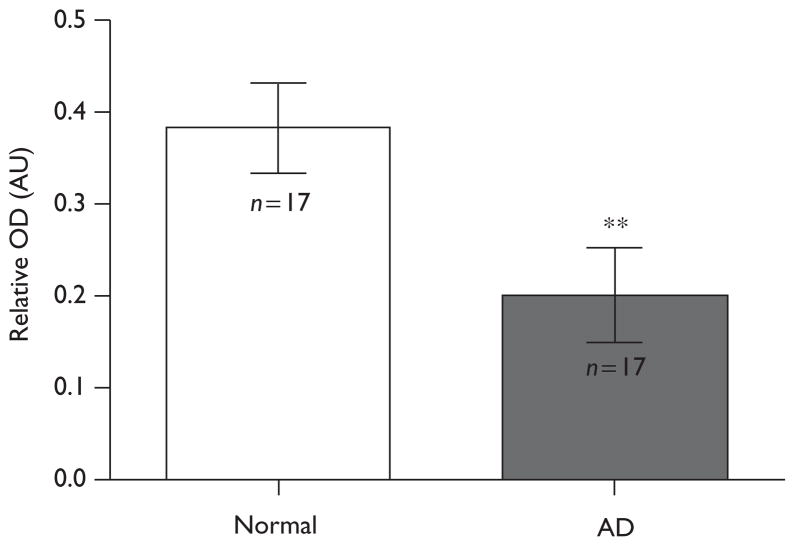
Tauopathies are a family of neurodegenerative diseases that have the pathological hallmark of intraneuronal accumulation of filaments composed of hyperphosphorylated tau proteins that tend to aggregate in an ultrastructure known as neurofibrillary tangles. The identification of mutations on the tau gene in familial cases of tauopathies underscores the pathological role of the tau protein. However, the molecular process that underlines tau-mediated neurodegeneration is not understood. Here, a proteomics approach was used to identify proteins that may be affected during the course of tau-mediated neurodegeneration in the tauopathy mouse model JNPL3. The JNPL3 mice express human tau proteins bearing a P301L mutation, which mimics the neurodegenerative process observed in humans with tauopathy. The results showed that the protein amphiphysin-1 (AMPH1) is significantly reduced in terminally ill JNPL3 mice. Specifically, the AMPH1 protein level is reduced in brain regions known to accumulate aggregates of hyperphosphorylated tau proteins. The AMPH1 protein reduction was validated in Alzheimer's disease cases. Taken together, the results suggest that the reduction of the AMPH1 protein level is a molecular event associated with the progression of tau-mediated neurodegeneration.
Conflict of interest statement
I.E.V. is currently receiving a grant (SC1) from NIH-NINDS, S.E.A. is currently receiving a grant (R01AG039478) from NIH-NIA, and H.D.J. and C.J.N. were supported by training grants from NIH-NIGMS. For the remaining authors there are no conflicts of interest.
Figures


References
-
- Morgan K. The three new pathways leading to Alzheimer’s disease. Neuropathol Appl Neurobiol. 2011;37:353–357. - PubMed
-
- Wu Y, Matsui H, Tomizawa K. Amphiphysin I and regulation of synaptic vesicle endocytosis. Acta Med Okayama. 2009;63:305–323. - PubMed
-
- Lewis J, McGown E, Rockwood J, Melrose H, Nacharaju P, Slegtenhorst MV, et al. Neurofibrillary tangles, amyotrophy and progressive motor disturbance in mice expressing mutant (P301L) tau protein. Nat Genet. 2000;25:402–405. - PubMed
Publication types
MeSH terms
Substances
Grants and funding
LinkOut - more resources
Full Text Sources
