. 2012 Dec 1;125(Pt 23):5887-96.
doi: 10.1242/jcs.113688. Epub 2012 Sep 12.

RIN1 orchestrates the activation of RAB5 GTPases and ABL tyrosine kinases to determine the fate of EGFR

Affiliations

RIN1 orchestrates the activation of RAB5 GTPases and ABL tyrosine kinases to determine the fate of EGFR

Kavitha Balaji et al. J Cell Sci. .

Abstract

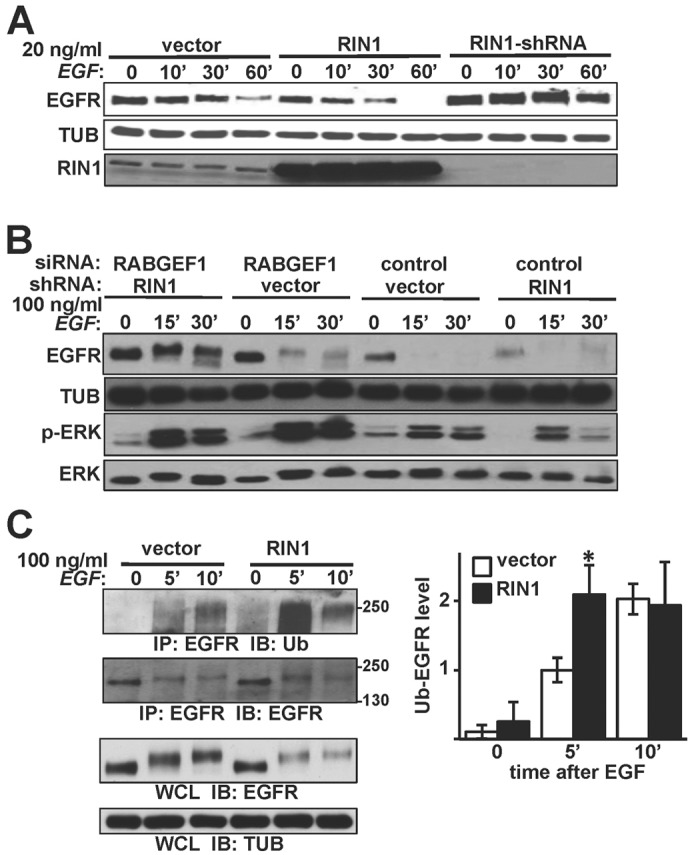
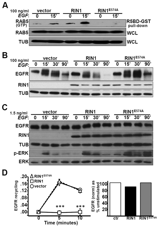
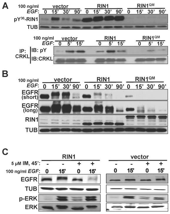
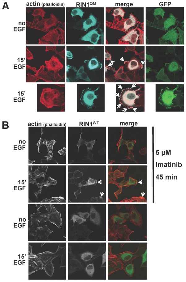
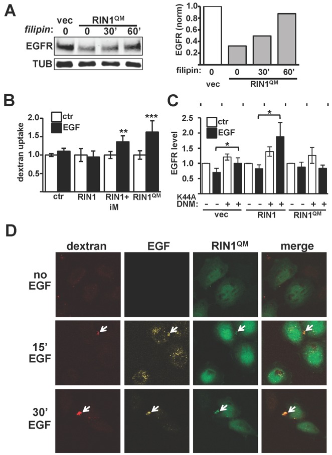
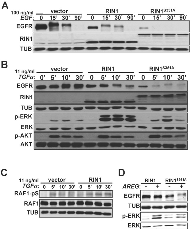
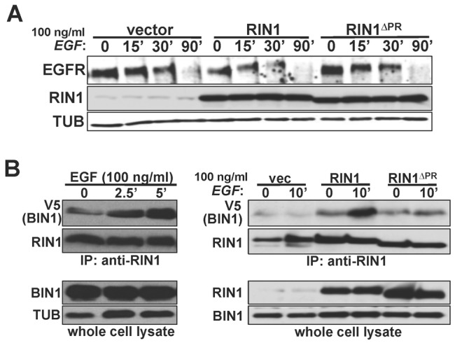
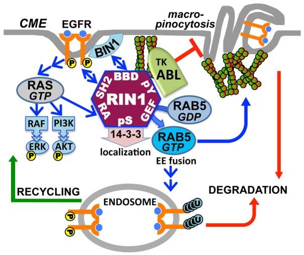
Stimulation of epidermal growth factor receptor (EGFR) initiates RAS signaling simultaneously with EGFR internalization. Endocytosed EGFR is then either recycled or degraded. EGFR fate is determined in part by the RAS effector RIN1, a guanine nucleotide exchange factor (GEF) for RAB5 GTPases. EGFR degradation was slowed by RIN1 silencing, enhanced by RIN1 overexpression and accelerated by RIN1 localization to the plasma membrane. RIN1 also directly activates ABL tyrosine kinases, which regulate actin remodeling, a function not previously connected to endocytosis. We report that RIN1-RAB5 signaling favors EGFR downregulation over EGFR recycling, whereas RIN1-ABL signaling stabilizes EGFR and inhibits macropinocytosis. RIN1(QM), a mutant that blocks ABL activation, caused EGF-stimulated membrane ruffling, actin remodeling, dextran uptake and EGFR degradation. An ABL kinase inhibitor phenocopied these effects in cells overexpressing RIN1. EGFR activation also promotes RIN1 interaction with BIN1, a membrane bending protein. These findings suggest that RIN1 orchestrates RAB5 activation, ABL kinase activation and BIN1 recruitment to determine EGFR fate.


Figures

Fig. 1.
Fig. 1.
RIN1 promotes EGFR degradation after EGF stimulation. (A) HeLa cells stably transduced with vector, RIN1 or RIN1-shRNA were treated with 20 ng/ml EGF for the indicated time (minutes) and lysates immunoblotted for EGFR, RIN1 or α-tubulin (TUB). (B) Vector and RIN1-shRNA cells transfected with control or RABGEF1 siRNA were treated with 100 ng/ml EGF for the indicated time and lysates immunoblotted as indicated. (C) Left: Vector- and RIN1-transduced HeLa cells were stimulated with 100 ng/ml EGF. EGFR immunoprecipitates were blotted for EGFR or ubiquitin. Whole cell lysates (WCL) were immunoblotted for EGFR and tubulin. Right: Ubiquitin-EGFR levels normalized to total immunoprecipitated EGFR using NIH ImageJ. Mean ± s.d. of three experiments; *P<0.05. EGFR activation was confirmed by p-ERK immunoblot in A and C (data not shown).
Fig. 2.
Fig. 2.
The RIN1→RAB5 signal pathway favors EGFR downregulation. (A) HeLa cells stably transduced with vector, RIN1 or RIN1E574A were treated with 100 ng/ml EGF for 0 or 15 minutes. Active RAB5 was isolated using a RAB5 binding domain fused to GST (R5BD-GST). Pull-down material and whole cell lysate (WCL) were immunoblotted with anti-RAB5 to quantify RAB5(GTP) and total RAB5, respectively. (B) Transduced HeLa cells (as in A) were stimulated with 100 ng/ml EGF for the time indicated and lysates immunoblotted for EGFR and RIN1. (C) Transduced HeLa cells (as in A) were stimulated with 1.5 ng/ml EGF for the time indicated and lysates immunoblotted for EGFR, RIN1, ERK1/2 and p-ERK1/2. (D) Left: Transduced HeLa cells were pulsed with 10 ng/ml EGF-Alexa Fluor 647 for 5 minutes. Cells were acid-stripped and chased in serum-free medium at 37°C for the time indicated. The data represent fluorescence of acid-stripped recycled EGF-AF647 normalized to bound EGF at time 0 (data from two independent experiments, each performed in duplicate); ***P<0.0001. Right: Total EGFR in transduced HeLa cells stimulated with 10 ng/ml EGF for 10 minutes. Final EGFR levels (normalized to tubulin) are shown as a percentage of starting EGFR (unstimulated). EGFR activation was confirmed by p-ERK immunoblot in all experiments (data not shown).
Fig. 3.
Fig. 3.
EGF activates RIN1→ABL signaling to regulate EGFR downregulation. (A) Top: HeLa cells transduced with vector, RIN1 or RIN1QM were stimulated with 100 ng/ml EGF for the indicated times, lysated and immunoblotted with anti-pY36-RIN1. Bottom: Cells were treated as shown and lysates subjected to anti-CRKL immunoprecipitation. After immunoblotting this material with pY36, the blot was stripped and reprobed with anti-CRKL. (B) Transduced HeLa cells were EGF-stimulated (as in A) and lysates immunoblotted. Two exposures of the EGFR blot are shown. (C) HeLa cells stably transduced with vector or RIN1 were pre-treated (or not) with 5 µM imatinib and stimulated (or not) with EGF. Cell lysates were immunoblotted for EGFR, ERK1/2, p-ERK1/2 and tubulin (TUB). EGFR activation was confirmed by p-ERK immunoblot in A, B and C (data not shown).
Fig. 4.
Fig. 4.
Strong RAB5 activation without ABL activation causes EGF-induced membrane ruffles. (A) HeLa cells transduced with RIN1QM were stimulated or not with 100 ng/ml EGF for 15 minutes. Fixed and permeabilized cells were stained with phalloidin (red) and anti-RIN1 (light blue). Arrows point to membrane ruffles. Untransfected cells in the same field (no anti-RIN1 or GFP signal) serve as internal controls. (B) RIN1-transduced HeLa cells were pre-treated, or not, with 5 µM imatinib for 45 minutes. and stimulated, or not, with 100 ng/ml EGF for 15 minutes. Fixed and permeabilized cells were stained with phalloidin (red) or anti-RIN1 (green). Arrows point to membrane ruffles.
Fig. 5.
Fig. 5.
Enhanced RAB5 signaling without ABL signaling induces macropinocytosis. (A) Left: HeLa cell transduced with vector or RIN1QM were treated, or not, with 5 µg/ml filipin for the indicated time and lysates probed with anti-EGFR. Right: EGFR levels normalized to TUB. (B) Fluorescent dextran uptake in transduced HeLa cells; iM indicates pre-treated with 5 µM imatinib for 45 minutes. Other samples were mock-treated. Values are means ± s.d. of 5–7 data points. (C) HeLa cells transduced with vector, RIN1 or RIN1QM were transfected with control vector (−) or dominant-negative dynamin (K44A-DNM). EGFR levels, normalized to TUB, were determined for control and EGF-treated (20 ng/ml, 60 minutes) cells. Values are means ± s.d. of three experiments. (D) RIN1QM-transduced HeLa cells were pulsed with 20 ng/ml EGF and 1 mg/ml dextran for 5 minutes and chased in serum-free medium for the indicated time. RIN1QM-transduced cells are GFP-positive (untransduced cells serve as internal controls). Arrows indicate co-localized dextran and EGF. Red, dextran; yellow, EGF; green, GFP. *P<0.05, **P<0.005, ***P<0.0005.
Fig. 6.
Fig. 6.
Plasma membrane localization of RIN1 promotes EGFR degradation in a ligand-independent manner. (A) HeLa cells transduced with vector, RIN1 or RIN1S351A were stimulated with 100 ng/ml EGF for the indicated times, and cell lysates immunoblotted for EGFR and RIN1. EGFR activation was confirmed by p-ERK immunoblot (data not shown). (B) HeLa cells transduced with vector, RIN1 or RIN1S351A were stimulated with 11 ng/ml TGFα for the indicated times, and lysates immunoblotted for EGFR, RIN1, tubulin, total ERK1/2, p-ERK1/2, total AKT and p-AKT. (C) Transduced HeLa cells were stimulated with 11 ng/ml TGFα for the indicated times, and lysates immunoblotted for total RAF1 and RAF1 phosphorylated at Ser338 (RAF1-pS). (D) HeLa cells transduced with RIN1 or RIN1S351A were transfected with a vector (−) or amphiregulin (AREG) construct (+). Cell lysates prepared 48 hours later were immunoblotted for EGFR, tubulin, p-ERK1/2 and total ERK1/2.
Fig. 7.
Fig. 7.
EGF triggers BIN1 recruitment to RIN1. (A) HeLa cells transduced with vector, RIN1 or RIN1ΔPR were stimulated with 100 ng/ml EGF for the time indicated. Cell lysates were immunoblotted for EGFR, RIN1 and tubulin (TUB). (B) Left: RIN1-transduced cells were transfected with BIN1-V5 and stimulated with 100 ng/ml EGF for the time indicated. Anti-RIN1 immunoprecipitates and whole cell lysates were blotted for V5 (BIN1) and RIN1. Right: HeLa cells transduced with vector, RIN1 or RIN1ΔPR were stimulated, or not, with EGF (100 ng/ml, 10 minutes). Anti-RIN1 immunoprecipitates and whole cell lysates were blotted for V5 (BIN1) and RIN1. RIN1ΔPR brought down 60% less BIN1 from unstimulated cells than did wild-type RIN1 (normalized to RIN1 in immunoprecipitate; n = 3; P<0.05). EGFR activation was confirmed by p-ERK immunoblot for A and B (data not shown).
Fig. 8.
Fig. 8.
Model of coordinated RIN1 signal pathways in EGFR endocytosis. Upon EGF stimulation, the RAS effector RIN1 activates RAB5 to drive early endosome fusion and promote EGFR degradation. Concomitant ABL activation regulates actin remodeling to inhibit macropinocytosis, which otherwise results from unchecked RAB5 signaling. Strong ABL activation in the absence of RAB5 activation favors EGFR stability and recycling. Recruitment of BIN1 to the BIN1 binding domain (BBD) facilitates membrane bending and endocytosis, whereas 14-3-3 binding inhibits plasma membrane access and endocytosis.

References

    1. Andoniou C. E., Thien C. B., Langdon W. Y. (1996). The two major sites of cbl tyrosine phosphorylation in abl-transformed cells select the crkL SH2 domain. Oncogene 12, 1981–1989 - PubMed
    1. Bache K. G., Raiborg C., Mehlum A., Stenmark H. (2003). STAM and Hrs are subunits of a multivalent ubiquitin-binding complex on early endosomes. J. Biol. Chem. 278, 12513–12521 10.1074/jbc.M210843200 - DOI - PubMed
    1. Barbieri M. A., Kong C., Chen P. I., Horazdovsky B. F., Stahl P. D. (2003). The SRC homology 2 domain of Rin1 mediates its binding to the epidermal growth factor receptor and regulates receptor endocytosis. J. Biol. Chem. 278, 32027–32036 10.1074/jbc.M304324200 - DOI - PubMed
    1. Barbieri M. A., Fernandez–Pol S., Hunker C., Horazdovsky B. H., Stahl P. D. (2004). Role of Rab5 in EGF receptor-mediated signal transduction. Eur. J. Cell Biol. 83, 305–314 10.1078/0171-9335-00381 - DOI - PubMed
    1. Bauer B., Bartfeld S., Meyer T. F. (2009). H. pylori selectively blocks EGFR endocytosis via the non-receptor kinase c-Abl and CagA. Cell. Microbiol. 11, 156–169 10.1111/j.1462-5822.2008.01246.x - DOI - PubMed

Publication types

MeSH terms

Substances