. 2012 Dec 1;125(Pt 23):5819-29.
doi: 10.1242/jcs.110825. Epub 2012 Sep 12.

The SUMO ligase PIAS1 regulates UV-induced apoptosis by recruiting Daxx to SUMOylated foci

Affiliations

The SUMO ligase PIAS1 regulates UV-induced apoptosis by recruiting Daxx to SUMOylated foci

Raghavi Sudharsan et al. J Cell Sci. .

Abstract

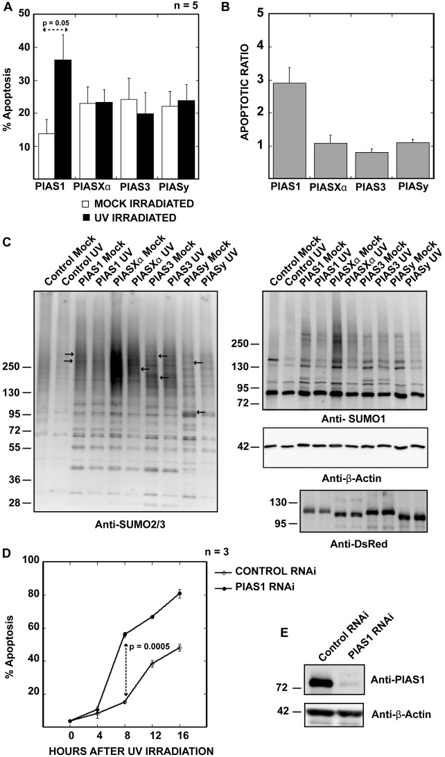
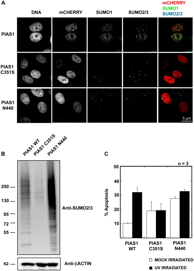
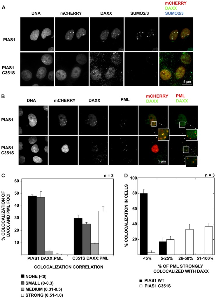
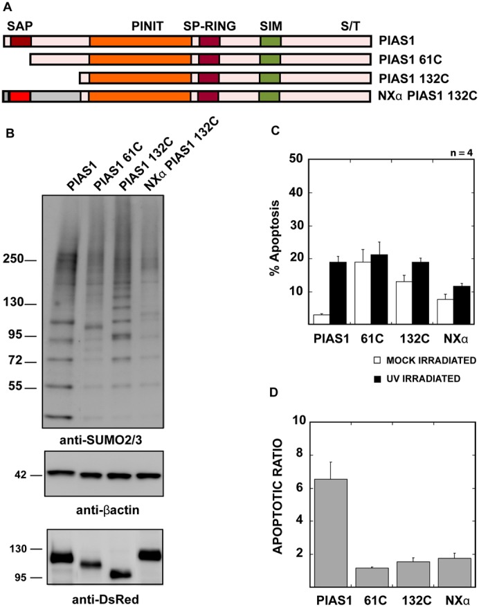
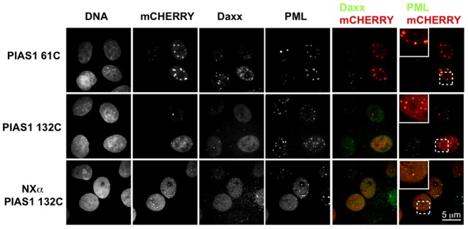
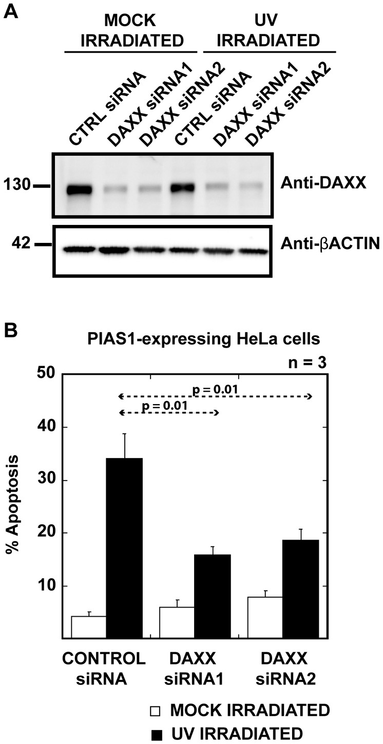
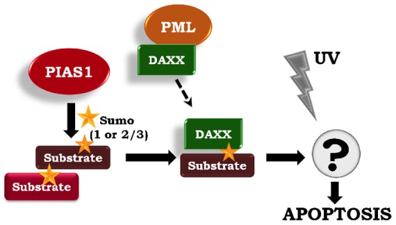
The small ubiquitin-like modifier (SUMO) ligase PIAS1 (Protein Inhibitor of Activated Stat-1) has been shown to play a role in cellular stress response by SUMOylating several proteins that are involved in DNA repair, apoptosis and transcription. In this paper, we show that PIAS1 regulates ultraviolet (UV)-induced apoptosis by recruiting Death-associated protein 6 (Daxx) to PIAS1-generated SUMO-foci. Cells that ectopically express PIAS1, but not other PIASes, show increased sensitivity to UV irradiation, suggesting that PIAS1 has a distinct function in UV-dependent apoptosis. Domain analysis of PIAS1 indicates that both PIAS1 SUMO-ligase activity and the specific localization of PIAS1 through its N-terminal and C-terminal domains are essential for UV-induced cell death. Daxx colocalizes with PIAS1-generated SUMOylated foci, and the reduction of Daxx using RNAi alleviates UV-induced apoptosis in PIAS1-expressing cells. PIAS1-mediated recruitment of Daxx and apoptosis following UV irradiation are dependent upon the Daxx C-terminal SUMO-interacting motif (SIM). Overall, our data suggest that the pro-apoptotic protein Daxx specifically interacts with one or more substrates SUMOylated by PIAS1 and this interaction leads to apoptosis following UV irradiation.


Figures

Fig. 1.
Fig. 1.
PIAS1 overexpression sensitizes HeLa cells to UV irradiation. (A) PIAS1-expressing cells undergo increased UV irradiation-induced apoptosis. HeLa cells were UV irradiated (30 J/m2) or mock irradiated 24 hours after transfection with the PIASes. After a 4-hour recovery period, the numbers of dead and surviving cells were counted based on their nuclear morphologies and Parp1 staining patterns. The plotted values are the average of five independent experiments. The error bars represent 1 SEM. (B) The differences between UV-irradiation induced apoptosis in various PIAS isoform-expressing cells are emphasized by plotting the ratio of the percentage of apoptotic cells after UV irradiation over the percentage of apoptotic cells in mock-irradiated samples, as calculated for A. (C) PIAS SUMO ligases have distinct substrates in cells. HeLa cells expressing mCherry-fused PIASes were sorted from untransfected cells, and SUMOylation was analyzed by immunoblotting using either the anti-SUMO-1 or the anti-SUMO-2/3 antibody. Some of the distinct SUMO-2/3 bands have been indicated by arrows. The anti-DsRed antibody marks the mCherry-tagged PIASes, and anti-β-actin antibody was used as a loading control. (D) PIAS1 RNAi-treated HeLa cells show increased apoptosis after UV irradiation. HeLa cells were treated with PIAS1 siRNA or control siRNA for 48 hours before treatment with 30 J/m2 UV irradiation. Apoptotic cells were counted by Annexin V staining every 4 hours. PIAS1 siRNA-treated HeLa cells show an almost 40% increase in apoptotic cells 8 hours after UV irradiation compared with control siRNA treated cells. The plotted values are an average of three independent experiments, and the error bars represent 1 SEM. (E) PIAS1 RNAi reduces the amount of PIAS1 in HeLa cells to less than 10% of the control RNAi treatment.
Fig. 2.
Fig. 2.
PIAS1's SUMOylation activity is required for UV sensitivity. (A) The ligase-dead mutant (PIAS1 C351S) and the SIM domain truncation mutant (PIAS N440) do not exhibit nuclear punctate localization that is shown by wild-type PIAS1. C-terminal mCherry-tagged PIAS1wt, PIAS1 C351S or PIAS1 N440 were expressed in HeLa cells for 24 hours. Fixed cells were stained with anti-SUMO-1 and anti-SUMO-2/3 antibodies. The images were acquired using a confocal microscope using a 100× objective. Scale bar: 5 µm. (B) Altered SUMOylation profile in PIAS1-mutant-expressing cells. The cells expressing mCherry-tagged PIAS1 C351S and PIAS1 N440 were sorted, and their SUMO-2/3 profile was compared with those of PIAS1-expressing cells by immunoblotting using the anti-SUMO-2/3 antibody. (C) Wild-type PIAS1, but neither PIAS1 C351S nor PIAS1 N440, promotes increased apoptosis upon UV irradiation. The plotted values are an average of three independent experiments. The error bars represent 1 SEM. The percentage of apoptotic cells was determined, as explained in Fig. 1A.
Fig. 3.
Fig. 3.
PIAS1 colocalizes with Daxx in cells and regulates its localization to PML bodies. (A) Daxx colocalizes with the SUMOylated foci in PIAS1-expressing cells, but not in PIAS1 C351S-expressing cells. PIAS1-mCherry and PIAS1 C351S-mCherry fusions were expressed in HeLa cells. The cells were stained with an anti-Daxx antibody 24 hours after transfection. The stained samples were imaged using confocal microscopy with a 100× objective. Scale bar: 5 µm. (B) PIAS1 expression prevents Daxx from colocalizing with PML bodies. PIAS1- or PIAS1 C351S-transfected cells were stained with anti-Daxx and anti-PML antibodies. Daxx colocalizes with PML bodies in PIAS1 C351S-expressing HeLa cells but not in PIAS1-expressing cells. Scale bar: 5 µm. PIAS1-Daxx and PIAS1-PML colocalization is enlarged (inset). (C) Quantitative measurement of colocalization correlation between Daxx and PML in PIAS1 versus PIAS1 C351S expressing cells. PIAS1- or PIAS1 C351S-expressing cells were stained with anti-Daxx and anti-PML antibodies. Over 90 images were acquired from three independent experiments using a confocal microscope. Pearson's correlation between Daxx and PML was calculated in the transfected cells from the images using CellProfiler v2.0 software. (D) A cell-by-cell count of PML and Daxx colocalization. The percentage of PML spots in each transfected nucleus showing a strong (>0.5) Pearson's correlation with Daxx was calculated using the data in (C) and plotted against the percentage of cells showing strong colocalization between PML and Daxx.
Fig. 4.
Fig. 4.
PIAS1 N-terminus SAP domain is essential for correct localization and UV hypersensitivity. (A) Diagrammatic representation of PIAS1 N-terminus truncation and domain swap mutant design. The truncation and the PIASXα domain swap mutants were designed with a C-terminal mCherry fusion, as described in the Materials and Methods section. (B) The PIAS1 N-terminus is required for correct substrate modification. PIAS1wt or truncation/swap mutant-expressing cells were sorted based on mCherry signal and SUMOylation profile was analyzed by immunoblotting with anti-SUMO-2/3 antibody. Anti-DsRed antibody marks the mCherry-tagged PIAS1 or PIAS1 truncation mutants and anti-β-actin antibody was used as a loading control. (C) PIAS1 mutants lacking PIAS1 N-terminus do not increase apoptosis on UV irradiation. The plotted values are an average of four independent experiments. The error bars represent 1 SEM. The percentage of apoptotic cells was determined, as explained in Fig. 1A. (D) The apoptotic ratio was determined as explained in Fig. 1B using the values calculated for C.
Fig. 5.
Fig. 5.
PIAS1 N-terminus mutants localize preferentially to PML-bodies but do not colocalize with Daxx. C-terminal mCherry-tagged PIAS1 61C, 132C and NXα 132C mutants were expressed in HeLa cells for 24 hours. Fixed cells were stained with anti-Daxx and anti-PML antibodies. Images were acquired using a confocal microscope using a 100× objective. Scale bar: 5 µm.
Fig. 6.
Fig. 6.
Daxx depletion alleviates UV sensitivity in PIAS1-expressing cells. (A) Treatment with either siRNA reduced cellular Daxx by nearly 70%. Daxx was depleted from HeLa cells using two different siRNAs against Daxx. mCherry-tagged PIAS1 was transfected into HeLa cells 24 hours after the Daxx siRNA treatment. siRNA-treated HeLa cell extracts were immunoblotted with anti-Daxx antibody. Anti-β-actin antibody was used as a loading control. The intensity of the Daxx signals was measured by Image station 400R (Kodak). (B) The expression of Daxx siRNA, but not the control siRNA, significantly reduced apoptosis in cells expressing PIAS1 after UV treatment. Daxx siRNA-treated, PIAS1-expressing cells were UV irradiated, and the apoptotic cells were counted, as described above, 48 hours after Daxx depletion. The plotted values are an average of three independent experiments. The error bars represent 1 SEM.
Fig. 7.
Fig. 7.
The Daxx C-terminal SIM domain is required for PIAS1-mediated UV hypersensitivity. (A) Diagrammatic representation of Daxx SIM domain truncation mutants. The truncation mutants were designed as described in the Materials and Methods section. The Daxx dNSIM mutant lacks the first 17 amino acids, dCSIM lacks the final 20 amino acids, and the dSIMs mutant lacks both the first 17 amino acids and the last 20 amino acids, thus eliminating both SIM domains from Daxx. All Daxx constructs have an N-terminal Myc-tag. (B) The Daxx C-terminal SIM domain is essential for proper Daxx localization. HeLa cells transfected with the bicistronic vector carrying PIAS1-mCherry and Myc-Daxx or Myc-Daxx truncations. Fixed cells were stained with anti-Myc-tag antibody. The images were acquired using a confocal microscope with a 100× objective. Scale bar: 5 µm. (C) The replacement of endogenous Daxx with the C-terminal SIM-deletion mutants rescues UV-hypersensitivity phenotype in PIAS1-expressing cells. Apoptotic cells were counted as described in Fig. 1A. The plotted values are an average of three independent experiments. The error bars represent 1 SEM. (D) Daxx does not appear to be a major SUMOylation substrate. HeLa cells were transfected with each of the PIAS isoforms. Mock- and UV-treated cells were sorted based on mCherry signal and the lysates were immunoblotted with anti-Daxx antibody. Anti-β-actin was used as a loading control.
Fig. 8.
Fig. 8.
PIAS1-directed SUMO-modified substrates recruit Daxx to mediate UV-hypersensitive apoptosis. Based on our data, we propose a model wherein the ectopic expression of PIAS1 promotes SUMOylation of specific substrates. One or more of these SUMOylated proteins recruit Daxx. The Daxx C-terminal SIM domain is necessary for this interaction. The Daxx-mediated corepression of certain transcription factors may then contribute to apoptosis after UV irradiation.

Similar articles

Cited by

References

    1. Azuma Y., Arnaoutov A., Anan T., Dasso M. (2005). PIASy mediates SUMO-2 conjugation of Topoisomerase-II on mitotic chromosomes. EMBO J. 24, 2172–2182 10.1038/sj.emboj.7600700 - DOI - PMC - PubMed
    1. Carpenter A. E., Jones T. R., Lamprecht M. R., Clarke C., Kang I. H., Friman O., Guertin D. A., Chang J. H., Lindquist R. A., Moffat J.et al. (2006). CellProfiler: image analysis software for identifying and quantifying cell phenotypes. Genome Biol. 7, R100 10.1186/gb-2006-7-10-r100 - DOI - PMC - PubMed
    1. Chang C. C., Naik M. T., Huang Y. S., Jeng J. C., Liao P. H., Kuo H. Y., Ho C. C., Hsieh Y. L., Lin C. H., Huang N. J.et al. (2011). Structural and functional roles of Daxx SIM phosphorylation in SUMO paralog-selective binding and apoptosis modulation. Mol. Cell 42, 62–74 10.1016/j.molcel.2011.02.022 - DOI - PubMed
    1. Chen L. Y., Chen J. D. (2003). Daxx silencing sensitizes cells to multiple apoptotic pathways. Mol. Cell. Biol. 23, 7108–7121 10.1128/MCB.23.20.7108-7121.2003 - DOI - PMC - PubMed
    1. Croxton R., Puto L. A., de Belle I., Thomas M., Torii S., Hanaii F., Cuddy M., Reed J. C. (2006). Daxx represses expression of a subset of antiapoptotic genes regulated by nuclear factor-kappaB. Cancer Res. 66, 9026–9035 10.1158/0008-5472.CAN-06-1047 - DOI - PubMed

Publication types

MeSH terms

Substances